 Research Article
Research of The Effect of Saliva and Hygiene Products for Oral Cavity with the Use of The Indicators of Mineralization of Hard Tooth Tissues of Various Functional Groups
Prikule DV, Alexandrov MT*, Kukushkin VI, Pashkov EP, Achmedov AN and Namiot ED
Scientific and Clinical Center for the Rehabilitation of Women’s Health, Russia
Alexandrov MT, Scientific and Clinical Center for the Rehabilitation of Women’s Health, Russia.
Received Date: March 04, 2019; Published Date: March 28, 2019
Abstract
Aim: In vitro using Raman-fluorescence spectroscopy study the effect of saliva and hygiene products for oral cavity on the mineralization indicators of various anatomical and topographic zones of the teeth for their various functional groups, justify its clinical expediency and effectivity.
Methods: In preclinical In vitro study on 90 model tooth test objects (incisors, premolars and molars) removed according to clinical indications, Raman-Fluorescence study of the degree of mineralization (Raman characteristics spectrum) was conducted and violation of the hygienic condition of the teeth (the presence of plaque and its fluorescence intensity) in various functional groups of teeth was also investigated. For the registration of the studied parameters APK “InSpectr M” with a wavelength probe radiation of 532 nm was used. Advantages of Raman Fluorescent spectroscopy for determining the degree of mineralization and hygienic condition tooth digital tissues are objectivity (digital technology), expressivity, non-invasive, simple and non-destructive degree control of mineralization / demineralization of the selected tooth tissues and its hygienic state, possibility of documenting and storing information (creating a database).
Result: During the study a qualitative and quantitative analysis of the effects of saliva and hygiene products for the oral cavity on mineralization and hygienic condition of various functional groups of teeth was performed.
Keywords: Enamel; Saliva; Mineralization, Anatomical Topographic Area; Hard Tooth Tissue; Oral Hygiene Products; Raman- Fluorescence Spectroscopy; Hygiene Assessment
Introduction
Currently, dental caries is one of the most common dental diseases among children and adults of the Russian Federation. One of local factors of caries occurrence are unsatisfactory hygienic oral care and changes in the quantitative and qualitative composition of the oral fluid [1]. In the system of caries prevention, the leading aspect is oral hygiene. The theoretical justification for the use of oral hygiene with the aim of remineralization in the prevention of caries and mineralization of hard tooth tissues are scientific studies confirming that the most important feature of enamel is permeability, which is ensured by the presence of micro-spaces in it, filled with water, which are able to permeate substances both inorganic and organic nature - depending on their polarity and size. Penetration and the movement of ions in the aqueous phase of the enamel is promoted by osmotic pressure, which is the main mechanism of the process of remineralization and demineralization of solid tooth tissues. In this physiological process, the decisive role is played by the oral fluid (saliva), which is the main source of income of substances in tooth structure [2-4]. However, the unexplored question of how to expressly, simultaneously and without changing the studied structures of the tooth hold the diagnostic measurements of its mineralization and the presence of plaque, and how oral hygiene products (toothpastes and powders of different mechanism of action) and methods of their use affect the mineralization of hard tooth tissues and their hygienic status.
Thus, it is of interest to investigate all three main factors affecting mineralization of hard dental tissues: saliva, hygiene products and methods of application remineralizing tooth powders and pastes. There is practically nothing written about this in the literature. Based on the presented issues, it should be noted that in recent years. There is an increasing interest in clinical and experimental medicine in innovative diagnostic methods to evaluate the mineralization of solid tissues of the tooth and bones, including when exposed to physical, chemical and biological factors at the level of molecular transformations and, most importantly, using the non-invasive way [5]. Such methods include Raman-fluorescent spectroscopy, developed methodologically by prof., laureate of the State Prize of the Russian Federation and Ph.D. Aleksandrov M.T. for scientific and clinical purposes. It is believed that Raman spectroscopy allows you to objectively evaluate spectral bands associated with specific chemical structures of solid tooth tissues [6,7]. Thus, this technology, at present, can be considered the most preferred in the study of the remineralization of teeth [8-14].
In this connection, the purpose of our study was a comprehensive study of the influence saliva, oral hygiene products (toothpastes, toothbrushes) on the indicators of mineralization and hygienic condition of various anatomical and topographic zones of the teeth of various functional groups by the method of Raman-fluorescence spectroscopy.
Summarizing, it should again be noted that the Ramanfluorescent technologies allowing real-time, simultaneous evaluation of both the hygienic condition of the tooth hard tissues and their degree mineralization are not presented in the literature. At the same time, such clinically important aspects as the influence of saliva and oral hygiene products for the studied indicators of hard tooth tissues are yet to study.
Materials and Methods
In the present preclinical In vitro study on 90 model tooth test objects (incisors, premolars and molars) removed according to clinical indications, the effect of saliva and hygiene products for oral cavity as well as acid-forming factor on the mineralization indicators and the presence of plaque of various anatomical and topographic zones of teeth was also studied with the use of Laser hardware-software complex Raman-fluorescent diagnostics “InSpectr M” with a wavelength of probe radiation of 532 nm.
The study was performed on the basis of carrying out preliminary experiments In vitro, where it was objectively revealed that the Raman spectra of the teeth (tooth enamel) were adequate to those of a reference hydroxylapatite sample - the HAP line. This result determined the choice of the presented methodology for solving the set goal and objectives of the study. With the help of AIC “InSpectr M” test - objects (enamel, dentin, cement of the studied teeth) were exposed to low-intensity laser radiation in the visible range of 532 nm. At the same time the received information was assembled and processed. The measurements were carried out in the contact-stable position of the object (tooth) to the radiation source. Each spectral measurement corresponded to the MSR of five hundred measurements, with a duration of one measurement - 100 μs (based on the assembled signal accumulation time required in the experiment for its visualization and measurement). The total time of one measurement corresponded to 2.5-3 minutes. A total of 90 dental test objects were used in the preclinical study, 30 from each functional group (incisors - 30, premolars - 30, molars - 30) removed for clinical reasons.
To quantify the intensity of Raman radiation at a wavelength of hydroxylappatite - 963 cm-1 (in absolute units), indicators were measured at the maximum and minimum of its spectral power. The obtained difference (relative units) was taken as the intensity of Raman, which characterizes the presence and concentration of hydroxylapatite in the enamel, dentin and cement of the studied teeth (M mid.). To quantify the intensity of fluorescence (as a characteristic of the presence of plaque) measured its performance in the maximum.
Colgate Total toothpaste was used as a hygiene product for cleaning the teeth, and fresh lemon juice was used as an acidic factor.
Results
The results are presented in tabular form (Table 1).
Table 1: Effect of saliva, hygiene products and acid-forming drugs on the degree of mineralization of enamel incisors.

From the obtained results it follows that the initial indicators of the intensity of Raman radiation, which characterize mineralization, in the presence of saliva increase and amount to 1,218 abs. units, at the initial level of 645 abs. units. The use of tooth powder in cleaning teeth leads to superficial demineralization (859 abs.).
When organic acids act on the enamel, it dissolves (216 abs. Units). Indicators of plaque increase with prolonged application of saliva (1 day.) and significantly reduce with a similar time application of organic acid almost doubled. Similar results were obtained for premolars, the initial intensity level of Raman radiation was 218 abs. Units, in the presence of saliva it increased (706 abs. Units), with the use of dental powder decreased to 321 abs. Units.
The action of acid caused deep demineralization of enamel (94 abs. Units). Fluorescence indicators had similar changes. For molars, the following results were obtained: the initial level of intensity of Raman radiation of molars was 365 abs. Units, in the presence of saliva it doubled (681 abs. Units), with the use of tooth powder decreased to 194 abs. The action of the acid caused a deep demineralization of enamel (191 abs. Units). In terms of fluorescence, it is the same (Table 2).
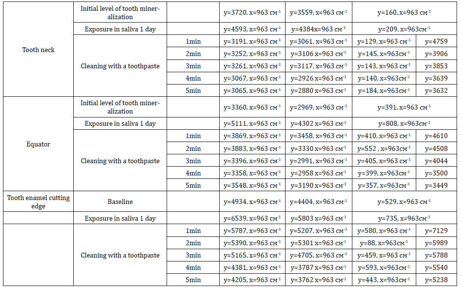
Table 2: Averaged indicators (M mid.) of the spectral characteristics of a tooth for various anatomical and topographic zones under the influence of saliva and hygiene products.

From the table below it follows that the enamel of the tooth (cutting edge) has the greatest mineralization (according to the intensity of the Raman line (y =) at the wavelength (x = 963 cm-1), the tooth neck is the least). It is shown that the application of saliva increases the mineralization of hard tooth tissues and at the same time increases the concentration of dental plaque (according to its fluorescence intensity). And, most importantly, in all cases, the use of a toothbrush and toothpaste leads to significant demineralization of hard tooth tissues (up to 50%). This phenomenon requires the revision of existing technologies for their hygienic processing and the additional use of remineralizing preparations for it to quickly restore the strength characteristics of a tooth (its mineralization).
Discussion
From the presented data it follows that in all groups of teeth (incisors, premolars, molars) there is an increase in the level of mineralization by two and three times in the presence of saliva and is 1218 - 706 - 681 abs. units, at the initial level of 645 - 218 - 365 abs. units The use of toothpaste in cleaning teeth leads to surface demineralization (859 - 321 - 194 abs. Units). When organic acids act on the enamel, its dissolution (demineralization) occurs, which is apparently accompanied by a change in the shape, size and orientation of hydroxyapatite crystals (216 - 94 - 191 abs. Units, which is more than 5 times lower than the level of salinity in the presence of saliva). The reason for the rapid penetration of organic acids into enamel is dislocations in the crystal lattice of the enamel, the so-called vacant places and defects - the absence of atoms or columns of atoms.
When assessing the intensity of fluorescence, which corresponds to the presence of plaque, it is established that the concentration of plaque and fluorescence intensity are dependent, and this dependence is close to linear. In addition, it is shown that when observing the studied parameters during the day, it can be concluded that the plaque does not prevent mineralization in the presence of saliva. In the presence of acid (citric acid), apparently, the mineralization of hard tooth tissues decreases, and microbial growth is suppressed. It also follows from the tables that the fluorescence intensity has increased in the process of cleaning the teeth, which apparently indicates the detection of a microbial plaque under dental plaque. Thus, Raman-fluorescence spectroscopy shows the integrated fluorescence in both plaque and dental plaque.
In general, the clinical significance of the results obtained In vitro explains the negative impact on the degree of tooth mineralization (decreasing): acids, toothpastes and the positive impact (decreasing) of saliva and toothpastes. Thus, the use of toothbrushes apparently does not contribute to mineralization, although they improve the hygienic condition of the oral cavity.
Conclusion
According to the literature, many researchers agree on the need for high-quality daily hygiene [15,16], remineralizing therapy [17], and others. As shown by our study, the use of toothpastes as oral hygiene, leads to a decrease in mineralization on the one hand, on the other hand there is a decrease in the intensity of fluorescence, which indicates the removal of soft dental deposits. The mineralizing potential of saliva also confirms our results. The data obtained indicate that Raman technologies allow express, practically online, “on-site” to assess both the degree of mineralization of hard tooth tissues, and simultaneously detect the presence and degree of activity of plaque, as well as conduct a comparative assessment of the effect of various different physical and biological factors affecting the mineralization / demineralization of teeth. It is shown that at the present time it is necessary to revise the existing technologies of hygienic processing of teeth and the additional use of remineralizing preparations for it in order to quickly restore the tooth’s strength characteristics (its mineralization).
Acknowledgement
None.
Conflict of Interest
Author declares no conflicts of interest.
References
- (2013) Therapeutic dentistry. Diseases of the teeth. Volkova, Yanushevich OO.
- Bezrukov SG, Galkina OP (2014) The prevalence and intensity of dental caries, depending on the functional properties of oral fluid in patients with juvenile rheumatoid arthritis. Modern dentistry 2 (59): 67-68.
- Leontiev VK, Pakhomov GN (2006) Prevention of dental diseases. pp. 416.
- Okushko VR (2008) Fundamentals of tooth physiology. Pp. 240.
- Krasnikova OV, Runova OA, Gordetsov AS (2015) The study of the crystal-chemical composition of tooth tissues by infrared spectroscopy. Collection of articles of the International Scientific and Practical Conference “Regularities and trends in the development of science.” - Ufa pp. 83-86.
- Tramini P, Bonnet B, Sabatier R, Maury L (2001) A method of age estimation using Raman micro spectrometry imaging of the human dentin. Forensic Sci Int 118(1): 1-9.
- Penel G, Leroy G, Rey C, Bres E (1998) Micro-Raman spectral study of the PO4 and CO3 vibrational modes in synthetic and biological apatites. Calcif Tissue Int 63(6): 475-481.
- Miyazaki M, Onose H, Moore BK (2002) Analysis of the dentin–resin interface by use of laser Raman spectroscopy Dent. Mater. 18(8): 576– 580
- ACT Ko, Hewko M, Sowa MG, Dong CC, Cleghorn B, et al. (2008) Early dental caries detection using a fiber-optic coupled polarization-resolved Raman spectroscopic system. Opt Express 16 (9): 6274–6284.
- Ko ACT, Choo Smith LP, Hewko M, Sowa MG, Dong CC et al. (2006) Detection of early dental caries using polarized Raman spectroscopy 14(1): 203-215.
- Shan Yang, Bolan Li, Anna Akkus, Ozan Akkus, Lisa Lang Wide-Field Raman Imaging of Dental Lesions School of Dental Medicine.
- Ionita I (2009) Diagnosis of tooth decay using polarized micro-Raman confocal spectroscopy Rom. Rep Phys 61: 567–574.
- Timchenko EV, Timchenko PE, Volova LT, Yu A Rosenbaum, A Yu (2016) Kulabukhova Jornal of Physies: Conference Series 769 Tooth tissue analysis using Raman spectroscopy.
- Penel G, Delfosse C, Descamps M, Leroy G(2005) Composition of bone and apatite biomaterials as revealed by intravital Raman microspectroscopy. Bone 36(5): 893-901.
- Mikheykina NI (2015) Analysis of indicators of the state of organs and tissues of the oral cavity of caries-resistant and caries-bearing individuals in the dynamics of preventive measures. Bulletin of the East- Siberian Scientific Center of the Siberian Branch of the Russian Academy of Medical Sciences 1(101): 29-33.
- Nagaitseva EA (2016) Oral hygiene as the prevention of dental diseases. International Student Science Journal 2: 41-47.
- Fattal RK, Ammaev MG, Melekhov SV (2015) The effectiveness of modern micro-invasive methods of treatment of initial dental caries depending on the level of oral hygiene of the patient. Dental forum 1(56): 5-8.
-
Prikule DV, Alexandrov MT, Kukushkin VI, Pashkov EP, Achmedov AN, Namiot ED. Research of The Effect of Saliva and Hygiene Products for Oral Cavity with the Use of The Indicators of Mineralization of Hard Tooth Tissues of Various Functional Groups. On J Dent & Oral Health. 1(5): 2019. OJDOH.MS.ID.000525.
-
Dental services, Oral health, Orthodontic, Restorative dentistry, Dentistry, Dental health, Dental treatments, Tooth brushing
-

This work is licensed under a Creative Commons Attribution-NonCommercial 4.0 International License.


