



 Research Article
Pressure-less Acoustic Immittance Measurements in Diagnosis of Otosclerosis
11Department of Otorhinolaryngology, Head and Neck Surgery, KMG Klinikum, Güstrow, Germany
22Department of Audiology, KMG Klinikum, Güstrow, Germany
Blanda Kamin: OCRID-ID: 0009-0006-6072-6630
Moayad Anbari: OCRID-ID: 0009-0003-5565-6437
Dr. Dörte Fischer: OCRID-ID: 0000-0003-3736-4441
Professor Tino Just: OCRID-ID: 0000-0003-2036-8569
Received Date: April 18, 2025; Published Date: May 08, 2025
Abstract
Objective: This preliminary study aims to determine whether pressure-less acoustic immittance (PLAI) measurements can be used to diagnose
otosclerosis (OS).
Methods: A total of 77 participants (40 men and 37 women) from the Department of Otorhinolaryngology, with a mean age of 50.5 ± 15.3 years,
were included in the study. The patient group comprised 10 patients with unilateral otosclerosis (3 men and 7 women) and a mean age of 47.5 ± 9.5
years. The control groups included 56 healthy subjects with bilateral healthy ears (32 men and 24 women), with a mean age of 50.7 ± 16.1 years, and
11 patients with unilateral myringosclerosis (5 men and 6 women) with a mean age of 52.4 ± 15.9 years.
In all patients, OS was confirmed through stapes surgery.
All participants underwent a comprehensive medical evaluation, including an ear, nose, and throat examination. Additionally, pneumatic
otoscopy was performed on all patients with presumed otosclerosis to exclude fixation of the malleus.
All participants underwent pure tone audiometry, pressure-less measurement of acoustic immittance using MedWave® (Neuranix, Italy), and
conventional single-frequency tympanometry.
Results: For all 10 ears with otosclerosis, an average resonance frequency of admittance (RF) of 502.2 Hz ± 25.1 Hz (ranging from 452.2 to 527.6
Hz) and an average peak of admittance of 2.28*10-2 mmho (ranging from 2.06 to 2.61*10-2 mmho) were determined. Overall, 40% of patients with
otosclerosis received a PLAI diagnosis of “normal,” while 60% were diagnosed with “OME/rigid”. For PLAI measures of RF and peak of admittance,
significantly higher values were found in patients with otosclerosis compared with healthy subjects (all p’s < 0.001). Correlational analyses revealed
a significant correlation between resonance frequency measurements and the air-bone gap (r10 = 0.73, p = 0.017). The greater the preoperative
conductive hearing loss, the higher the RF values measured.
Conclusion: PLAI provides reliable information for diagnosing advanced OS. RF and peak of admittance values are higher than those in healthy
controls and in patients with isolated myringosclerosis. However, a significant limitation of this preliminary study is the small number of OS cases
included. Further studies should incorporate all stages of OS as well as pre- and post-operative measurements to assess the PLAI measurements in
OS.
Introduction
Pressure-less acoustic immittance (PLAI) measurements refer to a method that determines the immittance of the outer ear canal and the tympanic membrane [1]. This method is based on microelectro- mechanical system (MEMS) microphone technology, which simultaneously measures the acoustic pressure wave and the corresponding acoustic velocity wave using a probe with two microphones.
A calibrated impedance probe with a known air volume is coupled with the outer ear canal through one end of the probe [1]. The probe sends a broadband exiting sound signal to the auditory canal. The two microphones directly determine the acoustic impedance based on the sound pressure (p) and the speed of the air molecules (v).
From the acoustic impedance or the admittance, all classical tympanometric parameters can be calculated, such as tympanic rigidity and the volume of the outer ear canal. These parameters are derived from the resonance curve obtained through the admittance measured in the outer ear canal. The separation of the air volume inside the outer ear canal from that inside the probe and the energy absorption by the tympanic membrane are obtained from the analysis of the shapes of the resonance curve of the acoustic admittance. Among many parameters, the resonance frequency of the admittance (RF) and the peak of the admittance are provided by PLAI measurements. Currently, the algorithm with the related dashboard does not include the OS data.
In preliminary studies, we demonstrated normative values for both parameters, RF and peak of the admittance, in healthy adult subjects [2, 3]. Some pathologies, such as atrophic tympanic membrane and mesotympanic retraction, revealed a lower RF compared with that of healthy ears. This can be explained by a loss of rigidity of the tympanic membrane. In contrast, calcification of the tympanic membrane and fixation of the ossicular chain led to higher values of RF and peak of admittance, indicating an increase in the rigidity of the tympanic membrane and middle ear.
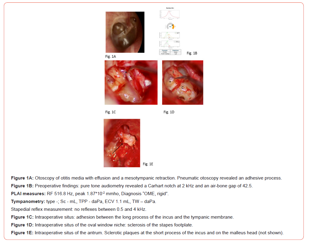
However, in most patients, various pathologies were found, such as tympanic membrane perforation, calcified and/or atrophic changes of the remnants of the tympanic membrane, and disconnection of the ossicular chain combined with fixation of the stapes [1, 2]. The presented values of RF and peak of admittance seem to be the results of many pathologies. Figure 1 demonstrates a complex case with ventilation disorder, otitis media with effusion, tympanosclerosis, and scars resulting in a fixation of the ossicular chain. Therefore, the values and diagnoses provided by the PLAI measuring device need to be interpreted cautiously.
Currently, the question of whether an isolated fixation of the ossicular chain can be detected with PLAI arose. Otosclerosis leads to calcification of the otic capsule [4], primarily affecting the stapes footplate, resulting in conductive or combined, and rarely sensorineural, hearing loss. The tympanic membrane, outer ear canal, and aeration of the middle ear are not affected by this disease. The hypothesis is that PLAI detects otosclerosis via changes in RF and peak of the admittance. If so, PLAI could be used for middle ear diagnosis in patients without a history of ear inflammation and with a normal outer ear canal, an apparently normal tympanic membrane, tympanogram type A, and conductive or combined hearing loss. This preliminary study included 10 patients with an initially presumed diagnosis of otosclerosis and performed audiometric, tympanometric, and PLAI measurements before stapes surgery. The data were compared with those on two age-matched control groups: healthy ears and myringosclerosis.
Material and Methods
Ethical considerations
The study design was approved by the Ethics Committee of the General Medical Council of Mecklenburg-West Pomerania (A2024- 0183). This prospective study was conducted in strict adherence to the revised version of the Helsinki Declaration and is registered at the German Register of Clinical Studies (DRKS 00035288).
Participants
Inclusion criteria for participation were healthy subjects and patients with otosclerosis or myringosclerosis aged 18–90 years. Patients who were younger than 18, had cochlea or middle ear implants, glomus tumors or cognitive disorders, or underwent previous ear surgeries were excluded.
A total of 77 participants (40 men and 37 women) from the Department of Otorhinolaryngology, with a mean age of 50.5 ± 15.3 years, were included. The patient group included 10 individuals with unilateral otosclerosis (3 men and 7 women) and a mean age of 47.5 ± 9.5 years, and the control group comprised 11 patients with unilateral myringosclerosis (5 men and 6 women) and a mean age of 52.4 ± 15.9 years.
Subjects with no history of inflammation or disease affecting the tympanic membrane or middle ear, recent hearing disabilities, aural symptoms and with normal tympanic membranes (without atrophy, scarring, retraction, or perforation) were defined as normal. The healthy control group consisted of 56 healthy subjects with bilateral healthy ears (32 men and 24 women), with a mean age of 50.7 ± 16.1 years.
A total of 133 ears from 56 healthy subjects (112 healthy ears), 10 ears with unilateral otosclerosis, and 11 ears with unilateral myringosclerosis were considered for further analysis.
All subjects were informed about the aims of the study and provided their written consent.
Procedure
Participants, including both patients with otosclerosis or myringosclerosis and healthy subjects, underwent a comprehensive medical evaluation, including an ear, nose, and throat examination. Video otoscopy was performed on all participants, and images and videos were stored. Additionally, pneumatic otoscopy was performed on all patients with presumed otosclerosis to exclude fixation of the malleus.
All participants underwent pure tone audiometry, pressure-less measurement of acoustic immittance using MedWave® (Neuranix, Italy), and conventional single-frequency tympanometry.
PLAI (Pressure-Less Acoustic Immittance)
The end of a MedWave® probe was inserted into the lateral portion of the external ear canal. This test system applied a signal ranging from 100 to 3000 Hz. Evaluating the responses resulted in the calculation of the complex acoustic admittance. MedWave® provides objective numerical values: the frequency at which the maximum value of the admittance curve is reached (Fr [Hz]) and peak admittance value (P [*10-2 mmho]).
Conventional tympanometry
Tympanometry was performed using the Madsen® Zodiac (type 1096; Otometrics) at 226 Hz from -400 to +200 daPa. The following measures were used for calculation: tympanometric peak pressure (TPP), tympanic width (TW), equivalent ear canal volume (ECV), and static peak compliance (SC).
Pure tone audiometry
All audiological assessments were conducted in an audiometric sound-attenuated room, using calibrated signals and equipment according to accepted ISO standards. For assignment of the participants (OS or healthy subject), a pure tone audiometry was performed measuring air conduction (AC) and bone conduction (BC) thresholds. The PTA4 was measured across the frequencies of 0.5, 1, 2, and 4 kHz, and the air-bone gap was calculated.
Statistical Analyses
Statistical tests (Kolmogorov test and Shapiro-Wilk test) indicated non-normal distribution for all parameters, with all p’s < 0.05. Nonparametric tests were performed for group comparisons whenever appropriate. Pearson statistics were used for correlational analyses.
The alpha level was set to 0.05. SPSS version 30.0 (SPSS Inc. Chicago, IL, USA) was used for statistical analyses.
Results
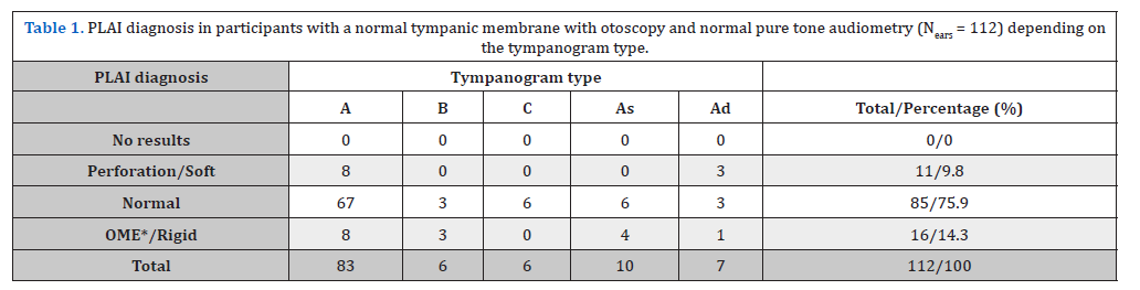
Overall, 112 ears (of 56 healthy subjects with bilateral normal ears) were classified as normal based on otoscopy and pure tone audiometry.
Of these, 83 ears had a type A tympanogram (74%), 6 ears (5%) had a type B tympanogram, and 6 ears (5%) had a type C tympanogram, while 10 (8.9%) and 7 (6%) ears exhibited tympanogram types As and Ad, respectively (Table 1).
Table 1:
OME = otitis media with effusion.
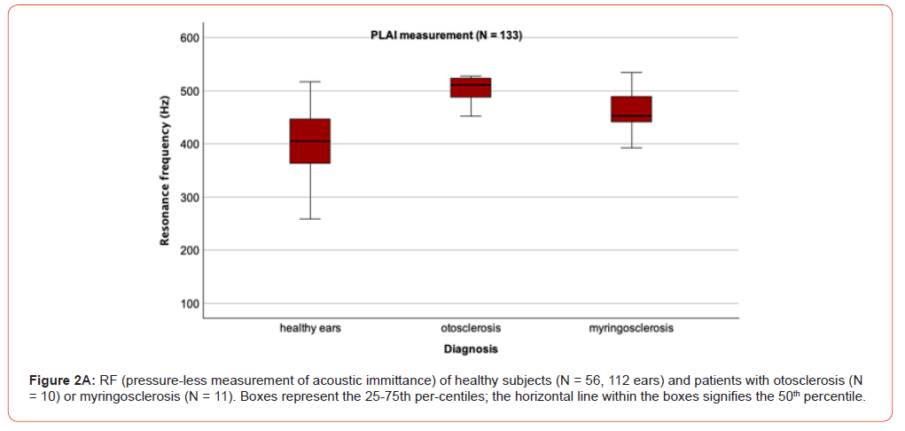
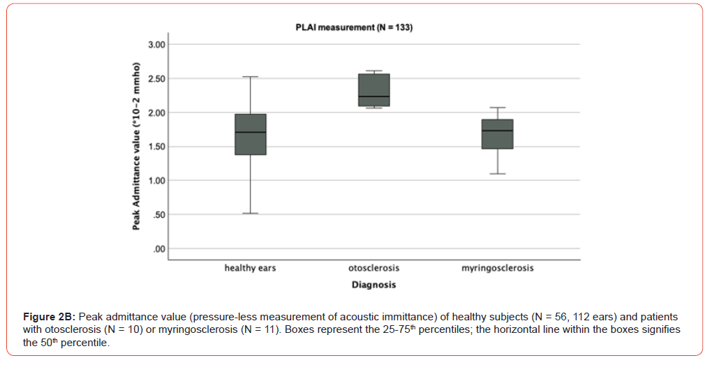
The demographic and audiometric data of patients with otosclerosis are presented in Table 2. Using the PLAI measurements for all 10 ears presenting otosclerosis, an average RF of 502.2 Hz ± 25.1 Hz (ranging from 452.2 to 527.6 Hz) and an average peak of admittance of 2.28*10-2 mmho (ranging from 2.06 to 2.61*10-2 mmho) were determined. Overall, 40% of patients with otosclerosis received a PLAI diagnosis of “normal”, while 60% were diagnosed with “OME/rigid” (Table 3).
Table 2:
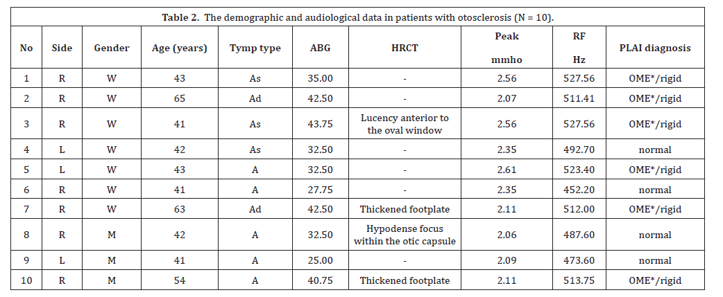
L = left ear; R = right ear; Tymp type = type of tympanogram; ABG = air-bone gap at 0.5, 1, 2, and 4 kHz;
Peak = peak of admittance; RF = resonance frequency of admittance; HRCT = high-resolution computed tomography.
Table 3:
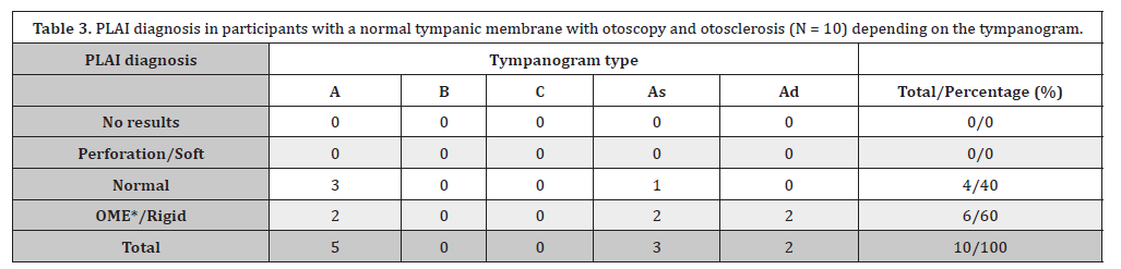
OME = otitis media with effusion.
For PLAI measures of RF and peak of admittance, significantly higher values were found between values of patients with otosclerosis compared with healthy subjects (all p’s < 0.001). Correlational analyses revealed a significant correlation between RF measurements and the air-bone gap (r10 = 0.73, p = 0.017). The greater the preoperative conductive hearing loss, the higher the RF values measured.
Figure 3A presents the CT scan of a 41-year-old woman with right-sided otosclerosis. The clinical correlate was a sclerotic stapes footplate with fixation of the stapes (Figure 3B). The preoperative RF and peak admittance were 527.6 Hz and 2.56*10-2 mmho (Figure 3C), respectively.
Two months after right-sided partial stapedectomy and insertion of a 0.4 x 4.5 mm titanium stapes prosthesis, a nearly complete closure of the air-bone gap was found. The RF and peak admittance decreased to 479.1 Hz and 0.96*10-2 mmho, respectively (Figure 3D). The contralateral ear demonstrated the beginning of otosclerosis, showing a Carhart notch and an air-bone gap of 12.5. The RF values ranged from 452 to 479 Hz (normal diagnosis).
For all 10 ears presenting myringosclerosis the PLAI measurements result in an average RF of 461.7 Hz ± 45.8 Hz (ranging from 393.0 to 534.7 Hz) and an average peak of admittance of 1.68*10-2 mmho (ranging from 1.10 to 2.07*10-2 mmho). Overall, 36% of patients with myringosclerosis received a PLAI diagnosis of “normal”, 54% were diagnosed with “OME/rigid”, and 9% received a diagnosis of “perforation/soft” (Table 4).
For PLAI measures of RF and peak of admittance, significantly higher values were found between values of patients with myringosclerosis compared with healthy subjects (p < 0.05) and for the parameter peak between patients with otosclerosis and myringosclerosis (p < 0.001).
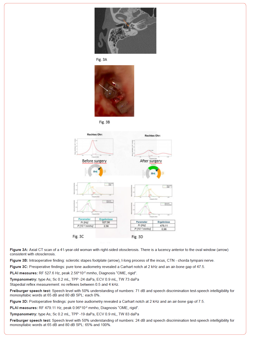
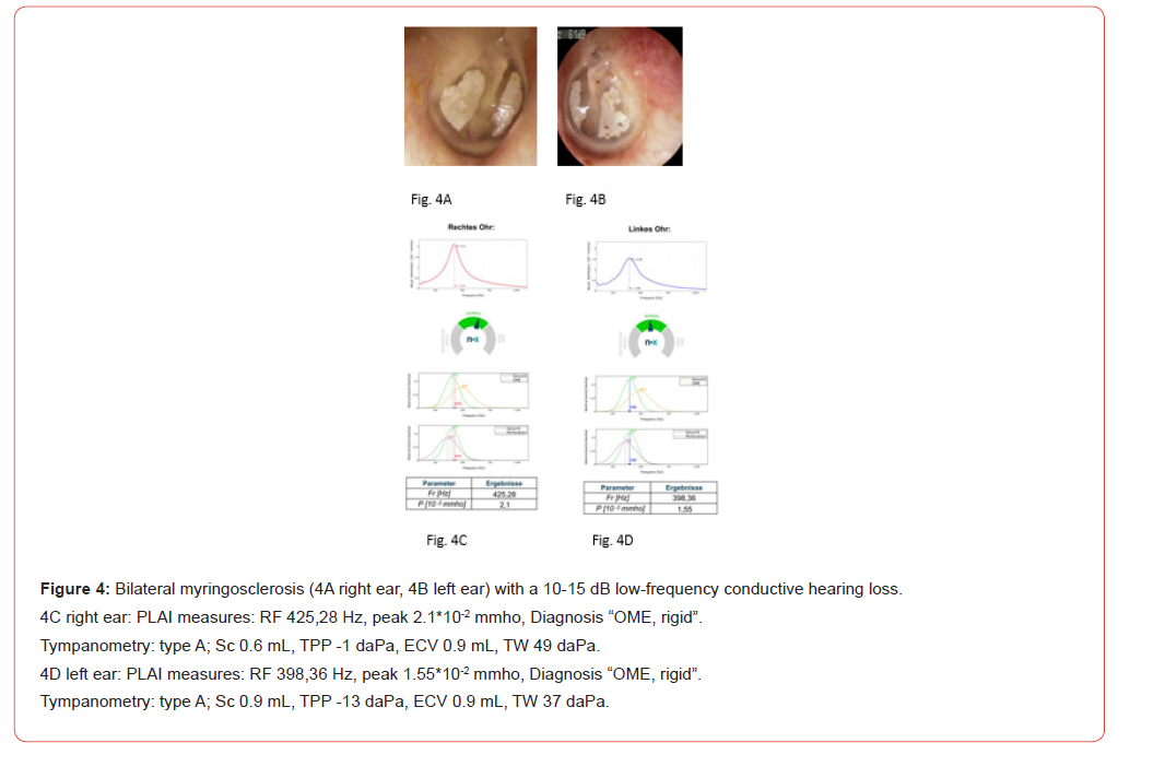
Table 4:
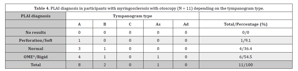
OME = otitis media with effusion
Discussion
This preliminary study focuses on whether PLAI measurements, particularly the RF and peak of admittance, can detect an isolated fixation of the stapes footplate in advanced OS. Otosclerosis was first described in 1741 in an autopsy by Valsalva [5] and primarily leads to a bony fixation of the anterior part of the stapes footplate, resulting in decreased stapes mobility. This condition leads to progressive conductive or mixed hearing loss and rarely sensorineural hearing loss. In OS, the tympanic membrane morphology remains unchanged. The presence of sclerotic plaques on the tympanic membrane could indicate tympanosclerosis, another cause of fixation of the ossicular chain. In very rare cases, a reddish blush is visible on the promontory, caused by abnormal vascular shunts between the otosclerotic focus and the vessels on the promontory, known as Schwartze’s sign [6, 7]. Another morphological change was described by Yamasoba et al. in 2025 [8], who noted a white spot with a clear boundary on the bone surface of the fissula ante fenestrum in about 41% of 27 OS cases. While Schwartze’s sign can rarely be detected via otoscopy, this white spot was identified during endoscopic stapes surgery. However, in most otosclerotic cases, the mucosa of the promontory appears normal.
The diagnosis of OS is based on medical history, physical examination with otoscopy and pneumatic otoscopy, and audiometric testing and is supported by imaging, primarily highresolution computed tomography. With simple pneumatic otoscopy, first described in 1978 [9], the presence of malleus fixation can be excluded before stapes surgery. Besides the tuning fork tests (Weber, Rinne, Bing, Schwabach, ABC - Absolute Bone Conduction, and Gellé), pure tone and speech audiometry, impedance audiometry with tympanometry and stapedial reflex measurements, otoacoustic emissions, and vestibular tests are needed. The Carhart’s notch is considered typical of OS [10], characterized by an increase of bone conduction threshold of about 5-15 dB between 500 Hz and 4 kHz. This mechanical artifact can also be found in stapes fixation due to other diseases, such as tympanosclerosis. In otosclerosis, increased or absent acoustic stapedial reflex thresholds and a lack of otoacoustic emissions are typically found [11]. Reflectance tests, parameterized by absorbance and group delay at ambient pressure, have been recommended to differentiate between otosclerotic and normal ears [11]. Besides the available test batteries for diagnosing OS, the definitive diagnosis of OS is only provided by stapes surgery.
The RF of the middle ear is known to increase in otosclerosis. The purpose is to determine whether the RF is also increased in OS and where PLAI can be used to diagnose otosclerosis. For the control group, patients with healthy ears or with isolated myringosclerosis were selected. It was assumed that in myringosclerosis and OS, the stiffness of the ossicular chain or the tympanic membrane increases, which is detected by PLAI.
Previous studies have shown that both PLAI parameters, RF and peak of admittance, change in various middle ear pathologies compared with healthy ears [2, 3]. While myringosclerosis and tympanosclerosis induce fixation of the ossicular chain, increasing the stiffness of the tympanic membrane and ossicular chain, tube ventilation disorders with atrophy of the tympanic membrane result in retraction or hyperectasis, leading to a hypermobility of the tympanic membrane. The latter finding leads to the PLAI diagnosis of “perforation/soft”. In many ears, various changes could be found, such as myringosclerosis, atrophic parts of the tympanic membrane, and fixation of the ossicular chain. Figure 1 demonstrated various pathological findings in one ear, and parameters are the result of measuring various pathologies. Therefore, in these cases, it seems challenging to interpret the PLAI diagnosis of “perforation/soft” or “OME/rigid”. Additionally, even in normal ears, artificially induced underpressure of the middle ear temporarily changes the RF [12].
In recent years, wideband absorbance measurements with commercially available software have been used for impedance and wideband tympanometry to diagnose OS [13, 14]. The measured frequency range was 0.226-8 kHz, with ear canal pressure varying from +200 to -300/-600 daPa. The stimulus was approximately 65 dB HL. Wideband absorbance and RF were calculated and compared between otosclerotic and normal ears. Sliwa et al. found significant differences in absorbance, especially at low and middle frequencies [13]. Besides energy reflectance measurements of WAI, Wang et al. analyzed the RF in otosclerotic patients and normal individuals, finding a slight but not significant increase in RF values in otosclerotic patients [14]. Similar findings were reported by Shahnaz and Polka [15], who concluded that energy reflectance measurements can distinguish otosclerotic ears from normal ears as well as from ears with other sources of conductive hearing loss. However, they also concluded that these measurements should be interpreted in conjunction with other audiometric tests. Wang et al. noted that due to the overlap between RF in OS and normal subjects, RF cannot serve as an effective and independent tool for diagnosing OS [14]. Other authors found lower values of RF in otosclerotic patients compared with controls [16]. The discrepancies in air-bone gap in OS could be attributed to the different stages of otosclerosis, resulting in variable changes in stapes footplate mobility [17].
Experimental determined absorbance and similar measures have demonstrated high variability in both healthy [18] and pathological ears [11]. It is assumed that natural inter-subject variability of acoustic properties [19] and random measurement errors of diverse origin [20-22] may explain this variability. The range of absorbance values results in an overlap of measures from normal and pathological ears, making differentiation between OS and normal ears challenging. Many immittance measure tests have been used to assess the effectiveness of diagnosing OS [11, 13-15, 23, 24]. Among these, ambient pressure or peak pressure absorbance averages in selected bands [11], RF or RF at π/2 phase shift of the middle ear admittance, and the group delay of energy reflectance [11] should be mentioned. For all tests, a reduced effectiveness has been noted in reliably diagnosing otosclerosis.
In our preliminary study, we demonstrated that the peak of admittance and RF increase in otosclerosis compared with control values. The greater the fixation rate of the stapes footplate, resulting in a higher air-bone gap, the higher the RF values measured. In all 10 patients with OS and an air-bone gap over 20, a fixation of the malleus head could be excluded. Therefore, the increases in RF values in these patients are solely based on fixation of the footplate. However, all selected cases in this study had advanced OS. The demonstrated OS case (contralateral ear with presumed otosclerosis; Figure 3) and unpublished data revealed that in cases of presumed OS without or with a slight Carhart’s notch and an air-bone gap under 20, the RF is not or only slightly increased. To validate these findings, further studies with a larger sample size are needed, especially with air-bone gaps lower than 20. It would be interesting to determine at which air-bone gap the RF is increased, i.e., RF values being 450-470 Hz and higher.
Interestingly, in the selected case, the preoperative increased RF value decreased after stapes surgery. This implies that PLAI could be helpful in monitoring the success of the stapes surgery. Therefore, pre- and post-operative measurements must be incorporated in further studies.
Conclusion
PLAI provides reliable information for diagnosing advanced OS. RF and peak of admittance values are higher in OS than in healthy controls and in patients with isolated myringosclerosis. However, a significant limitation of this preliminary study is the small number of OS cases included. Further studies should incorporate all stages of OS as well as pre- and post-operative measurements to assess the PLAI measurements in OS.
Funding
This research received no external funding.
Conflict of Interest
The authors declare no conflict of interest.
Acknowledgement
The authors thank Axel Monien and Anna Bäumer for help with data collection.
References
- Graffigna CE, Stanzial D, inventors (2021) Apparatus and method for measuring clinical-audiometric parameters. Italy 2021 European patent 4 125 577 B1.
- Anbari M, Zhumabaeva S, Beishenova M, Fischer D, Sulaimanov S, Just T (2024) Comparison of conventional single-frequency tympanometry with pressure-less acoustic immittance measurements of pathological and normal middle ears. On J Otolaryngol & Rhinol 7(2):1-11.
- Anbari M, Zhumabaeva S, Beishenova M, Fischer D, Sulaimanov S, Just T (2025) Pressure-less acoustic immittance measurements of patients with persistent ventilation disorders. On J Otolaryngol & Rhinol 7(3): 1-11.
- Merchant SN, Nadol JB. Schuknecht’s pathology of the ear. (3rd), Merchant SN, Nadol JB (Eds.), Shelton, CT, USA: People’s Medical Pub. House; 2010.
- Valsalva AM. Opera: hoc est, tractatus de aure humana editione has quarta accuratissime descriptus. Venice: Apud Franciscum Pitteri; 1741.
- Koenen L, Gupta G (2025) Schwartze Sign. 2023 Sep 4. In: StatPearls [Internet]. Treasure Island (FL): StatPearls Publishing.
- Peng KA, House JW (2018) Schwartze sign. Ear Nose Throat J 97(3): 54.
- Yamasoba T, Uranaka T, Koyama H, Kashio A (2025) A white spot around the fissula ante fenestrum: A new diagnostic indicator for otosclerosis. J Clin Med 14(3).
- Moon CN Jr, Hahn M (1978) Pneumatic otoscopy and impedance studies in middle ear diagnosis. Laryngoscope 88(9 Pt 1): 1439-1448.
- Carhart R (1950) Clinical application of bone conduction audiometry. Arch Otolaryngol (1925). 51(6): 798-808.
- Keefe DH, Archer KL, Schmid KK, Fitzpatrick DF, Feeney MP, et al. (2017) Identifying otosclerosis with aural acoustical tests of absorbance, group delay, acoustic reflex threshold, and otoacoustic emissions. J Am Acad Audiol 28(9): 838-860.
- Just T, Anbari M, Monien A, Fischer D (2025) Changes of the resonance frequency of the middle ear without opening of the Eustachian tube. Br J Cancer Res 8(1): 742-746.
- Sliwa L, Kochanek K, Jedrzejczak WW, Mrugala K, Skarzynski H (2020) Measurement of wideband absorbance as a test for otosclerosis. J Clin Med 9(6).
- Wang S, Hao W, Xu C, Ni D, Gao Z, Shang Y (2019) A study of wideband energy reflectance in patients with otosclerosis: Data from a Chinese population. Biomed Res Int 2019: 2070548.
- Shahnaz N, Bork K, Polka L, Longridge N, Bell D, et al. (2009) Energy reflectance and tympanometry in normal and otosclerotic ears. Ear Hear 30(2): 219-233.
- Zhao F, Wada H, Koike T, Ohyama K, Kawase T, Stephens D (2002) Middle ear dynamic characteristics in patients with otosclerosis. Ear Hear 23(2): 150-158.
- Cherukupally SR, Merchant SN, Rosowski JJ (1998) Correlations between pathologic changes in the stapes and conductive hearing loss in otosclerosis. Ann Otol Rhinol Laryngol 107(4): 319-326.
- Feeney MP, Keefe DH, Hunter LL, Fitzpatrick DF, Garinis AC, et al. (2017) Normative wideband reflectance, equivalent admittance at the tympanic membrane, and acoustic stapedius reflex threshold in adults. Ear Hear 38(3): e142-e60.
- Abur D, Horton NJ, Voss SE (2014) Intrasubject variability in power reflectance. J Am Acad Audiol 25(5): 441-448.
- Feeney MP, Stover B, Keefe DH, Garinis AC, Day JE, Seixas N (2014) Sources of variability in wideband energy reflectance measurements in adults. J Am Acad Audiol 25(5): 449-461.
- Groon KA, Rasetshwane DM, Kopun JG, Gorga MP, Neely ST (2015) Air-leak effects on ear-canal acoustic absorbance. Ear Hear 36(1): 155-163.
- Voss SE, Horton NJ, Woodbury RR, Sheffield KN (2008) Sources of variability in reflectance measurements on normal cadaver ears. Ear Hear 29(4): 651-665.
- Shahnaz N, Longridge N, Bell D (2009) Wideband energy reflectance patterns in preoperative and postoperative otosclerotic ears. Int J Audiol 48(5): 240-247.
- Wegner I, Shahnaz N, Grolman W, Bance ML (2017) Wideband acoustic immittance measurements in assessing crimping status following stapedotomy: A temporal bone study. Int J Audiol 56(1): 1-7.
-

This work is licensed under a Creative Commons Attribution-NonCommercial 4.0 International License.




