 Case Report
Osseous Heart- A Rare Case of Metastatic Osteosarcoma to the Heart
Arnaldo A Nieves Ortiz1*, Kyomara Hernández Moya1, Arnaldo Rojas Figueroa1, Vanessa Fonseca Ferrer1, Hernan Gonzalez Monroig2, Jonathan Martinez Feshold1, William Marrero1, Gabriela Torres Torres1, Alexis Cruz Chacón2, Rafael Rivera Berrios3 and Teodosio Rucabado4
1Internal Medicine Department, San Juan City Hospital, Puerto Rico
2Hematology/Oncology Fellowship, San Juan City Hospital/VA Caribbean Healthcare System, Puerto Rico
3Cardiovascular Department Fellowship, Saint Luke’s Episcopal Medical Center, Puerto Rico
4Cardiovascular Department, San Juan City Hospital, Puerto Rico
Arnaldo A Nieves Ortiz, Internal Medicine Department, San Juan City Hospital, San Juan, Puerto Rico.
Received Date:July 21, 2021; Published Date:August 05, 2021
Abstract
Osteosarcomas are uncommon malignant tumors of the bone that arise from the production of osteoid or immature bone by the malignant cells. In rare occasions this tumor metastasizes to other organs including the brain and lungs. Although hematogenous spread of osteosarcoma is well known, cardiovascular metastasis is seldomly reported. Most cases of metastatic osteosarcoma to the heart involve the myocardium rather than the pericardium. The right side of the heart is affected in 20–30% of cases and the left side in 10–33% of reported cases. Only 5% of cases affect the endocardium or chamber cavities. Clinical signs and symptoms of metastatic cardiac neoplasms include dyspnea, cough, chest pain, palpitations, superior vena cava syndrome, right ventricular outflow tract obstruction and pericardial effusion [1].
Keywords:Cardiac Osteosarcomas; GM-CSF autoantibodies; Autoimmune
Case Presentation
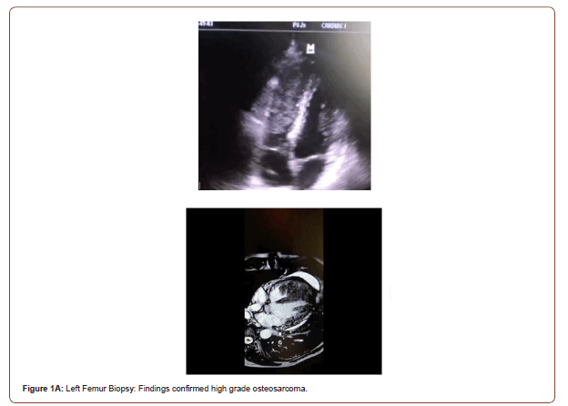
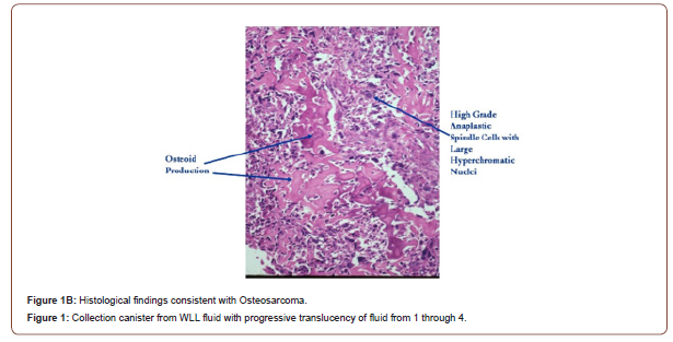
We present the case of a 48 y/o male with history of osteosarcoma of the left femur s/p 5 cycles of neoadjuvant chemotherapy with Doxorubicin and Cisplatin on May 2020, who presented with complains of left femur pain. Upon review of system, patient denied chest pain, SOB, dyspnea on exertion, palpitations, or syncope. Physical exam remarkable for left knee decreased range of motion and strength 4/5. Laboratories showed normocytic normochromic anemia and mild thrombocytopenia. Left leg X-ray showed a 9.9 cm sclerotic lesion with associated cortical thickening and prominent sunburst reaction at the femoral shaft to the metaphysis worrisome for aggressive osteosarcoma. Knee CT scan without IV contrast shows sunburst periosteal reaction measuring 13.9cm long x 6.1 cm transverse x 5.8 cm. On December 2020, Chest CT shows enlarged pulmonary nodules consistent with metastasis. Patient was referred to osteosarcoma specialist who recommended Etoposide, Ifosfamide, Mesna and high dose methotrexate. Prior to initiation of induction chemotherapy, transthoracic echocardiography was done and showed a 11.25cm mass filling the right ventricle with preserved EF. Cardiac MRI results showed homogenously enhancing intracavitary mass centered at the apex of the right ventricle measuring approximately 3.2 x 4.2 cm x 5.0 cm, an internal and eccentric non-enhancing hypointense signal representative of an ossifying focus characteristic of osteosarcoma metastasis. Bone marrow biopsy showed no evidence of metastatic osteosarcoma. Patient was initiated on 2 cycles of chemotherapy, with improved clinical response (Figures 1-3).


Conclusion
Osteosarcomas are malignant tumors that most frequently involves the long bones of young adults within the second and third decades of life. The clinical characteristics involve hemodynamic compromise or precordial abnormality, with magnetic resonance imaging emerging as the gold standard for diagnosis. Osteosarcoma metastasis most commonly occur in the lungs. Osteosarcoma very rarely metastasizes to the heart’s right ventricle, all of them in the RV outflow tract. Although surgery is generally contraindicated in the presence of metastatic disease, the role of surgical palliative of intracardiac tumor to relieve obstruction and to prevent embolization is considered. However, even with successful surgical resection with or without chemotherapy, the prognosis of patients with osteosarcoma is very poor [2]. Despite being extremely rare to find antemortem involvement in the heart, 20% of autopsies show involvement in the heart. In this case, the enhanced resolution of echocardiogram and Chest CT scan lead to detection of cardiovascular involvement.
Acknowledgment
We would like to acknowledge the department of Internal Medicine Program, San Juan City Hospital. We have no conflict of interest to declare. This statement is to certify that all authors have seen and approved the manuscript being submitted.
Conflict of Interest
No Conflict of Interest.
References
- Maleki M, Mozaffari K, Givtaj N, Tatina A, Bahadorian B (2013) Metastatic osteosarcoma and heart: a rare involvement in an unusual cardiac location. Heart Asia 5(1): 120-121.
- Abdelfatah Elasfar, Ahmed Khalifa, Abdulaziz AlGhamdi, Rizwan Khalid, Mohamed Ibrahim, et al. (2013) Asymptomatic metastatic osteosarcoma to the right ventricle: Case report and review of the literature. J Saudi Heart Assoc 25(1): 39-42.
-
Arnaldo A Nieves Ortiz, Kyomara Hernández Moya, Arnaldo Rojas Figueroa, Vanessa Fonseca Ferrer etc all,... Osseous Heart- A Rare Case of Metastatic Osteosarcoma to the Heart. On J Cardio Res & Rep. 5(3): 2021. OJCRR.MS.ID.000615
-
Cardiac Osteosarcomas, GM-CSF autoantibodies, Autoimmune, Endocardium, Cardiac neoplasms, Dyspnea, Cough, Chest pain, Palpitations
-

This work is licensed under a Creative Commons Attribution-NonCommercial 4.0 International License.


