


 Research Article
In Vitro and In Vivo Genotoxicity Assessment of Piper longum L. Powder and Hot-Water Extract
Received Date:October 26, 2026; Published Date:November 03, 2026
Abstract
Piper longum L. is used in traditional medicine, but genotoxicity evidence must be related to the preparation tested. This study evaluated P. longum powder (PL-P) and a freeze-dried hot-water extract (PL-W) using bacterial reverse mutation, in vitro chromosomal aberration, and mouse bone marrow micronucleus assays. Four chemical markers were quantified by high-performance liquid chromatography. The main bacterial experiment tested five strains at 12.346–1000 g/plate with and without rat liver S9. Chinese hamster lung cells were treated for 6 h with or without S9 or for 22 h without S9. Male ICR mice received 500, 1000, or 2000 mg/kg by oral gavage on two consecutive days. All four marker contents were lower in PL-W than in PL-P; piperine contents were 0.414% and 2.427%, respectively. Neither preparation produced a biologically relevant bacterial mutagenic response or a statistically significant increase in structural chromosomal aberrations. Relative population doubling at the highest scored PL-P concentrations was 45–49%, and PL-W produced a value of 50% after prolonged exposure at 1600 g/mL. No statistically significant increase in bone marrow micronuclei was detected, and both in vivo positive controls produced significant responses. These results provide preparation-specific evidence of no detected genotoxic response under the conditions tested. Precipitation limits interpretation of dissolved in vitro exposure, and bone marrow exposure was not demonstrated. The findings do not establish safety for long-term human use.
Keywords:Piper longum; Genotoxicity; Bacterial reverse mutation; Chromosomal aberration; Micronucleus assay
Introduction
Piper longum L. (Piperaceae), or long pepper, is used as a spice and in traditional medicine [1]. Its extracts and constituents have attracted interest for their immunomodulatory, antitumor, and other biological activities [2, 3]. However, pharmacological activity and a history of use do not establish the genotoxic safety of a defined preparation. Guidance for herbal products emphasizes the relationship between the material tested and the preparation being assessed [4].
Chemical characterization is therefore essential. Piperine content varies among P. longum populations and tissues [1], and chromatographic studies demonstrate variation in piperamide profiles and support multicomponent analysis [3, 5, 6]. Piperine can also inhibit human P-glycoprotein and CYP3A4 in experimental systems, illustrating its potential to affect biological exposure [7]. Powdered material and hot-water extracts should consequently be evaluated as distinct mixtures rather than assumed to be toxicologically interchangeable.
Evidence for individual constituents is informative but cannot replace testing of whole preparations. Negative bacterial and mouse micronucleus findings have been reported for piperine [8], and subsequent work found no micronucleus induction in Chinese hamster ovary cells or mice under the conditions examined [9]. Studies reporting protection by piperine or P. longum extracts against chemically induced genetic damage address a different question from intrinsic genotoxicity [10, 11]. Conversely, piperlongumine induced DNA damage in breast cell lines [12]. These findings emphasize the importance of chemical identity, exposure conditions, and the biological model when interpreting constituent-level evidence.
Complementary assays address different forms of genetic damage: bacterial reverse mutation detects gene mutations, in vitro chromosomal aberration testing detects structural chromosome damage, and the in vivo micronucleus assay detects chromosomal damage or mitotic disturbance in erythroblasts [13–15]. We therefore evaluated P. longum powder (PL-P) and a freeze-dried hotwater extract (PL-W) using these three assays and characterized their piperine, pellitorine, pipernonaline, and guineensine contents by HPLC. The objective was to assess their genotoxic potential under defined experimental conditions and provide preparationspecific evidence for nonclinical safety assessment.
Materials and Methods
Chemicals and reagents
Sodium azide (SA), 2-nitrofluorene (2-NF), 9-aminoacridine (9-AA), 4-nitroquinoline-N-oxide (4-NQO), benzo[a]pyrene (BP), 2-aminoanthracene (2-AA), mitomycin C (MMC), cyclophosphamide monohydrate (CPA), colchicine, dimethyl sulfoxide were obtained from Sigma-Aldrich (USA). Sterile distilled water was used as the vehicle control unless otherwise specified. Metabolic activation in the bacterial reverse mutation and in vitro chromosomal aberration assays was provided by a liver S9 fraction obtained from Aroclor 1254-induced male Sprague–Dawley rats (Molecular Toxicology, Inc., USA; cat. no. 11-01L). The S9 mix was prepared immediately before use by combining the S9 fraction with cofactors (Wako Pure Chemical Industries, Ltd., Japan; cat. no. 309-50611) and was kept chilled until use. Both in vitro assays included treatment conditions with and without metabolic activation.
Test articles and chemical characterization
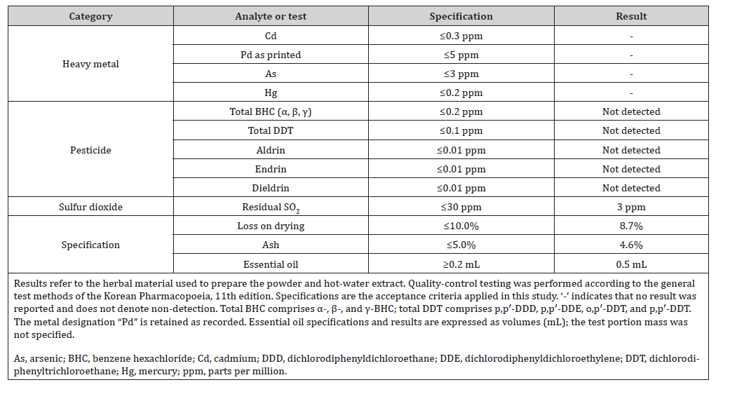
Dried fruit spikes of Piper longum L. (Piperaceae) were authenticated by a botanical identification advisory committee through morphological examination based on the botanical origin and macroscopic characteristics described in the Korean Herbal Pharmacopoeia. Quality-control specifications and results for the starting material are summarized in Supplementary Table S1.
Suplementary Table S1:Quality control specifications and results for the Piper longum starting material.
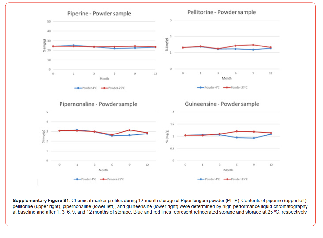
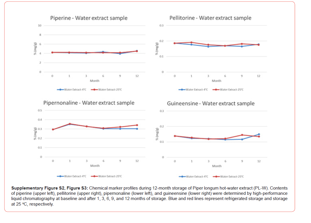
Two test articles were prepared from the authenticated material: a powdered preparation (PL-P) and a hot-water extract (PL-W). The particle size of PL-P was assessed against the finepowder specification of the Korean Pharmacopoeia. PL-W was prepared by hot-water extraction followed by freeze-drying. Sterile distilled water served as the vehicle for both preparations in the genotoxicity assays. Chemical characterization was performed by high-performance liquid chromatography (HPLC) to quantify four marker compounds: piperine, pellitorine, pipernonaline, and guineensine. Their contents and retention times are presented in Table 1. Marker contents were also measured at baseline and after 1, 3, 6, 9, and 12 months of storage at room temperature (25 °C) and under refrigeration. The corresponding profiles for PL-P and PL-W are presented in Supplementary Figures S1 and S2, respectively.
Table 1:Chemical marker contents of Piper longum powder and hot-water extract.

Animals, ethical approval, and husbandry
Male specific-pathogen-free ICR mice (CrlOri:CD1; Orient Bio, Korea) were obtained at six weeks of age and acclimatized for six days before dosing. Animals were housed in polycarbonate cages (180 × 240 × 143 mm), with up to four mice per cage during acclimatization and up to three per cage during the predosing and dosing periods. Room conditions were at 22 ± 3 °C and 40–75% relative humidity under a 12-hour light/dark cycle, with illumination of 150–300 lux and 10–20 air changes per hour. A standard laboratory diet (LabDiet #5053, PMI Nutrition International, USA) and filtered, ultraviolet-treated tap water were provided ad libitum. Mice were approximately seven weeks old at the start of treatment.
All animal procedures were approved by the Institutional Animal Care and Use Committee of the Korea Institute of Toxicology. The approval numbers were 2007-0235 for the Piper longum powder (PL-P) study and 2007-0234 for the hot-water extract (PLW) study.
The bacterial reverse mutation, in vitro chromosomal aberration, and mouse bone marrow micronucleus assays were conducted in compliance with Good Laboratory Practice (GLP).
Bacterial reverse mutation assay
The bacterial reverse mutation assay was performed using the plate-incorporation method [13, 16]. Salmonella typhimurium TA100, TA1535, TA98, and TA1537 and Escherichia coli WP2uvrA were obtained from Molecular Toxicology, Inc. (Boone, NC, USA). Strain characteristics, including amino acid requirements, UV sensitivity, the rfa phenotype, R-factor retention, and spontaneous reversion, were verified as applicable to each strain. Cultures were grown in 2.5% nutrient broth at 37 ± 1 °C with shaking at 120– 200 rpm for approximately 10 h. Ampicillin was added for strains carrying pKM101. Bacterial suspensions were used at a density of at least 1 × 10⁹ cells/mL. PL-P and PL-W were tested in the presence and absence of metabolic activation. Preliminary concentrationrange- finding experiments evaluated 8, 40, 200, 1000, 3000, and 5000 μg/plate, with one plate per strain, concentration, and metabolic activation condition. Based on these experiments, the main assay evaluated 12.346, 37.037, 111.111, 333.333, and 1000 μg/plate, with four replicate plates per strain, concentration, and metabolic activation condition.
For each plate, 0.1 mL of test-article preparation or vehicle, 0.1 mL of bacterial suspension, and 0.5 mL of either 0.2 M sodium phosphate buffer or S9 mix containing 5% (v/v) S9 were mixed with 2.0 mL of molten top agar maintained at approximately 45 °C. The top agar was supplemented with histidine and biotin for the S. typhimurium strains or tryptophan for E. coli WP2uvrA. The mixture was poured onto minimal glucose agar plates and allowed to solidify. Plates were then inverted and incubated at 37 ± 1 °C for approximately 48 h, after which revertant colonies were counted manually. Precipitation, turbidity, and changes in the bacterial background lawn were recorded.
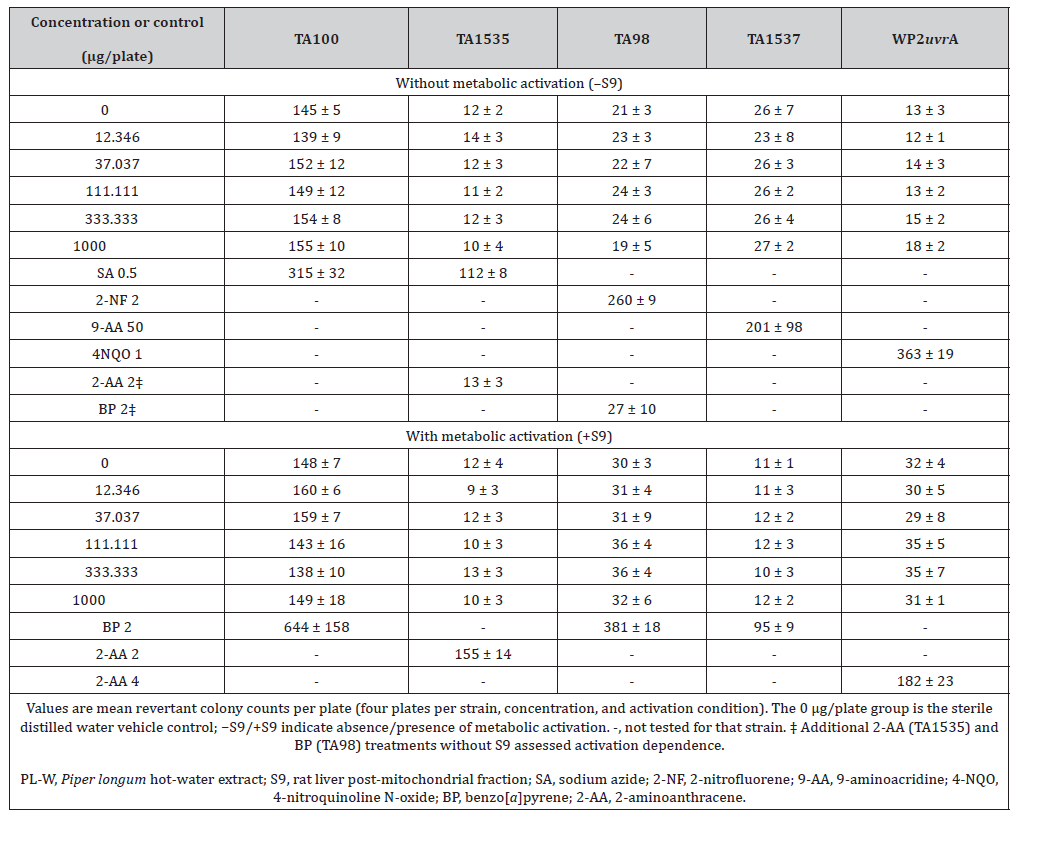
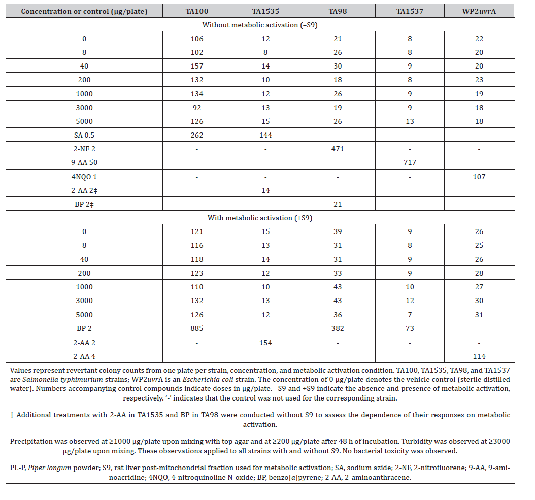
Sterile distilled water served as the vehicle control for both test articles. Strain-specific positive controls were included under the corresponding metabolic activation conditions, as detailed in Tables 2 and 3. Additional treatments with 2-aminoanthracene (2- AA) in TA1535 and benzo[a]pyrene (BP) in TA98 were included without S9 to assess the dependence of their mutagenic responses on metabolic activation.
Suplementary Table S2:Concentration range-finding results for Piper longum powder (PL-P) in the bacterial reverse mutation assay
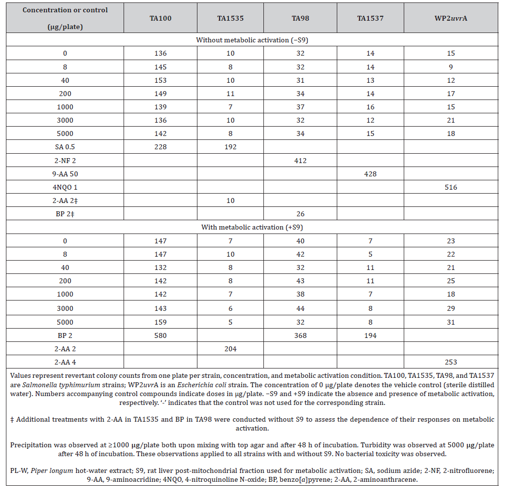
Table 2:Bacterial reverse mutation assay results for Piper longum powder (PL-P) with and without metabolic activation.
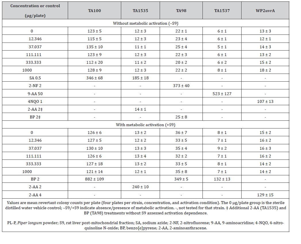
Table 3:Bacterial reverse mutation assay results for Piper longum hot-water extract (PL-W) with and without metabolic activation.
In vitro chromosomal aberration assay
The assay was performed according to OECD Test Guideline 473 [14]. Chinese hamster lung (CHL) cells obtained from the American Type Culture Collection (Manassas, VA, USA) were maintained at 37 ± 1 °C and 5% CO2 in minimum essential medium supplemented with 10% fetal bovine serum, 2 mM L-glutamine, 100 units/mL penicillin, and 100 μg/mL streptomycin. The cells had a modal chromosome number of 25 and an approximately 15-h cell cycle and were screened for mycoplasma contamination by PCR before use. Cells were seeded at 4 × 10⁴ cells per 25-cm2 flask in 5.0 mL of medium and cultured for approximately three days before treatment.
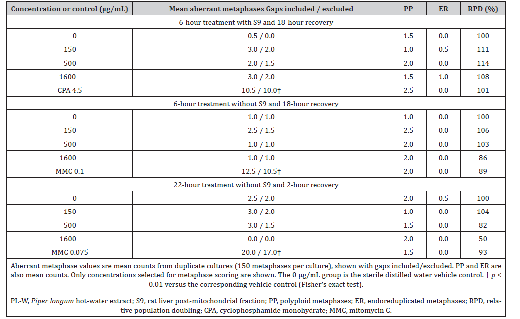
PL-P and PL-W were evaluated under three conditions: 6-h treatment with S9 followed by 18-h recovery, 6-h treatment without S9 followed by 18-h recovery, and 22-h treatment without S9 followed by 2-h recovery. Concentrations were selected from preliminary concentration-range-finding experiments (Supplementary Tables S4–S7). Results for concentrations selected for metaphase scoring are presented in Tables 4 and 5. Two replicate cultures were used per concentration and treatment condition. The final S9 concentration in activated cultures was 2% (v/v). Sterile distilled water served as the vehicle control. CPA at 4.5 μg/mL served as the positive control with S9, whereas MMC at 0.1 and 0.075 μg/mL served as the positive control for the 6-h and 22-h treatments without S9, respectively.
Table 4:Chromosomal aberrations and relative population doubling in Chinese hamster lung cells treated with Piper longum powder (PL-P)
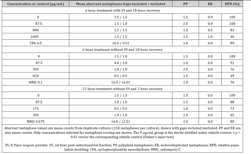
Table 5:Chromosomal aberrations and relative population doubling in Chinese hamster lung cells treated with Piper longum hot-water extract (PLW).
Suplementary Table S3:Concentration range-finding results for Piper longum hot-water extract (PL-W) in the bacterial reverse mutation assay.
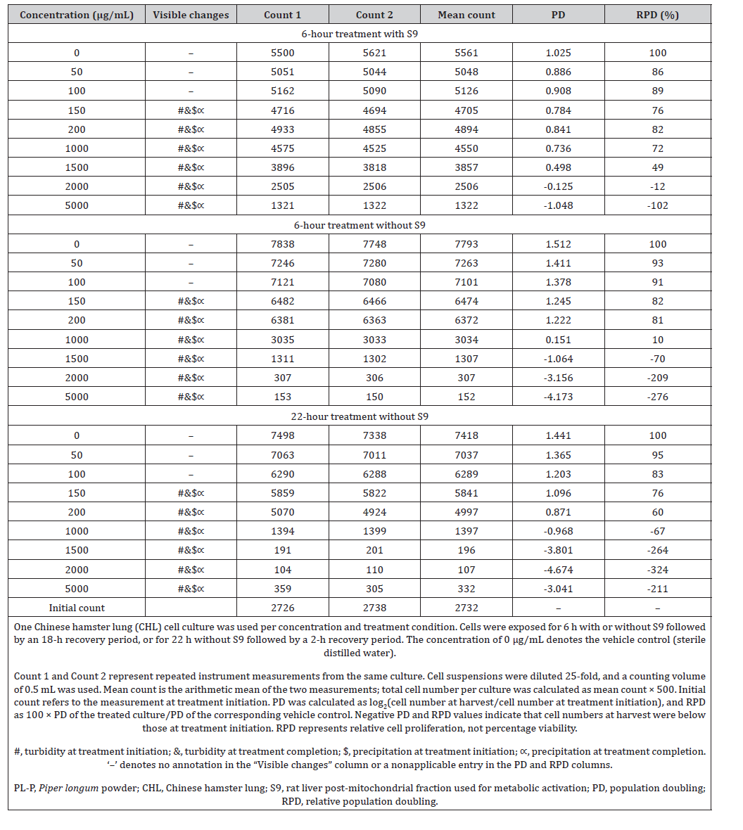
Suplementary Table S4:Cell proliferation in the first concentration range-finding experiment with Piper longum powder (PL-P) for the in vitro chromosomal aberration assay.
Suplementary Table S5:Cell proliferation in the second concentration range-finding experiment with Piper longum powder (PL-P) for the in vitro chromosomal aberration assay.
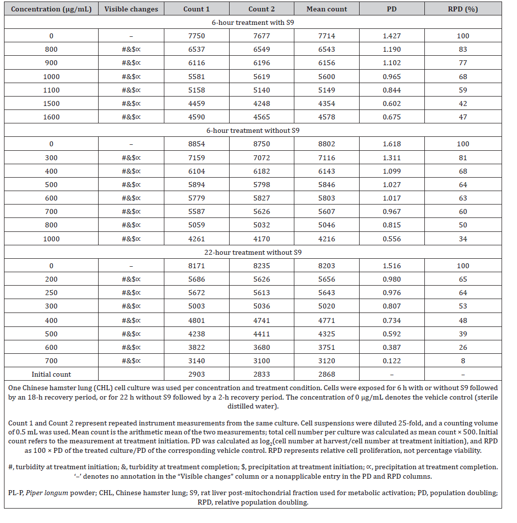
Suplementary Table S6:Cell proliferation in the first concentration range-finding experiment with Piper longum hot-water extract (PL-W) for the in vitro chromosomal aberration assay.
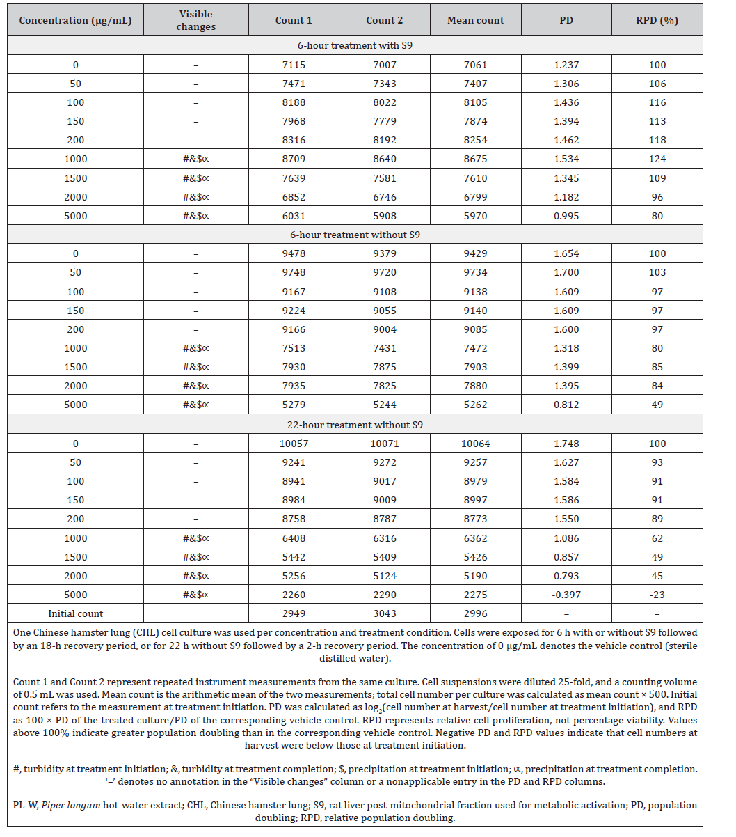
Suplementary Table S7:Cell proliferation in the second concentration range-finding experiment with Piper longum hot-water extract (PL-W) for the in vitro chromosomal aberration assay.
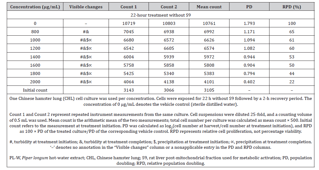
Precipitation and turbidity were assessed visually at the beginning and end of treatment. During preliminary range-finding experiments, medium pH and osmolality were measured in the vehicle control and the highest-concentration group (5000 μg/mL) at both time points. At the end of treatment, cultures were washed once with calcium- and magnesium-free Dulbecco’s phosphate-buffered saline and incubated in fresh medium for the designated recovery period. Colchicine was added at a final concentration of 1 μM approximately 22 h after treatment initiation, and cells were harvested 2 h later. Cell numbers were determined using an automated cell counter. Population doubling (PD) was calculated as log2(N final/N initial), where N initial and N final denote cell numbers at treatment initiation and harvest, respectively. Relative population doubling (RPD) was calculated as 100 × PD treated/PD vehicle control. Cells were treated with 75 mM potassium chloride for 10 min at room temperature and fixed with methanol and glacial acetic acid (3:1, v/v). Two slides were prepared per culture and stained with 3% (v/v) Giemsa solution in Gurr buffer for 7 min. One suitable slide per culture was selected and coded before microscopic evaluation. For structural aberration assessment, 150 well-spread metaphases containing 23–27 chromosomes were examined per culture at ×1000 magnification. Chromatid- and chromosome-type gaps, breaks, exchanges, and multiple aberrations were recorded. Gaps were recorded separately, and aberrant metaphases were tabulated both including and excluding gaps. An additional 150 metaphases per culture were examined for polyploidy (PP; ≥37 centromeres) and endoreduplication (ER), irrespective of the presence of structural aberrations.
Mouse bone marrow micronucleus assay
The mouse bone marrow micronucleus assay was conducted according to OECD Test Guideline 474 [15]. PL-P and PL-W were evaluated in separate experiments. For each preparation, six mice were allocated to each of five groups: a vehicle control, three treatment groups receiving 500, 1000, or 2000 mg/kg, and a positive- control group.
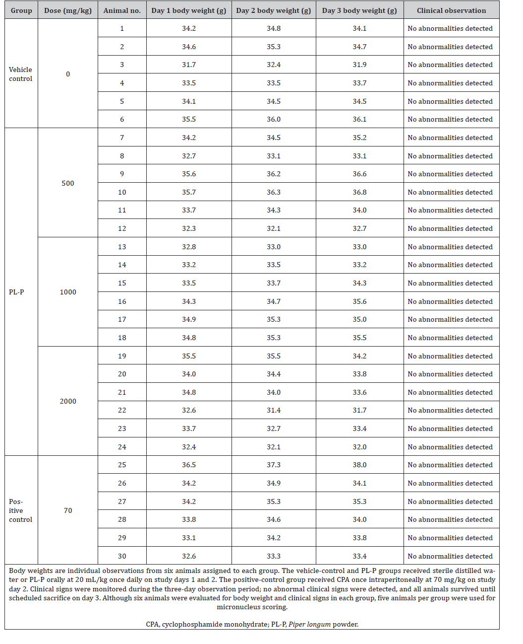
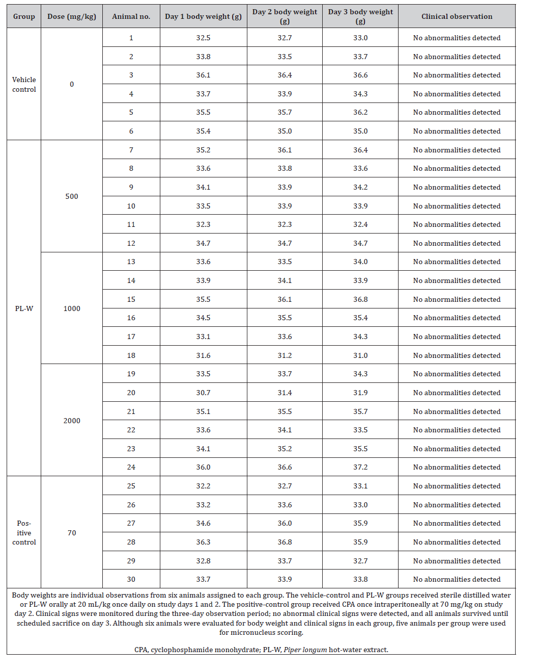
The test articles or vehicle (sterile distilled water) were administered orally gavage at 20 mL/kg once daily for two consecutive days. CPA, prepared in sterile saline, was administered to the positive-control group as a single intraperitoneal dose of 70 mg/ kg at 10 mL/kg at the time of the second test-article administration. Clinical signs and body weights were monitored in all six mice per group through Day 3 (Supplementary Table S8 and S9). Body weights were recorded before dosing on Days 1 and 2 and on Day 3. The stability, homogeneity, and concentration of the dosing formulations were not analytically verified.
Suplementary Table S8:Individual body weights and clinical observations in male ICR mice administered Piper longum powder (PL-P) in the bone marrow micronucleus assay
Suplementary Table 9:Individual body weights and clinical observations in male ICR mice administered Piper longum hot-water extract (PL-W) in the bone marrow micronucleus assay
Approximately 23 h after the final administration, mice were euthanized with CO2, and femoral bone marrow was collected. Bone marrow was flushed with 2 mL of fetal bovine serum and filtered through an α-cellulose/Sigmacell cellulose column (1:1, v/v). After the addition of 1 mL of Hanks’ balanced salt solution, the suspension was centrifuged at 1000 rpm for 5 min at room temperature. The supernatant was removed, and two smears were prepared per animal from the remaining cell suspension. Smears were air-dried, fixed in methanol for 5 min, and stained with 5% (v/v) Giemsa solution prepared in Gurr buffer.
The first five animals in animal-number order within each group were evaluated for micronuclei. One suitably stained smear per animal was selected and randomly coded before microscopic evaluation. For each animal, micronucleated polychromatic erythrocytes (MNPCEs) were counted among 4000 polychromatic erythrocytes (PCEs). The endpoint was expressed as the number of MNPCEs per 4000 PCEs. In addition, 500 erythrocytes per animal were classified as PCEs or normochromatic erythrocytes (NCEs), irrespective of micronucleus status. The proportion of immature erythrocytes, calculated as PCE/(PCE + NCE), was used as an indicator of bone marrow cytotoxicity.
Statistical Analysis
For the bacterial reverse mutation assay, no inferential statistical tests were performed. Mean revertant colony counts were compared descriptively with the corresponding vehicle controls, with interpretation based on the magnitude and concentration dependence of the responses and their biological relevance.
For the in vitro chromosomal aberration assay, aberrant metaphases including and excluding gaps and the combined polyploid and endoreduplicated metaphases endpoint (PP + ER) were analyzed using SAS. Test-article groups were initially compared with the vehicle control using a chi-square test. When p < 0.05, Fisher’s exact test and the Cochran–Armitage trend test were applied to assess group differences and concentration-related trends, respectively. Positive-control comparisons used Fisher’s exact test. Interpretation of structural chromosomal damage emphasized aberrations excluding gaps.
For the mouse bone marrow micronucleus assay, MNPCE counts were summarized as means ± standard deviations for five mice per group. Test-article groups were compared with the concurrent vehicle control using the Kruskal–Wallis test and Dunn’s rank-sum test. The protocol specified a Cochran–Armitage trend test following a significant result; this test was not performed because the initial comparisons yielded p ≥ 0.05. Positive-control MNPCE counts were compared with the vehicle control using the Mann–Whitney U-test. PCE/(PCE + NCE) ratios were arcsine square-root transformed before analysis. Test-article comparisons used one-way analysis of variance, followed by Dunnett’s test when significant; positivecontrol comparisons used Student’s t-test. The individual mouse was the experimental unit, and cells scored within an animal were subsamples.
Body weights were analyzed for six mice per group. Homogeneity of variances was assessed using Bartlett’s test. Homogeneous data were evaluated using one-way analysis of variance followed by Dunnett’s test; heterogeneous data were evaluated using the Kruskal–Wallis test followed by Dunn’s ranksum test. In vivo analyses used Pristima (version 7.4) and/or SAS/ STAT (version 9.4). A p value < 0.05 was considered statistically significant for inferential analyses. Historical control data were considered separately when evaluating assay performance.
Results and Discussion
Chemical differences between the preparations
HPLC analysis revealed differences in the contents of the four measured marker compounds between PL-P and PL-W (Table 1). Piperine was the most abundant of these markers in both preparations, with contents of 2.427% in PL-P and 0.414% in PL-W. The corresponding contents of pellitorine, pipernonaline, and guineensine were 0.138%, 0.301%, and 0.115% in PL-P and 0.008%, 0.021%, and 0.008% in PL-W, respectively. Thus, PL-W contained lower concentrations of all four markers than PL-P.
These compositional differences support evaluating the two preparations separately, because equal doses of PL-P and PL-W do not contain equivalent amounts of the measured constituents. However, marker concentrations in the finished preparations do not establish extraction recoveries relative to the starting herbal material or distinguish incomplete extraction from chemical transformation during processing. Furthermore, the four-marker analysis characterizes only part of the chemical composition of these preparations. The genotoxicity findings therefore apply to the tested preparations as mixtures and cannot be attributed to individual markers solely on the basis of their concentrations. Quality testing of the source herbal material showed no detectable residues of the pesticides examined and a sulfur dioxide content of 3 ppm. Loss on drying and ash content were 8.7% and 4.6%, respectively (Supplementary Table S1).
Bacterial reverse mutation findings
Neither PL-P nor PL-W produced a biologically relevant increase in revertant colony counts in Salmonella typhimurium TA100, TA1535, TA98, and TA1537 or Escherichia coli WP2uvrA over the main-study concentration range of 12.346–1000 μg/plate, with or without S9 metabolic activation (Tables 2 and 3). Colony counts generally remained close to the corresponding vehicle-control values. Small numerical increases occurred in some treatment groups; for example, WP2uvrA counts without S9 increased from 13 colonies/plate in the vehicle controls to 18 colonies/plate at 1000 μg/plate for both preparations. Considered together with the concentration profiles and preliminary findings, these changes did not indicate a biologically relevant mutagenic response.
The strain-specific positive controls increased revertant colony counts in both activation conditions. In addition, 2-aminoanthracene in TA1535 and benzo[a]pyrene in TA98 produced pronounced responses with S9, whereas their responses without S9 remained close to the corresponding vehicle-control values. These findings supported the responsiveness of the bacterial strains and the functional activity of the metabolic activation system.
No background-lawn thinning, microcolony formation, or reduction of at least 50% in revertant colony counts was observed with either preparation. Precipitation nevertheless occurred during the main experiment at PL-P concentrations of 333.333 μg/plate and above and at 1000 μg/plate for PL-W, irrespective of S9 activation. The preliminary experiments extended to 5000 μg/ plate and likewise showed no clear mutagenic response or overt bacterial toxicity (Supplementary Tables S2 and S3). However, these experiments used one plate per concentration and condition, whereas the main experiment used four plates. Consequently, the replicated evidence supporting the negative findings extends to 1000 μg/plate.
The results support the absence of detectable bacterial mutagenic activity for both preparations under the conditions examined. OECD TG 471 identifies solubility and cytotoxicity as relevant considerations in concentration selection and emphasizes biological relevance in interpreting colony-count changes [13]. Here, precipitation indicates that nominal preparation concentrations cannot be assumed to represent fully dissolved exposure. The negative findings are also consistent with the negative bacterial mutagenicity result for piperine reported by the National Toxicology Program [17]. However, this constituent-level evidence does not establish the contributions of piperine or other compounds to the responses of the whole preparations. Because bacterial mutation assays do not reproduce all aspects of mammalian uptake and metabolism, these findings should be considered together with the chromosomal aberration and micronucleus results.
Chromosomal aberrations in CHL cells
Neither PL-P nor PL-W significantly increased the frequency of metaphases with structural chromosomal aberrations excluding gaps under any of the three treatment conditions (Tables 4 and 5). Mean numbers of aberrant metaphases excluding gaps ranged from 0.5 to 3.0 per culture for PL-P and from 0.0 to 2.0 per culture for PL-W, with 150 metaphases examined per culture. No consistent concentration-related increase was evident. The combined frequency of polyploid and endoreduplicated metaphases (PP + ER) also showed no statistically significant increase. In comparison, CPA and MMC increased structural chromosomal aberrations in the corresponding treatment conditions (p < 0.01), demonstrating the responsiveness of the assay.
The preparations differed in their effects on cell proliferation. For PL-P, the highest concentrations evaluated for chromosomal aberrations were 1600 μg/mL after 6-h treatment with S9, 820 μg/mL after 6-h treatment without S9, and 350 μg/mL after 22-h treatment without S9. The corresponding RPD values were 46%, 49%, and 45%. For PL-W, the highest evaluated concentration was 1600 μg/mL under all three conditions, with RPD values of 108%, 86%, and 50%, respectively. Thus, PL-P inhibited proliferation under all three conditions, whereas pronounced inhibition by PL-W occurred after prolonged exposure without S9. The RPD values of 45–50% were within the range corresponding to the cytotoxicity target recommended by OECD TG 473 when cytotoxicity determines the highest concentration [14]. In these cultures, substantial inhibition of proliferation was not accompanied by an increase in structural chromosomal damage.
Visible turbidity and precipitation occurred at all evaluated PL-P concentrations at the beginning and end of treatment. For PLW, precipitation was observed initially at concentrations of 500 μg/ mL and above, while both turbidity and precipitation were present at 1600 μg/mL at the beginning and end of treatment. Preliminary testing at 5000 μg/mL showed no vehicle-relative changes of ≥1 pH unit or ≥50 mOsm/kg in osmolality. Nevertheless, precipitation introduces uncertainty regarding the dissolved concentrations available to cells. The observed inhibition of proliferation demonstrates a cellular response to treatment but does not establish intracellular exposure to each constituent.
Published studies of individual Piper constituents provide context for these findings. Piperine was negative in an in vitro micronucleus assay in Chinese hamster ovary cells [9], whereas piplartine (piperlongumine) induced chromosomal aberrations and micronuclei in V79 cells [18]. These studies involved isolated compounds and different cellular systems, and their outcomes cannot be directly extrapolated to PL-P or PL-W. Piplartine was also not among the four markers quantified in the present study. The present findings therefore support the absence of detectable clastogenic activity of the tested preparations in CHL cells under the conditions examined. The absence of an increase in PP + ER provides supplementary information on ploidy changes but does not exclude aneugenic activity, which is not comprehensively assessed by this assay [14].
Bone marrow micronuclei in mice
Oral administration of PL-P or PL-W at 500, 1000, and 2000 mg/kg for two consecutive days did not significantly increase bone marrow MNPCE counts relative to the corresponding vehicle controls (Table 6). Mean counts for PL-P were 6.40, 6.80, and 6.00 MNPCEs per 4000 PCEs, respectively, compared with 6.00 in the vehicle control. Corresponding values for PL-W were 5.60, 5.00, and 5.00, compared with 5.00 in its vehicle control. Neither preparation showed a consistent dose-related pattern of increasing MNPCE counts.
Table 6:Bone marrow micronucleus assay results in male ICR mice administered Piper longum powder (PL-P) or hot-water extract (PL-W).
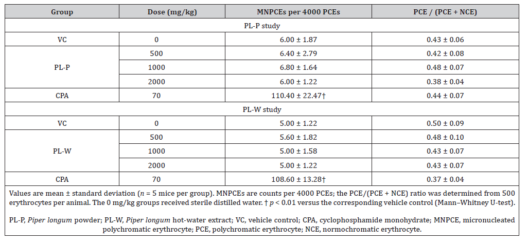
The proportion of immature erythrocytes, expressed as PCE/ (PCE + NCE), did not differ significantly between the treatment groups and their respective vehicle controls. Mean proportions ranged from 0.38 to 0.48 for PL-P and from 0.43 to 0.48 for PL-W, compared with vehicle-control values of 0.43 and 0.50, respectively. No deaths or abnormal clinical signs were observed in the testarticle groups, and body weights did not differ significantly from those of the vehicle controls. Individual body weights and clinical observations are provided in Supplementary Tables S8 and S9.
CPA significantly increased MNPCE counts in both experiments (p < 0.01), with means of 110.40 ± 22.47 for PL-P and 108.60 ± 13.28 for PL-W, compared with vehicle-control means of 6.00 ± 1.87 and 5.00 ± 1.22 per 4000 PCEs, respectively. Both concurrent vehicle-control means were within the laboratory’s historical male vehicle-control range of 2–12 MNPCEs per 4000 PCEs (2015–2019; data not shown). These observations support the responsiveness of the micronucleus assay.
The absence of increased MNPCE counts is consistent with the findings of Thiel et al., who observed no increase in bone marrow micronuclei in mice treated with isolated piperine up to the maximum tolerated dose [9]. However, constituent-level findings cannot establish the in vivo genotoxicity profile of whole herbal preparations, whose composition and disposition may differ from those of an isolated compound.
A further limitation is that bone marrow exposure was not established for either preparation. The absence of a significant reduction in the immature erythrocyte proportion provides no evidence of marrow toxicity through this endpoint, but it does not demonstrate adequate exposure of erythroid precursor cells. OECD TG 474 and EFSA guidance identify evidence of targettissue exposure as an important consideration when interpreting a negative micronucleus result [15, 19]. Accordingly, the present observations support the absence of detectable micronucleus induction in the sampled bone marrow under the dosing conditions examined. They do not provide a definitive exclusion of in vivo genotoxicity, given the unestablished target-tissue exposure and uncertainty in the PL-P positive-control response.
Implications and limitations
The combined chemical characterization and genotoxicity findings provide complementary evidence for assessing PL-P and PL-W. Neither preparation produced a biologically relevant bacterial reverse mutation response or a statistically significant increase in structural chromosomal aberrations in CHL cells under the conditions examined. The differences in marker contents and effects on cell proliferation support interpreting these findings in relation to each preparation. Lower marker concentrations in PL-W do not, by themselves, establish lower genotoxic potential.
Precipitation during the in vitro experiments introduces uncertainty regarding dissolved concentrations, and nominal concentrations do not establish intracellular exposure to individual constituents. Chemical characterization was restricted to four markers and did not comprehensively describe the mixtures or identify the constituents responsible for inhibition of cell proliferation. In the micronucleus experiments, dosing formulations were not analyzed for stability, homogeneity, or concentration, adding uncertainty to characterization of the administered preparations.
The in vivo experiments showed no statistically significant increase in bone marrow micronuclei, but target-tissue exposure was not established. Unchanged immature erythrocyte proportions cannot independently confirm adequate marrow exposure [15, 19]. The use of male mice also limits direct assessment of responses in females. These limitations qualify the biological scope of the findings despite the significant responses in both positive-control groups.
These findings apply to the tested preparations and experimental conditions. Their generalizability to other P. longum products depends on comparability of botanical material, processing, and chemical composition. The study does not establish safety during long-term human use, exclude non-genotoxic mechanisms of carcinogenesis, or define a clinical dose or quantitative human safety margin. Additional evidence concerning systemic and bone marrow exposure to relevant constituents or metabolites would strengthen interpretation of the in vivo findings.
Conclusion
Neither PL-P nor PL-W produced a biologically relevant bacterial mutagenic response or a statistically significant increase in structural chromosomal aberrations in CHL cells. No statistically significant increase in mouse bone marrow micronuclei was detected. These findings provide preparation-specific evidence for the genotoxicity assessment of P. longum under the conditions tested. Interpretation remains limited by precipitation in vitro and the absence of demonstrated bone marrow exposure in vivo. The results do not establish safety for long-term human use.
Acknowledgements
We would like to thank Professor Ozkan Ozden (Kafkas University, Turkiye) for English language editing and scientific comments.
Funding
This research was supported by the Korea Institute of Toxicology (KK-2603-02, KS-2601-02).
Conflict of Interest
The authors declare no competing interests.
Data Availability
The data supporting the findings of this study are included in this article and its supplementary materials. Additional datasets generated and/or analyzed during the current study are available from the corresponding author upon reasonable request.
Author Contributions
N.-Y.K.: Writing–original draft, Formal analysis, Data curation. S.-M.B.: Writing–original draft, Investigation, Formal analysis, Conceptualization, Methodology. D.Y.K.: Writing–original draft, Investigation, Formal analysis, Conceptualization, Methodology. N.-Y.J.: Investigation, Data curation. K.-H.H: Investigation, review & editing Y.-B.K.: Investigation, review & editing. S.-H.P.: Writing– original draft, review & editing, Supervision, Investigation, Conceptualization, Funding acquisition.
- Biswas P, Pandey DK, Shekhawat MS, Dey A, Malik T (2024) Tissue-specific variations of piperine in ten populations of Piper longum L.: bioactivities and toxicological profile Scientific Reports 14(1).
- Sunila ES, Kuttan G (2004) Immunomodulatory and antitumor activity of Piper longum Linn. and piperine. Journal of Ethnopharmacology 90(2-3): PP 339-346.
- Luca SV, Gaweł-Bęben K, Strzępek-Gomółka M, Czech K, Trifan A, et al. (2021) Insights into the phytochemical and multifunctional biological profile of spices from the genus Piper. Antioxidants 10(10): 1642.
- European Medicines Agency (2008) Guideline on the assessment of genotoxicity of herbal substances/preparations. EMEA/HMPC/107079/2007. Committee on Herbal Medicinal Products, London.
- Sivaranjani R, George JK, Saji KV (2019) Evaluation of chemo-diversity in major Piper species for three piperamides using validated RP-HPLC method. Genetic Resources and Crop Evolution 66 (3): PP 1635-1641.
- Xu R, Chen X, Wang X, Yu L, Zhao W, et al. (2019) Development and validation of an ultra-high performance supercritical fluid chromatography-photodiode array detection-mass spectrometry method for the simultaneous determination of 12 compounds in Piper longum L. Food Chemistry 298: 125067.
- Bhardwaj RK, Glaeser H, Becquemont L, Klotz U, Gupta SK, et al. (2002) Piperine, a major constituent of black pepper, inhibits human P-glycoprotein and CYP3A4. Journal of Pharmacology and Experimental Therapeutics 302(2): PP 645-650.
- Karekar VR, Mujumdar AM, Joshi SS, Dhuley J, Shinde SL, et al. (1996) Assessment of genotoxic effect of piperine using Salmonella typhimurium and somatic and germ cells of Swiss albino mice. Arzneimittel-forschung 46(10): PP 972-975.
- Thiel A, Buskens C, Woehrle T, Etheve S, Schoenmakers A, et al. (2014) Black pepper constituent piperine: genotoxicity studies in vitro and in vivo. Food and Chemical Toxicology 66: PP 350-357.
- Selvendiran K, Padmavathi R, Magesh V, Sakthisekaran D (2005) Preliminary study on inhibition of genotoxicity by piperine in mice. Fitoterapia 76(3-4): PP 296-300.
- Yadav V, Krishnan A, Zahiruddin S, Ahmad S, Vohora D (2023) Amelioration of cyclophosphamide-induced DNA damage, oxidative stress, and hepato- and neurotoxicity by Piper longum extract in rats: the role of γH2AX and 8-OHdG. Frontiers in Pharmacology 14: 1147823.
- Baranoski A, Semprebon SC, Biazi BI, Zanetti TA, Corveloni AC, et al. (2024) Piperlongumine inhibits antioxidant enzymes, increases ROS levels, induces DNA damage and G2/M cell cycle arrest in breast cell lines. Journal of Toxicology and Environmental Health, Part A 87(7): PP 294-309.
- OECD (2020) Test No. 471: Bacterial Reverse Mutation Test. OECD Guidelines for the Testing of Chemicals, Section 4. OECD Publishing, Paris, France.
- OECD (2016) Test No. 473: In Vitro Mammalian Chromosomal Aberration Test. OECD Guidelines for the Testing of Chemicals, Section 4. OECD Publishing, Paris, France.
- OECD (2016) Test No. 474: Mammalian Erythrocyte Micronucleus Test. OECD Guidelines for the Testing of Chemicals, Section 4. OECD Publishing, Paris, France.
- Maron DM, Ames BN (1983) Revised methods for the Salmonella mutagenicity test. Mutat Res 113(3-4): 173-215.
- National Toxicology Program. Piperine (94-62-2). Chemical Effects in Biological Systems (CEBS). Genetic Toxicity Evaluation of Piperine in Salmonella/E. coli Mutagenicity Test or Ames Test, Study A60806. Research Triangle Park, NC: National Toxicology Program.
- Bezerra DP, Vasconcellos MC, Machado MS, Villela IV, Rosa RM, et al. (2009). Piplartine induces genotoxicity in eukaryotic but not in prokaryotic model systems. Mutation Research, 677(1-2): PP8-13.
- EFSA Scientific Committee (2017). Clarification of some aspects related to genotoxicity assessment. EFSA Journal, 15(12): e05113.
-

This work is licensed under a Creative Commons Attribution-NonCommercial 4.0 International License.