 Case Report
Image in Surgery Diverticula of The Thoracic Esophagus Discover on a Gastroduodenal Transit
Wael Ferjaoui*, Mohamed Wejih Dougaz, Mohamed Ali Chaouch, Hichem Jerraya, Ibtissem Bouasker and Ramzi Nouira
Department B of surgery, Charles Nicolle Hospital, Tunisia
Wael Ferjaoui, Department B of surgery, Charles Nicolle Hospital, Tunisia.
Received Date: April 19, 2020; Published Date: May 01, 2020
Case Presentation
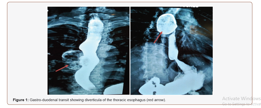
We present the case of a 49-year-old female patient with no past medical history. She presented with dysphagia and regurgitation for 8 months before admission. Physical examination was unremarkable. Blood tests were normal. Eso-gastro-duodenal fibroscopy was made and showed a huge diverticulum 31 cm from the dental arches. In order to better exploit this diverticulum, we completed with a transit which showed a large diverticulum at the level of 1/3 of the middle of the esophagus (Figure 1). The patient was operated bay right thoracoscopy and a diverticulectomy was done. The post-operative period was uneventful.

Diverticula of the thoracic esophagus are anatomically classified into diverticula of the proximal esophagus, the midthoracic esophagus and the distal esophagus [1]. Most of these diverticula are asymptomatic [2]. However, they may symptomatic. They are manifested by chest pain, dysphagia, and regurgitation. Eso-gastro-duodenal fibroscopy, the gastro-duodenal transit and the computed tomography of the chest are essential for the diagnosis. The treatment is surgical.
References
- Duranceau AC (1988) Diverticula of the oesophageal body. In: Jamieson GG (edr), Surgery of the oesophagus, Churchill Livingstone, UK, pp. 489-500.
- Fékété F, Vons C (1992) Surgical management of esophageal thoracic diverticula. Hepatogastroenterology 39(2): 97-99.
-
Wael Ferjaoui*, Mohamed Wejih Dougaz, Mohamed Ali Chaouch, et al., Image in Surgery Diverticula of The Thoracic Esophagus Discover on a Gastroduodenal Transit. Anaest & Sur Open Access J. 1(5): 2020. ASOAJ.MS.ID.000525.
-
Surgery, Thoracic esophagus, Gastroduodenal transit, Physical examination, dental arches, Diverticula, Diagnosis, Patient
-

This work is licensed under a Creative Commons Attribution-NonCommercial 4.0 International License.


