

 Review Article
Respiratory Muscle Effort Monitoring in Neurocritical Care: Moving Beyond Brain-Centric Management
Received Date: July 20, 2026; Published Date:July 28, 2026
Introduction
For decades, neurocritical care management has been predominantly “brain-centric.” Protocols strictly prioritize the maintenance of cerebral perfusion pressure (CPP), tight control of intracranial pressure (ICP), and prevention of secondary brain injury. However, the lungs and the brain exist in a continuous, bidirectional dialogue [1]. Respiratory failure is a major non-neurological complication in neurocritical patients, often requiring invasive mechanical ventilation (IMV).
While protective ventilation strategies (low tidal volumes and controlled driving pressures) are standard in general intensive care units [2-3], applying them to brain-injured patients presents unique challenges. Respiratory drive in these patients is unpredictable, ranging from severe hypoventilation to intense, central-driven hyperventilation [4]. Consequently, monitoring respiratory muscle effort is no longer a luxury; it is a critical necessity to prevent both lung and brain secondary injuries.
The Pathophysiological Dilemma: P-SILI vs. Secondary Brain Injury
In neurocritical patients capable of triggering the ventilator, excessive respiratory drive leads to high inspiratory effort, resulting in Patient-Self-Induced Lung Injury (P-SILI) [5-6]. The large transpulmonary pressure swings created by strong diaphragmatic contraction can generate excessive transpulmonary driving pressure, regional hyperinflation, and occult pendelluft [7], compounding pulmonary inflammation.
Conversely, suppressing this effort via heavy sedation or neu romuscular blockade is not a benign intervention. Sedatives can alter neurological examinations, the cornerstone of neurocritical monitoring, and worsen hemodynamics. Furthermore, excessive respiratory muscle suppression induces rapid diaphragm atrophy, ventilator-induced diaphragmatic dysfunction (VIDD) [8].
Finding the “sweat spot” where the patient breathes enough to maintain diaphragmatic tone without generating harmful transpulmonary pressure swings requires precise, dynamic monitoring tools [6-9].
The Toolkit: Advanced Markers of Respiratory Effort
To individualize mechanical ventilation in neurocritical care, clinicians must move beyond standard bedside airway pressures (Ppeak, Pplat) and look directly at respiratory effort markers:
Airway Occlusion Pressure (P0.1)
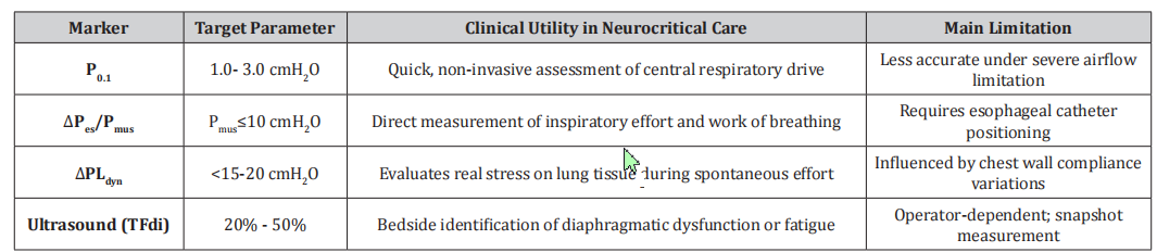
P0.1 reflects the drop in airway pressure during the first 100 milliseconds of an occluded inspiratory effort [10]. It serves as a reliable surrogate for central respiratory drive. In neurocritical patients, where brainstem signals or cortical disinhibition can cause extreme drive variation [4], P0.1 is an invaluable, non-invasive bedside tool. Normal values range from -1.0 to -3.0 cmH₂O; values consistently above 3.5–4.0 cmH₂O signal excessive respiratory drive and impending fatigue or P-SILI risk [6,9].
Esophageal Manometry (Pes) and Muscle Pressure (Pmus)
Esophageal balloon catheterization remains the clinical gold standard for assessing pleural pressure dynamics [11].
Muscular Pressure (Pmus): Calculated from esophageal pressure swings (ΔPes) taking into account chest wall elastance, Pmus directly quantifies the mechanical work performed by the respiratory muscles [11]
Dynamic Transpulmonary Pressure (ΔPLdyn): Measured as Paw – Pes, dynamic transpulmonary pressure isolates the stress applied directly to the lung parenchyma during active breathing. Keeping ΔPLdyn within safe limits (typically < 15- 20 cmH₂O) protects against P-SILI, even in the presence of strong spontaneous efforts [2,7].
Diaphragmatic Ultrasound and Electromyography (Eadi)
Non-invasive bed-side ultrasound allows evaluation of diaphragm excursion and thickening fraction (TFdi), offering a morphological and functional assessment of work of breathing [12]. Neurally Adjusted Ventilatory Assist (NAVA) catheter technology measuring electrical activity of the diaphragm (Eadi) provides real-time neural drive monitoring, bridging the gap between neural output and mechanical delivery [6] (Table 1).
Table 1:Main markers of respiratory effort target parameters, clinical utility, and limitations in neurocritical patients.
Special Considerations in Brain-Injured Populations
Monitoring muscle effort in neurocritical care is complicated by specific physiological interactions: 1. Intracranial Pressure (ICP) Fluctuations: Large negative intrathoracic pressure swings generated by severe inspiratory effort increase venous return transiently, but can impair cerebral venous drainage, leading to spikes in ICP and drops in CPP [1,4].
2. PaCO2 and Cerebral Blood Flow: Patients with elevated drive often develop respiratory alkalosis, inducing cerebral vasoconstriction and ischemia. Conversely, under-ventilation worsens hypercapnia, raising ICP [4,13]. Effort markers help guide mechanical support adjustments to stabilize arterial blood gases without over-sedation.
Future Directions and Call to Action
The future of neurocritical mechanical ventilation lies in multimodal monitoring that integrates neuro-monitoring (ICP, brain tissue oxygenation) with advanced respiratory mechanics (P0.1, Pes, Pmus) [4,13].
We must move away from the binary protocol of “full suppression vs. unmonitored spontaneous breathing.” Clinical trials are urgently needed to establish targeted effort ranges specifically for brain-injured patients, evaluating whether effort-tailored ventilation protocols reduce ICU length of stay, weaning failure, and longterm neurological disability [13].
Conclusion
Optimizing mechanical ventilation in neurocritical patients demands a delicate balance between neuroprotection and lung protection. Respiratory muscle effort markers—such as P0.1, Pmus and PLdyn provide the necessary compass to navigate this tension. Incorporating these tools into routine neuro-intensive care is an essential step toward safer, personalized, and truly holistic patient management.
Acknowledgments
None.
Conflict of Interest
No Conflict of interest.
References
- Pelosi P, Rocco PR (2011) The lung and the brain: a dangerous liaison. Crit Care 15(3): 168.
- Brochard L, Slutsky A, Pesenti A (2017) Mechanical Ventilation to Minimize Progression of Lung Injury in Acute Respiratory Failure. Am J Respir Crit Care Med 195(4): 438-442.
- Goligher EC, Ferguson ND, Brochard LJ, et al. (2018) Mechanical Ventilation Strategies for Intensive Care Unit Patients: A Primer for Clinicians. JAMA 319(17): 1805-1816.
- Rola P, Miralles-Aguiar F, Fresno M (2020) Respiratory drive and effort in the neurocritical care patient: A narrative review. Neurocrit Care 33(2): 520-531.
- Gattinoni L, Tonetti T, Quintel M (2017) How the lung gets injured. Curr Opin Crit Care 23(1): 3-8.
- Telias I, Damiani F, Brochard L (2018) The patient-ventilator interaction: drives, efforts, and breathlessness. Curr Opin Crit Care 24(1): 1-8.
- Yoshida T, Torsani V, Gomes S, Roberta R De Santis, Marcelo A Beraldo, et al. (2013) Spontaneous effort causes occult pendelluft during mechanical ventilation. Am J Respir Crit Care Med 188(12): 1420-1427.
- Mauri T, Yoshida T, Bellani G, et al. (2016) Patient-ventilator dyssynchrony: clinical impact and management in acute respiratory failure. Lancet Respir Med 4(10): 782-791.
- Ferreira LL, Lunardi AC (2023) Non-invasive monitoring of drive and respiratory muscle effort in patients under mechanical ventilation: an integrative review. ASSOBRAFIR Cien 14: e723.
- Whitelaw WA, Derenne JP (1993) Airway occlusion pressure. J Appl Physiol 75(5): 2337-2347.
- Akoumianaki E, Maggiore SM, Valenza F, Giacomo Bellani, Amal Jubran, et al. (2014) The application of esophageal pressure measurement in patients with respiratory failure. Am J Respir Crit Care Med 189(5): 520-531.
- Umbrello M, Chiumello D (2018) Diaphragm assessment by ultrasound in ICU patients: how and why? Ann Intensive Care 8(1): 84.
- Stevens RD, Lazaridis C, Chalela JA (2019) Mechanical ventilation in neurocritical care: A consensus statement and clinical practice guidelines. Neurocrit Care. 31(1): 125-140.
-

This work is licensed under a Creative Commons Attribution-NonCommercial 4.0 International License.