




 Review Article
Prognostic Stratification in Heart Failure: Emerging Biomarkers and Explainable Artificial Intelligence in Real-World Care
Received Date:February 10, 2026; Published Date:February 17, 2026
Abstract
Heart Failure (HF) remains a leading cause of morbidity, mortality, and healthcare utilization worldwide, driven by population ageing,
multimorbidity, and improved survival after acute cardiovascular events. Although Left Ventricular Ejection Fraction (LVEF) remains central to
classification and therapeutic decision-making, it incompletely captures the biological and clinical heterogeneity of HF-particularly in heart failure
with preserved and mildly reduced ejection fraction. Traditional prognostic markers such as natriuretic peptides, renal function, sodium, and anaemia
underpin established risk scores but may underestimate vulnerability in older, frail, and socially disadvantaged populations. Emerging markers of
biological reserve, systemic inflammation, and nutritional vulnerability-including clinical frailty scales, temporal muscle thickness, neutrophil-tolymphocyte
ratio, and nutritional indices-have gained increasing attention as accessible, low-cost predictors with potential incremental prognostic
value.
In parallel, Artificial Intelligence (AI) and machine-learning approaches offer opportunities to integrate multidimensional clinical data,
identify phenotypic clusters, and improve prediction of mortality and readmission. However, their clinical adoption requires methodological rigor,
transparency, interpretability, and validation in real-world settings. This narrative state-of-the-art review synthesizes contemporary evidence across
HF phenotypes, classical and emerging prognostic markers, multidisciplinary HF care models, and explainable AI-enabled prognostication. We
highlight key knowledge gaps and discuss clinical and organizational implications for implementing integrated, parsimonious, and interpretable risk
stratification tools within multidisciplinary HF units. Together, these dimensions support a translational pathway from multimodal data integration
to deployable prognostic tools capable of informing individualized care and optimizing resource allocation in real-world HF management.
Keywords:Heart failure; prognosis; frailty; temporal muscle thickness; inflammation; neutrophil-to-lymphocyte ratio; artificial intelligence; multidisciplinary care
Abbreviations:HF: heart failure; LVEF: left ventricular ejection fraction; HFrEF: heart failure with reduced ejection fraction; HFmrEF: heart failure with mildly reduced ejection fraction; HFpEF: heart failure with preserved ejection fraction; CFS: Clinical Frailty Scale; TMT: temporal muscle thickness; NLR: neutrophil-to-lymphocyte ratio; AI: artificial intelligence
Case Description
Heart Failure (HF) is a complex clinical syndrome and a major public health challenge, associated with high prevalence, recurrent hospitalizations, and persistent mortality despite major therapeutic advances. Its burden continues to rise globally due to population ageing, improved survival after acute cardiovascular events, and increasing multimorbidity. Prognostic stratification is therefore central to HF management, guiding therapeutic intensity, followup strategies, and allocation of healthcare resources, particularly during vulnerable transition periods after hospitalization. Although classification based on Left Ventricular Ejection Fraction (LVEF) remains clinically useful and therapeutically actionable, it only partially reflects the underlying pathophysiology and fails to explain substantial within-phenotype heterogeneity. This limitation is particularly evident in HF with preserved and mildly reduced ejection fraction, where systemic comorbidities, inflammation, and biological vulnerability play a dominant role.
In recent years, HF has increasingly been conceptualized as a multisystem syndrome, in which frailty, sarcopenia, inflammation, and nutritional status substantially influence outcomes. However, these domains remain under-integrated into routine prognostic assessment and clinical risk models. At the same time, advances in Artificial Intelligence (AI) and machine learning have enabled the integration of complex, multidimensional clinical data, offering potential improvements in phenotyping and risk prediction. The challenge lies in translating these advances into clinically interpretable and deployable tools. This narrative review provides a clinically oriented synthesis of evidence on prognostic stratification in HF, focusing on the integration of classical and emerging biomarkers, organizational models of care, and explainable AI approaches, with emphasis on real-world applicability within multidisciplinary HF units [1-20].
Discussion
Epidemiology and Burden of Heart Failure
HF affects approximately 1–2% of the adult population and exceeds 10% among individuals older than 70 years. It remains a leading cause of hospitalization and healthcare expenditure, with recurrent admissions representing a major driver of disease burden. In Portugal and other ageing regions, HF prevalence and outcomes are further shaped by multimorbidity, social vulnerability, and geographic dispersion, underscoring the need for structured and proactive care models [21-30].
Pathophysiology and LVEF-Based Phenotypes
Contemporary HF classification recognizes HFrEF, HFmrEF, HFpEF, and HF with recovered ejection fraction. While HFrEF is predominantly characterized by systolic dysfunction and neurohormonal activation, HFpEF is increasingly understood as a multisystem disorder driven by cardiometabolic comorbidities, systemic inflammation, endothelial dysfunction, and impaired reserve. HFmrEF represents a heterogeneous and dynamic phenotype, while recovered EF denotes improvement with therapy but persistent residual risk. These distinctions highlight the limitations of LVEF as a sole prognostic descriptor [31-40].
Prognosis Across Ejection Fraction Phenotypes
Although prognosis varies across LVEF categories, substantial overlap exists. Mortality and rehospitalization remain clinically significant across all phenotypes, particularly in the early postdischarge period. Importantly, non-cardiovascular events contribute substantially to outcomes in HFpEF, reflecting systemic disease burden. These observations support the need for prognostic models that transcend LVEF-based classification [41-50].
Classical Prognostic Predictors
Classical prognostic markers-such as natriuretic peptides, renal dysfunction, hyponatraemia, anaemia, age, and comorbidity burden-form the backbone of existing risk scores. Tools such as the MAGGIC risk score and the Seattle Heart Failure Model integrate these variables to estimate survival. However, their performance may be suboptimal in older, frail, and socially vulnerable populations, where biological reserve and functional status are critical determinants of outcome [51-60].
Emerging Biomarkers of Biological Vulnerability
Frailty and Sarcopenia
Frailty is highly prevalent among HF patients and consistently associated with mortality, rehospitalization, and reduced tolerance to guideline-directed therapies. Sarcopenia and cachexia reflect advanced HF biology and catabolic imbalance, conferring additional risk through inflammatory and metabolic pathways [61-70].
Temporal Muscle Thickness
Temporal Muscle Thickness (TMT), measured opportunistically on routine cranial CT imaging, has emerged as an objective surrogate of sarcopenia and biological reserve. Evidence across clinical contexts supports its association with mortality and adverse outcomes, with good reproducibility and feasibility for automated assessment. In HF populations, TMT offers a pragmatic avenue to quantify vulnerability beyond traditional metrics [71-80].
Systemic Inflammation and Neutrophil-To-Lymphocyte Ratio
Chronic low-grade inflammation is a hallmark of HF, particularly HFpEF. The neutrophil-to-lymphocyte ratio (NLR) integrates innate immune activation and physiological stress and has shown consistent associations with mortality and adverse events in HF. Its low cost and universal availability make it an attractive candidate for routine risk stratification [81-90].
Nutritional Vulnerability
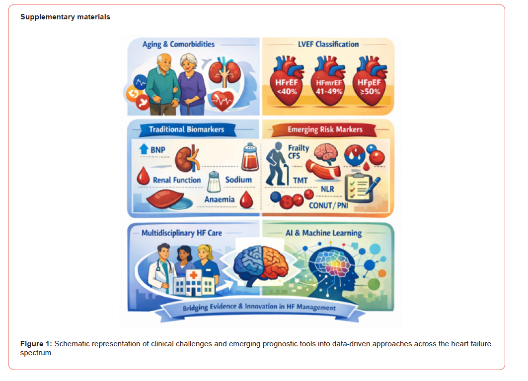
Nutritional indices such as CONUT and the prognostic nutritional index capture malnutrition and inflammatory burden, both independently associated with adverse outcomes in HF. Their integration may be particularly relevant in frail and advanced disease states (Figure 1).
Multidisciplinary Heart Failure Units and Organization of Care
Dedicated multidisciplinary HF units reduce rehospitalizations and improve outcomes, particularly during post-discharge transitions. These programs facilitate coordinated optimization of therapy, patient education, and structured follow-up. In regions characterized by ageing and social vulnerability, such models may mitigate inequities and prevent avoidable decompensation (Tables 1&2).
Table 1: Classical Prognostic Markers in Heart Failure.

Table 2: Classical Prognostic Markers in Heart Failure.

CT – Computed Tomography
Artificial Intelligence and Explainable Prediction in HF
AI and machine-learning approaches enable the integration of complex multimodal data, supporting phenotyping and risk prediction through non-linear modelling. However, clinical translation requires careful attention to overfitting, bias, calibration, and generalizability. Explainable AI techniques may enhance clinician trust and interpretability but must be applied within rigorous methodological frameworks aligned with reporting standards such as TRIPOD and PROBAST [91-100].
Knowledge Gaps and Research Agenda
Key gaps include under-integration of frailty and sarcopenia into routine risk assessment, limited incorporation of social determinants, and insufficient external validation of AI-based models. Future research should prioritize parsimonious, explainable models evaluated within multidisciplinary care pathways and focused on clinically actionable endpoints [101-110].
Limitations of this Review
As a narrative review, this work does not follow a formal systematic review protocol, and selection bias cannot be fully excluded. Nevertheless, emphasis was placed on high-level evidence, methodological rigor, and real-world applicability (Tables 3&4).
Table 3: Traditional Risk Scores vs AI-Based Prognostic Models.

Table 4: Clinically Relevant Prognostic Endpoints in Contemporary HF Care

Conclusion
HF remains a major clinical and organizational challenge characterized by rising prevalence, multimorbidity, and recurrent hospitalization. While LVEF-based phenotyping remains useful, it is insufficient for precise individual prognostication. Classical biomarkers are essential but do not fully capture systemic vulnerability. Emerging markers of frailty, sarcopenia, inflammation, and nutrition-particularly TMT and NLR-offer promising, accessible additions with potential incremental value. Multidisciplinary HF care models provide a compelling framework to operationalize risk-stratified care, while explainable AI approaches may further enhance prognostic precision. Together, these dimensions support the development of integrated, interpretable prognostic tools capable of informing personalized care and optimizing outcomes in real-world HF management.
Acknowledgements
The authors acknowledge the Multidisciplinary Heart Failure Unit (UMIC) team at Hospital de Faro for their clinical and organizational support.
Conflict of Interest
The authors declare no conflicts of interest related to this manuscript.
- McDonagh TA, Metra M, Adamo M, Gardner RS, Baumbach A, et al. (2021) 2021 ESC guidelines for the diagnosis and treatment of acute and chronic heart failure. Eur Heart J 42(36): 3599-3726.
- Heidenreich PA, Bozkurt B, Aguilar D, Allen LA, Byun JJ, et al. (2022) 2022 AHA/ACC/HFSA guideline for the management of heart failure. Circulation 145(18): e895-e1032.
- Bozkurt B, Coats AJS, Tsutsui H, Yancy C, Zhang J, et al. (2021) Universal definition and classification of heart failure. Eur J Heart Fail 27(4): 387-413.
- Savarese G, Becher PM, Lund LH, Seferovic P, Rosano GMC, et al. (2023) Global burden of heart failure: a comprehensive and updated review of epidemiology. Heart Fail Rev 28(5): 1451-1465.
- Virani SS, Alonso A, Aparicio HJ, Benjamin EJ, Bittencourt MS, et al. (2021) heart disease and stroke statistics-2021 Update. Circulation 143(8): e254-e743.
- Braunwald E. (2015) The war against heart failure. Lancet 385(9970): 812-824.
- Roth GA, Mensah GA, Johnson CO, Addolorato G, Ammirati E, et al. (2020) Global burden of cardiovascular diseases and risk factors, 1990-2019. J Am Coll Cardiol 76(25): 2982-3021.
- Ziaeian B, Fonarow GC. (2016) Epidemiology and aetiology of heart failure. Nat Rev Cardiol 13(6): 368-378.
- Roger VL (2013) Epidemiology of heart failure. Circ Res 113(6): 646-659.
- Dunlay SM, Roger VL, Redfield MM (2017) Epidemiology of heart failure with preserved ejection fraction. Nat Rev Cardiol 14(10): 591-602.
- Ambrosy AP, Fonarow GC, Butler J, Chioncel O, Greene SJ, et al. (2014) Global health and economic burden of hospitalizations for heart failure. J Am Coll Cardiol 63(12): 1123-1133.
- Gheorghiade M, Vaduganathan M, Fonarow GC, Bonow RO (2013) Rehospitalization for heart failure: problems and perspectives. J Am Coll Cardiol 61(4): 391-403.
- Chioncel O, Lainscak M, Seferovic PM, Anker SD, Crespo-Leiro MG, et al. (2017) Epidemiology and one-year outcomes in chronic heart failure. Eur J Heart Fail 19(12): 1574-1585.
- Ceia F, Fonseca C, Mota T, Morais H, Matias F, et al. (2002) Prevalence of chronic heart failure in Portugal (EPICA study). Eur J Heart Fail 4(4): 531-539.
- Fonseca C, Araújo I, Moreira I, et al. (2020) The Portuguese Heart Failure Study (PORTHOS). Rev Port Cardiol 39(3): 139-148.
- Lam CSP, Solomon SD (2022) Classification and pathophysiology of heart failure phenotypes. Circ Res 131(10): 827-846.
- Paulus WJ, Tschöpe C. (2013) A novel paradigm for heart failure with preserved ejection fraction. J Am Coll Cardiol 62(4): 263-271.
- Solomon SD, Vaduganathan M, Claggett B, et al. (2021) Heart failure with mildly reduced ejection fraction. Eur Heart J 42(17): 1672-1681.
- Dunlay SM, Roger VL (2020) Heart failure with recovered ejection fraction. Circulation 141(13): 1085-1095.
- Wilcox JE, Fang JC, Margulies KB, Mann DL. (2020) Heart failure with recovered left ventricular ejection fraction: JACC Scientific Expert Panel. J Am Coll Cardiol 76(6): 719-734.
- Halliday BP, Wassall R, Lota AS, Khalique Z, Gregson J, et al. (2019) Withdrawal of pharmacological treatment for heart failure in patients with recovered dilated cardiomyopathy (TRED-HF). Lancet 393(10166): 61-73.
- Maisel A (2017) Use of natriuretic peptides in heart failure. J Am Coll Cardiol 70(19): 243-256.
- Tang WHW, Kitai T (2017) Cardiorenal syndrome in heart failure. Circ J 81(11): 1626-1634.
- Liu M, Chan CP, Yan BP (2020) Hyponatremia in heart failure. Int J Cardiol 302: 1-7.
- Clark AL, Fonarow GC, Horwich TB (2014) Anaemia in heart failure. Eur J Heart Fail 16(8): 848-855.
- Pocock SJ, Ariti CA, McMurray JJV, Maggioni A, Kober L, et al. (2013) Predicting survival in heart failure (MAGGIC). Eur Heart J 34(19): 1404-1413.
- Levy WC, Mozaffarian D, Linker DT, Sutradhar SC, Anker SD, et al. (2006) The Seattle Heart Failure Model. Circulation 113(11): 1424-1433.
- Damman K, Valente MAE, Voors AA, O’Conner CM, Veldhhuisen DJV, et al. (2014) Renal impairment and outcome in heart failure. Eur Heart J 35(7): 455-469.
- Gheorghiade M, Rossi JS, Cotts W, et al. (2007) Persistent hyponatremia in severe heart failure. Arch Intern Med 167(18): 1998-2005.
- Anand IS, Gupta P (2018) Anemia and iron deficiency in heart failure. Circulation 138(1): 80-98.
- Anker SD, Comin Colet J, Filippatos G, Willenheimer R, Dickstein K, et al. (2009) Ferric carboxymaltose in patients with heart failure and iron deficiency. N Engl J Med 361(25): 2436-2448.
- Jankowska EA, Rozentryt P, Witkowska A, Nowak J, Hartmann O, et al. (2010) Iron deficiency in systolic chronic heart failure. Eur Heart J 31(15): 1872-1880.
- Ponikowski P, Kirwan BA, Anker SD, McDonagh T, Dorobantu M, et al. (2020) Ferric carboxymaltose after acute heart failure (AFFIRM-AHF). Lancet 396(10266): 1895-1904.
- McDonagh T, Damy T, Doehner W, et al. (2022) Screening, diagnosis and treatment of iron deficiency in chronic heart failure. Eur J Heart Fail 24(9): 1453-1463.
- Aimo A, Januzzi JL Jr, Vergaro G, et al. (2019) High-sensitivity troponin in chronic heart failure. Eur J Heart Fail 21(10): 1271-1283.
- Bayes-Genis A, Zhang Y, Ky B (2015) ST2 and galectin-3 in heart failure. J Am Coll Cardiol 65(19): 1988-2002.
- Felker GM, Hasselblad V, Tang WHW, Hernandez AF, Armstrong PW, et al. (2004) Troponin I in acute decompensated heart failure. J Am Coll Cardiol 44(6): 1075-1082.
- Murphy SP, Kakkar R, McCarthy CP, Januzzi JL Jr (2020) Inflammation in heart failure. J Am Coll Cardiol 75(11): 1324-1340.
- Mann DL (2015) Innate immunity and the failing heart. Circ Res 116(7): 1254-1268.
- Castillo EC, Vázquez-Garza E, Yee-Trejo D, et al. (2020) Role of inflammation in heart failure. Curr Cardiol Rep 22(11): 139.
- Mesquita T, Lin Y, Ibrahim A (2021) Chronic low-grade inflammation in HFpEF. Aging Cell. 20(9): e13453.
- Gullestad L, Ueland T, Vinge LE, Finsen A, Yndestad A, et al. (2012) Inflammatory cytokines in heart failure. Cardiology 122(1): 23-35.
- Zahorec R (2001) Ratio of neutrophils to lymphocytes as a marker of stress. Bratisl Lek Listy 102(1): 5-14.
- Vakhshoori M, Nemati S, Sabouhi S, Yavari B, Shakarami M, et al. (2023) Neutrophil-to-lymphocyte ratio in heart failure. BMC Cardiovasc Disord 23(1):345-555.
- Delcea C, Buzea CA, Dobrev D, Dan GA (2024) Prognostic roles of leukocyte ratios in heart failure. Int J Cardiol Heart Vasc 54: 101502.
- Angkananard T, Anothaisintawee T, McEvoy M, Attia J, Thakkinstain A, et al. (2018) Neutrophil-to-lymphocyte ratio and cardiovascular risk. Clin Cardiol 41(2): 236-245.
- Rawat V, Goyal P, Ahsan SA, Surya Srivyshnavi KS, Asghar AH, et al. (2025) Prognostic significance of neutrophil-to-lymphocyte ratio in transcatheter aortic valve implantation. Cureus 17(1): e77909.
- Lee JS, Kim NY, Na SH, Youn YH, Shin CS (2018) Reference values of neutrophil-to-lymphocyte ratio. Int J Lab Hematol 40(6): e83-e86.
- Forget P, Khalifa C, Defour JP, Latinne D, Van Pel MC, et al. (2017) What is the normal value of the neutrophil-to-lymphocyte ratio? BMC Res Notes 10: 12.
- Templeton AJ, McNamara MG, Šeruga B, Vera-Badillo FE, Aneja P, et al. (2014) Prognostic role of neutrophil-to-lymphocyte ratio in solid tumors. J Natl Cancer Inst 106(6): dju124.
- Zinellu A, et al. (2020) Neutrophil-to-lymphocyte ratio in systemic lupus erythematosus. Autoimmun Rev 19(6): 102543.
- Shen XN, Niu LD, Wang YJ, Cao XP, Liu Q, et al. (2019) Inflammatory markers and cognitive decline in Alzheimer’s disease. J Alzheimers Dis 68(3): 1081-1092.
- Cruz-Jentoft AJ, Bahat G, Bauer J, Boirie J, Bruyere O, et al. (2019) Sarcopenia: revised European consensus (EWGSOP2). Age Ageing 48(1): 16-31.
- von Haehling S, Ebner N, Dos Santos MR, Anker SD (2020) Sarcopenia and cachexia in heart failure. ESC Heart Fail 7(3): 1072-1084.
- Dent E, Chapman I, Piantadosi C, et al. (2022) Frailty and ageing. Age Ageing 51(1): afab244.
- Rockwood K, Song X, MacKnight C, Bergman H, Hogan DB, et al. (2005) A global clinical measure of fitness and frailty. CMAJ 173(5): 489-495.
- Fried LP, Tangen CM, Walston J, Newman AB, Hirsch C, et al. (2001) Frailty in older adults. J Gerontol A Biol Sci Med Sci 56(3): M146-M156.
- Ranganathan P, et al. (2022) Temporal muscle thickness as a marker of frailty. J Cachexia Sarcopenia Muscle 13(2): 948-957.
- Furtner J, Genbrugge E, Gorlia T, et al. (2019) Temporal muscle thickness as prognostic marker. Eur Radiol 29(8): 3889-3897.
- Furtner J, Berghoff AS, Albtoush OM, et al. (2018) Temporal muscle thickness is an independent prognostic marker in patients with brain metastases. Radiology 287(2): 593-603.
- Maeda K, Ishida Y, Nonogaki T, et al. (2021) Temporal muscle thickness as nutritional marker. Clin Nutr 40(4): 2053-2060.
- Swartz JE, Pothen AJ, Wegner I, Smid EJ, Swart KMA, et al. (2016) Feasibility of using head and neck CT imaging to assess skeletal muscle mass. Oral Oncol 62: 28-33.
- Ignacio de Ulíbarri J, González-Madroño A, de Villar NGP, Gonzalez P, Gonzalez B, et al. (2005) CONUT score. Nutr Hosp 20(1): 38-45.
- Onodera T, Goseki N, Kosaki G (1984) Prognostic nutritional index. Nihon Geka Gakkai Zasshi 85: 1001-1005.
- Anker SD, Ponikowski P, Varney S, Chua TP, Clark AL, et al. (1997) Wasting as a risk factor in chronic heart failure. Lancet 349(9058): 1050-1053.
- Havranek EP, Mujahid MS, Barr DA, Blair IV, Cohen MS, et al. (2015) Social determinants of cardiovascular outcomes. Circulation 132(9): 873-898.
- White-Williams C, Rossi LP, Bittner VA, Driscoll A, Durant RW, et al. (2020) Addressing social determinants in heart failure care. Circulation 141(22): e841-e863.
- Marmot M, Allen J, Goldblatt P, et al. (2010) Fair society, healthy lives. Lancet 376(9756): 1644-1661.
- Berkman LF, Glass T (2000) Social integration and health. In: Berkman LF, Kawachi I, editors. Social epidemiology. Oxford: Oxford University Press PP: 137-173.
- Holt-Lunstad J, Smith TB, Baker M, Harris T, Stephenson D, et al. (2015) Loneliness and mortality. Perspect Psychol Sci 10(2): 227-237.
- Peterson PN, Shetterly SM, Clarke CL, Bekelman DB, Chan PS, et al. (2011) Health literacy and outcomes among patients with heart failure. JAMA 305(16): 1695-1701.
- Riegel B, Moser DK, Buck HG, Dickson VV, Dunbar SB, et al. (2017) Self-care for prevention and management of cardiovascular disease. J Cardiovasc Nurs 32(3): 204-212.
- Lainscak M, Blue L, Clark AL, Dahlstrom U, Dickstein K, et al. (2011) Self-care management of heart failure. Eur J Heart Fail 13(2): 115-126.
- McAlister FA, Stewart S, Ferrua S, McMurray JJV (2004) Multidisciplinary management of heart failure. J Am Coll Cardiol 44(4): 810-819.
- Takeda A, Taylor SJC, Taylor RS, et al. (2012) Disease management programmes for heart failure. Cochrane Database Syst Rev 9: CD002752.
- Allen LA, Stevenson LW, Grady KL, Goldstein NE, Matlock DD, et al. (2012) Decision making in advanced heart failure. Circulation 125(15): 1928-1952.
- Koehler F, Koehler K, Deckwart O, Prescher S, Wegscheider K, et al. (2018) Telemedical interventional management in heart failure II (TIM-HF2). Lancet 392(10152): 1047-1057.
- Ong MK, Romano PS, Edgington S, Aronow HU, Auerbach AD, et al. (2016) Remote patient monitoring after heart failure discharge. JAMA Intern Med 176(3): 310-318.
- Abraham WT, Adamson PB, Bourge RC, Aaron MF, Costanzo MR, et al. (2011) Wireless pulmonary artery haemodynamic monitoring. Lancet 377(9766): 658-666.
- Desai AS, Bhimaraj A, Bhatt K, Jermyn R, Bhatt K, et al. (2017) Ambulatory hemodynamic monitoring in heart failure. J Am Coll Cardiol 69(19): 2357-2365.
- National Institute for Cardiovascular Outcomes Research. National heart failure audit: annual report. London: NICOR; 2015-2024.
- Ahmad T, Pencina MJ, Schulte PJ, et al. (2018) Machine learning-defined heart failure phenotypes. J Am Coll Cardiol 71(18): 2369-2379.
- Mortazavi BJ, Downing NS, Bucholz EM, Dharmarajan K, Manhapra A, et al. (2016) Machine learning techniques for heart failure readmissions. Circ Cardiovasc Qual Outcomes 9(6): 629-640.
- Kwon JM, Kim KH, Jeon KH, et al. (2020) Deep learning-based algorithm for detecting heart failure using electrocardiography. Eur Heart J 41(26): 1995-2003.
- Shadmi E, Korem M, Peri N, et al. (2015) Predicting 30-day readmissions with machine learning models. Med Care 53(10): 829-835.
- Rajkomar A, Dean J, Kohane I (2019) Machine learning in medicine. N Engl J Med 380(14): 1347-1358.
- Topol EJ (2019) High-performance medicine. Nat Med 25(1): 44-56.
- Esteva A, Robicquet A, Ramsundar B, Kuleshov V, DePristo M, et al. (2019) A guide to deep learning in healthcare. Nat Med 25(1): 24-29.
- Steyerberg EW, Vergouwe Y (2014) Towards better clinical prediction models. Eur Heart J 35(29): 1925-1931.
- Steyerberg EW (2019) Clinical prediction models. 2nd ed. Cham: Springer.
- Riley RD, Ensor J, Snell KIE, Burke DL, Harrell FE, et al. (2019) Minimum sample size for developing prediction models. BMJ 368: m441.
- Collins GS, Reitsma JB, Altman DG, Moons KGM (2015) Transparent reporting of multivariable prediction models (TRIPOD). Ann Intern Med 162(1): 55-63.
- Moons KGM, Wolff RF, Riley RD, Whiting PF, Westwood M, et al. (2019) PROBAST tool. Ann Intern Med 170(1): 51-58.
- Page MJ, McKenzie JE, Bossuyt PM, Boutron I, Hoffmann TC, et al. (2020) PRISMA 2020 statement. BMJ 372: n71.
- Hayden JA, van der Windt DA, Cartwright JL, et al. (2013) QUIPS tool. Ann Intern Med 158(4): 280-286.
- Vasan RS, Benjamin EJ (2018) The future of heart failure epidemiology. Circulation 137(1): 1-3.
- Hsu JJ, Ziaeian B, Fonarow GC (2017) Heart failure with mid-range ejection fraction. JACC Heart Fail 5(11): 763-771.
- Dharmarajan K, Hsieh AF, Lin Z, Bueno H, Ross JS, et al. (2013) Diagnoses and timing of 30-day readmissions after hospitalization for heart failure. JAMA 309(4): 355-363.
- Krumholz HM, Wang Y, Mattera JA, Wang Y, Han LF, et al. (2006) An administrative claims model for profiling hospital performance in heart failure. Circulation 113(13): 1693-1701.
- Greene SJ, Fonarow GC, Solomon SD, et al. (2018) Contemporary outcomes and readmission in heart failure. JACC Heart Fail 6(11).
- Rich MW (2019) Heart failure in the older adult. J Am Coll Cardiol 74(2).
- Taylor RS, Sagar VA, Davies EJ, Briscoe S, Coats AJS, et al. (2014) Exercise-based rehabilitation for heart failure. Cochrane Database Syst Rev 2014(4): CD003331.
- Schjødt I, Johnsen SP, Strömberg A, Kristensen NR, Logstrup BB, et al. (2019) Socioeconomic factors and outcomes in heart failure. JACC Heart Fail 7(9): 746-755.
- Witte KK, Patel PA, Walker AMN, Schechter CB, Drozd M, et al. (2018) Socioeconomic deprivation and outcomes in chronic heart failure. Heart 104(12): 993-998.
- Hawkins NM, Jhund PS, McMurray JJV, Capewell S (2012) Heart failure and socioeconomic status. Eur J Heart Fail 14(2): 138-146.
- van Deursen VM, Urso R, Laroche C, Damman K, Dahlstrom U, et al. (2014) Comorbidities in heart failure. Eur J Heart Fail 16(1).
- World Health Organization (2008) Closing the gap in a generation. Geneva: WHO.
- World Health Organization (2010) Social determinants of health: conceptual framework. Geneva: WHO.
- Direção-Geral da Saúde (2015-2024) Normas e relatórios nacionais relevantes para a insuficiência cardí Lisboa: DGS.
- Ministério da Saúde (2015-2024) Programas nacionais de saúde cardiovascular e envelhecimento. Lisboa: Ministério da Saú
-

This work is licensed under a Creative Commons Attribution-NonCommercial 4.0 International License.




