 Short Communication
Proposed Acute Coronary Syndrome (ACS) Chest Pain Pathway Using Quantitative FMTVDM Nuclear Imaging
Richard M Fleming1*, Matthew R Fleming1, Tapan K Chaudhuri2 and Andrew McKusick1,3
1FHHI-Omnific Imaging-Camelot, USA
2Eastern Virginia Medical School, USA
3Sebec Consulting & Media, North Carolina
Richard M Fleming, FHHI-Omnific Imaging-Camelot, El Segundo, CA, USA.
Received Date: June 24, 2019; Published Date: June 28, 2019
Short Communication
There are an estimated 8 to 10 million emergency room visits each year for the evaluation and treatment of acute coronary syndrome; i.e. ischemic coronary artery disease; with potential consequential damage to the heart muscle itself (infarction). The standard diagnostic evaluation has routinely included a series of tests looking for evidence of concurrent tissue injury happening at the time of evaluation. This includes a standard 12-lead (perhaps more depending upon specific clinical acumen and equipment) electrocardiogram (ECG) looking for evidence of ST elevations of greater than 1 mm in two or more contiguous leads or in some instances V4 alone for distal LAD disease. Additional information is occasionally gleaned by reciprocal changes. To compare changes over time, patients routinely undergo more than one ECG. In addition to the use of ECGs (also called EKGs to prevent confusion with the term EEG), looking for membrane conduction and ion transport abnormalities caused by damaged myocyte and conduction tissue resulting from insufficient coronary blood flow for the needs of the tissue, clinicians also routinely obtain a series of blood tests looking for enzymes and proteins which have been released from the myocytes themselves. The sequence with which these “cardiac enzymes” appear in the blood are directly related to the fact that ischemic membrane damage will initially result in the leakage of smaller molecules followed by progressively larger molecules. The classic sequence of released molecules is consequently, based upon our current level of knowledge, myoglobin, creatine kinase (CK), Troponin I (TnI) and Troponin T (TnT), serum glutamicoxaloacetic transaminase (SGOT) which is also known as aspartate aminotransferase (AST) and finally lactate dehydrogenase (LDH).
As already mentioned, the larger the molecule, the longer it will take for sufficient membrane damage to occur to allow sufficient quantities of these enzymes and proteins to appear in the blood in greater than “normalized” levels. It is also true, that once a thrombus within a coronary artery has been lysed either enzymatically or mechanically, the sooner these released compounds can be flushed from the coronary circulation into the peripheral circulation, where they can be measured. Taken together this means time is required to “flush out” the evidence of tissue damage. Another approach is the immediate use of cardiac catheterization intervention and while this will undoubtedly provide an immediate answer to the question, at least three points are worth considering here: (1) not every ACS needs immediate mechanical intervention and such interventions should be saved for those who would benefit most from immediate intervention, (2) there are considerable costs for taking everyone to the Cath lab, including the availability of manpower and equipment, raising certain economic factors along with the cost versus benefit, and finally (3) what about the patients who are not having an actual ACS? Interventional procedures are not without risk and the risk versus benefit ratio should always be considered. All of this leaves us with the issue of time, costs, benefits, and life.
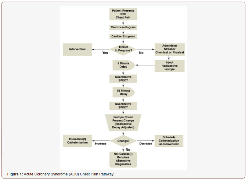
A final possible alternative involves the use of myocardial perfusion imaging (MPI). Until recently, it was thought that most MPI required a delay of at least 1-hour prior to actual imaging of the heart. Clearly if this were true, then based upon timing itself, MPI would be of little or no value in the ACS setting. Recent work with “quantification” [1-4] has demonstrated that a number of technetium-99m isotopes (in addition to other isotopes) are available which undergo redistribution beginning as early as 5-minutes post isotope injection under “stress” conditions. When “stress” imaging was initially conducted with Thallous-201, patients were literally “stressed”; however, the correct use of the term “stress” is actually the use of a method which promotes “relaxation” of coronary arteries, actually “enhancing” rather than “reducing” coronary blood flow and augmenting regional blood flow [5,6] differences thereby exposing areas of impaired (ischemic or occluded) coronary blood flow (CAD) (Figure 1).

The use of this protocol as shown in Figure 1, allows for ACS patients to undergo diagnostic MPI beginning within minutes of evaluation in the Emergency Department of most major medical centers, with comparison redistribution images no later than 55-minutes later. The “measurement” of MPI using FMTVDM [1-4] makes it possible to rapidly triage patients to appropriate therapy; saving time, money and lives.
Acknowledgment
None.
Conflict of Interest
FMTVDM patent was issued to primary author. All figures reproduced by expressed consent of primary author.
References
- Fleming RM (2017) The Fleming Method for Tissue and Vascular Differentiation and Metabolism (FMTVDM) using same state single or sequential quantification comparisons. Patent Number 9566037.
- Fleming RM, Fleming MR, Harrington G, McKusick A, Chaudhuri T (2018) USVAH Study demonstrates statistically significant improvement in diagnosis and care of U.S. Veterans using FMTVDM-FHRWW ©℗ “Quantitative” Nuclear Imaging. The era of truly quantitative stress-first, stress-only imaging has begun! J Nucl Med Radiat Ther S9: 006.
- Fleming RM, Fleming MR, McKusick A, Chaudhuri T (2018) Multi-Center Clinical Trial Confirms FMTVDM©℗ MPI in Seven Modern Clinical Laboratories in the U.S.A. and Asia. Artificial Intelligence (AI) with True Quantification. J Nucl Med Radiat Ther 9(4): 372.
- Fleming RM (2018) FMTVDM©℗ Provides the First True SPECT and PET Quantification and Not Virtual Guesstimation Produced by SUV and Extraction Fraction, Yielding First Accurate Theranostic Method. J Nucl Med Radiat Ther 9(5): e118.
- Fleming RM, Fleming MR, McKusick A, Chaudhuri TK (2018) FMTVDM©℗ Nuclear Imaging Artificial (AI) Intelligence but first we need to clarify the use of (1) Stress, (2) Rest, (3) Redistribution and (4) Quantification. Biomed J Sci & Tech 7(2): 1-4..
- Fleming RM, Fleming MR, McKusick A, Chaudhuri TK, Dooley WC, et al. (2018) FMTVDM©℗ stress-first/stress-only imaging is here! But first we need to clarify the use of what (1) Stress, (2) rest, (3) redistribution and (4) quantification, really mean. J Nucl Med Radiat Ther S9: 005.
-
Richard M F, Matthew R F, Tapan K C, Andrew M. Proposed Acute Coronary Syndrome (ACS) Chest Pain Pathway Using Quantitative FMTVDM Nuclear Imaging. Adv Can Res & Clinical Imag. 1(5): 2019. ACRCI.MS.ID.000525
-
Acute coronary syndrome, Heart muscle, Membrane conduction, Cardiac enzymes, Myoglobin, Creatine kinase, Coronary blood
-

This work is licensed under a Creative Commons Attribution-NonCommercial 4.0 International License.


