

 Research Article
Refractive Status of Diabetic Patient at BSMMU
Received Date: March 17, 2025; Published Date: March 25, 2025
Summary
Background: Diabetes mellitus is the most common metabolic disorder worldwide, leading to complications such as diabetic nephropathy, neuropathy, and various ocular diseases. Among these, refractive errors are frequently encountered and are a common cause of visual impairment in ophthalmic outpatient settings.
Objective: This study aims to assess the refractive status of diabetic patients by identifying the prevalence and types of refractive errors and evaluating their association with blood glucose levels. Additionally, it seeks to determine the impact of diabetes on visual acuity and the effectiveness of refractive correction.
Methods: A cross-sectional study will be conducted at the Department of Ophthalmology, Bangabandhu Sheikh Mujib Medical University (BSMMU). A total of 200 diabetic patients of both genders requiring refractive error correction will be randomly selected. Refractive errors will be assessed using retinoscopy, a low vision set, and slit-lamp examination. Diabetes will be diagnosed through laboratory biochemical tests and clinical examinations. Data analysis will be performed using SPSS.
Conclusion: The findings of this study will help identify the refractive status and visual acuity of diabetic patients attending BSMMU. This will facilitate comprehensive ocular examinations and appropriate refractive management for diabetic individuals.
Keywords: Diabetes malitus; Myopia; Hypermetropia; Retinoscopy; Astigmatism
Introduction
Introduction
Diabetes mellitus is a complex metabolic disorder that involves blood glucose levels often causing widespread damage to tissues, including the eyes, Ocular complications arise approximately 10 to 20 years after onset despite apparently adequate diabetic control. Improved treatment lengthened the life span of diabetics patient resulted in a marked increase in the incidence of retinopathy and other ocular complications.
Due to fluctuation of the blood sugar level in diabetic patient the refractive index of the human lens changed. Increase blood sugar level causing change of aqueous humor osmolality and there is a change of sorbitol metabolism. Due to increase sorbitol level within the lens, lens become osmotically active. Water imbibation within the lens followed by increase lens volume, lens thickness lens curvatures. After controlling of the blood sugar the opposite action happen even the refractive status of the lens can be changed, even causing diabetic related other metabolic complications like cataract.
The change of refractive status may be associated with other ocular complications like diabetic retinopathy and cataract. So, after getting any refractive change like Myopia, Hypermetropia. It is very much important to examine details of eye to exclude other ocular complications like retinopathy, neuropathy, cataract and glaucoma.
Methodology
This study aimed to identify the refractive status of diabetic patients attending the Ophthalmology Department at BSMMU. It will be a cross-sectional study conducted among 200 adult diabetic patients of both genders requiring refractive error correction, selected through a random sampling method. Diagnosed diabetic patients confirmed through biochemical laboratory tests and clinical examinations will be included, while those with significant ocular pathology other than diabetic complications or a history of ocular trauma or prior ocular surgeries affecting refraction was excluded. Refractive errors were measured using retinoscopy, a low vision set, and slit lamp examination. A structured data collection form was used to record demographic information, diabetes duration, glycemic control status, and refractive error measurements. Data analysis was performed using SPSS software, with descriptive statistics summarizing demographic and refractive data, and statistical tests evaluating associations between refractive errors and diabetes-related factors. All data were collected by one author (ASM). The findings will help identify the refractive status and visual acuity of diabetic patients presenting at BSMMU, facilitating early detection and appropriate correction of refractive errors. Additionally, comprehensive ocular examinations following refractive correction will contribute to the early diagnosis and management of other diabetic eye complications.
Results
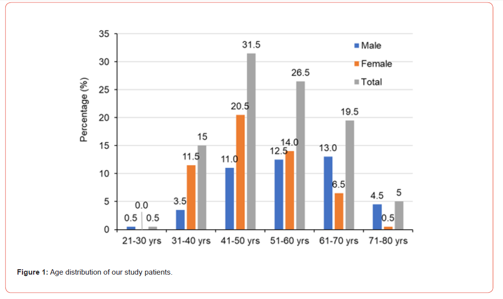
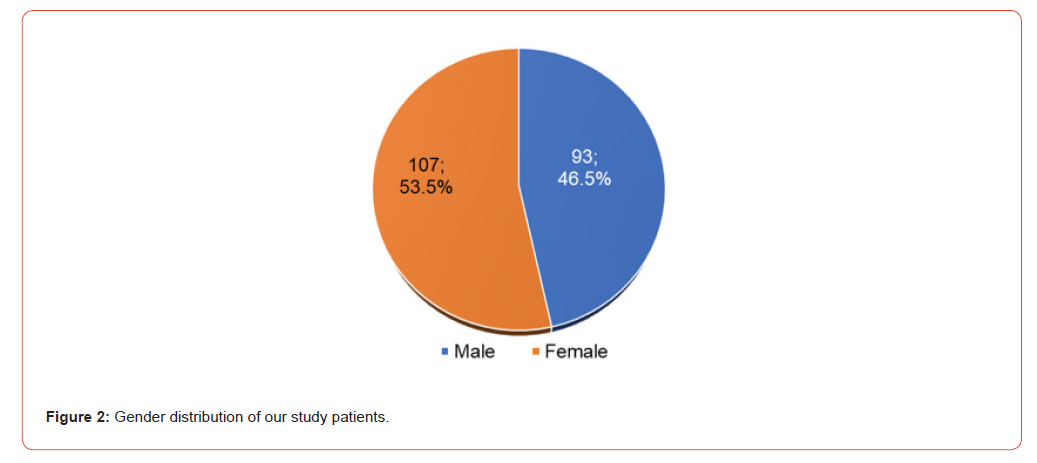
Age and gender distribution of the studied patients: The majority of the studied patients were in the 41-50 years (31.5%) and 51-60 (26.5) years old age groups (Figure 1). The percentage of female patients was higher than male patients with the age ranges between 31 to 60 years were female. A negligible percentage of patients were noticed in those below 30 years old age (Figure 1). The overall variation between male and female patients was not too high. About 53.5% of patients were male and 46.5% of patients were female (Figure 2).
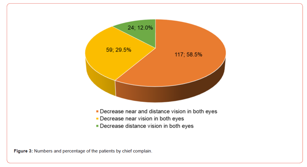
Patients with chief complaints and diabetes conditions: We used three categories to divide the studied patients to understand their major complaints, and these are (1) Decreased near and distance vision in both eyes, (2) Decreased near vision in both eyes, and (3) Decrease distance vision in both eye. 58.5% of patients complained that they decreased near and distance vision in both eyes, 29.5% of patients complained about a decrease of near vision in both eyes and only 12% of patients complained about a decrease in distance vision in both eyes (Figure 3).
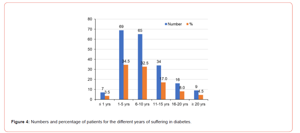
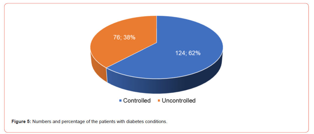
Most of the 200 patients were suffering from diabetes for a long duration. Only 3.5% of patients had less than one year of diabetes duration, 34.5% of patients had diabetes for 1-5 years, 32.5% of patients had diabetes for 6-10 years, 17% of patients had diabetes for 11-15 years, 8% patients had diabetes for 16-20 years, and 4.5% patients had diabetes for more than 20 years (Figure 4). However, it was also recorded that the conditions of diabetics were either controlled or uncontrolled. The majority percentage (62%) of the patients were able to control their diabetes while it was noticed that 38% of patients had uncontrolled diabetes (Figure 5).
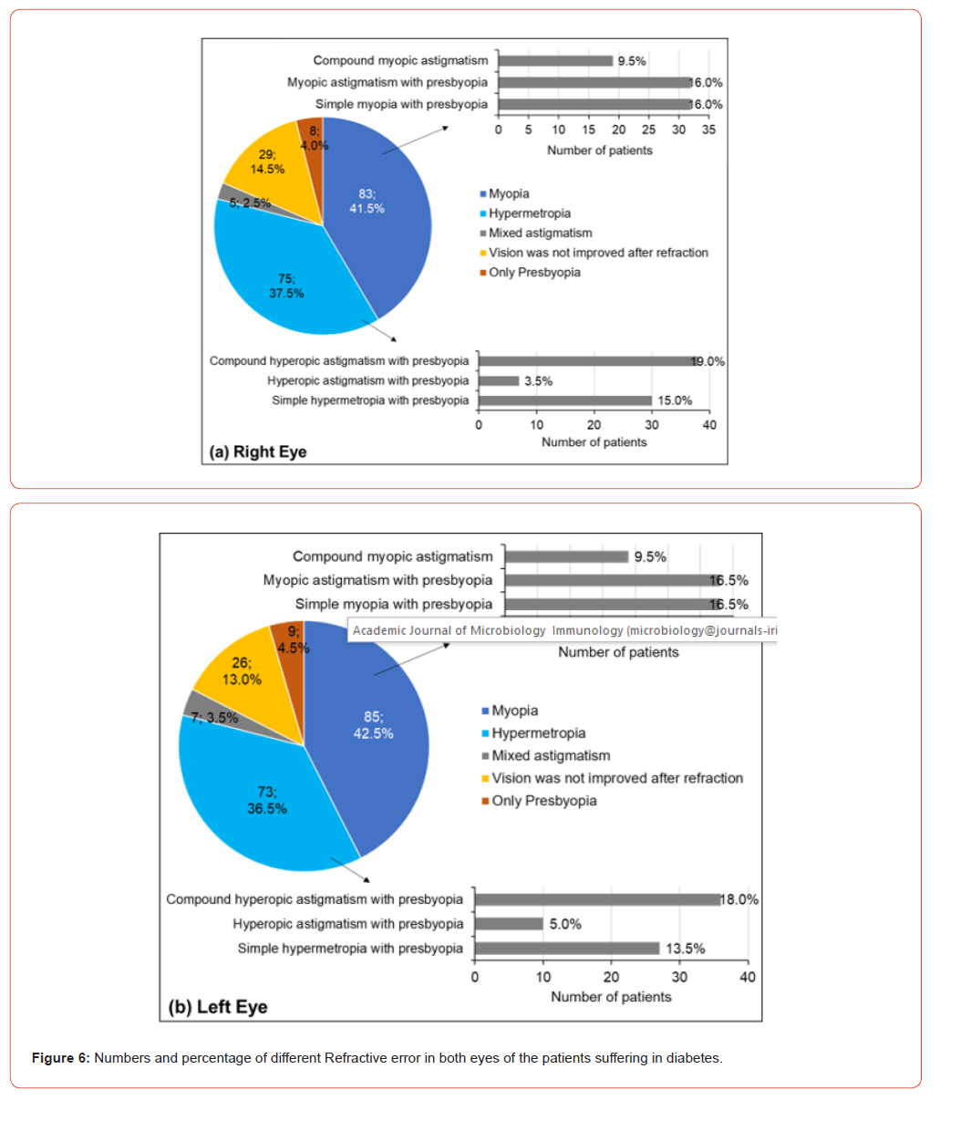
Refractive error in both eyes of the patients suffering from diabetes: Refractive error data were collected from the studied patients. 41.5% and 42.5% of the studied patients showed myopia in the right eye and left eye, respectively (Figure 6). Myopia was subcategorized into (1) Simple myopia with presbyopia, (2) Myopic astigmatism with presbyopia, and (3) Compound myopic astigmatism. Of 41.5% of patients with myopia in their right eye, 9.5% (of total patients) showed compound myopic astigmatism, and 16% of patients showed both myopic astigmatism with presbyopia and simple myopia with presbyopia (Figure 6, upper panel). Of 42.5% of patients with myopia in their left eye, 9.5% (of total patients) showed compound myopic astigmatism, and 16.5% of patients showed both myopic astigmatism with presbyopia and simple myopia with presbyopia (Figure 6, bottom panel).
37.5% and 36.5% of the studied patients showed hypermetropia, respectively, in their left and right eye (Figure 6). Hypermetropia was again sub categorized into (1) Simple hypermetropia with presbyopia, (2) Hyperopic astigmatism with presbyopia, and (3) Compound hyperopic astigmatism with presbyopia. Of 37.5% of patients with hypermetropia in their right eye, 19%, 3.5%, and 15% (of total patients) showed compound hyperopic astigmatism with presbyopia, hyperopic astigmatism with presbyopia, and simple hypermetropia with presbyopia, respectively (Figure 6, upper panel). Of 36.5% of patients with hypermetropia in their left eye, 18%, 5%, and 13.5% (of total patients) showed compound hyperopic astigmatism with presbyopia, hyperopic astigmatism with presbyopia, and simple hypermetropia with presbyopia, respectively (Figure 6, bottom panel).
In the right eye, 2.5%, 14.5%, and 4.0% of the total studied patients showed mixed astigmatism, vision was not improved after refraction, and only presbyopia, respectively (Figure 6, upper panel). In the left eye, 3.5%, 13.0%, and 4.5% of the total studied patients showed mixed astigmatism, vision was not improved after refraction, and only presbyopia, respectively (Figure 6, bottom panel).
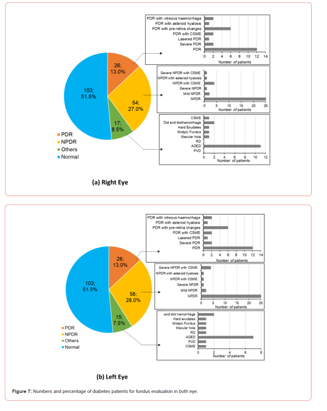
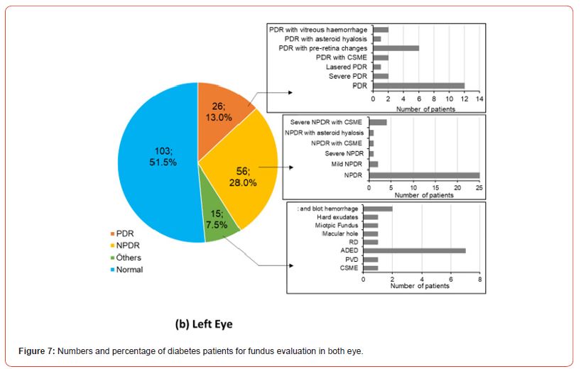
Fundus evaluation in both eyes of the patients suffering from diabetes: The results of fundus evaluation in both eyes were reported in Figure 7. Half of the patients showed normal on their fundus evaluation. The associated complication with PDR (13% in both eyes) was vitreous hemorrhage, CSME, pre-retina changes, and asteroid hyalosis (Figure 7). On the other hand, associated complications with NPDR (27% in right eye, and 28% in left eye) were CSME, and asteroid hyalosis (Figure 7). Some of the studied patients showed CSME, dot and blot haemmorage, hard exudates, mioptic fundus, macular hole, retinal detachment, ADED, and PVD. However, the number of patients with ADED was higher in number in both eyes (Figure 7).
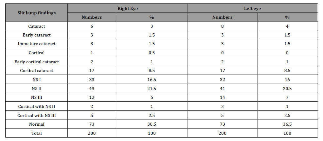
Slit lamp findings of the studied patients: Slit lamp findings data of the studied patients were categorized into cataract, early cataract, immature cataract, cortical, early cortical cataract, cortical cataract, NS I, NS II, NS III, cortical with NS II, and cortical with NS III (Table 1). 36.5% of the total patients showed normal in the slit lamp findings (Table 1). The highest percentage of the slit lamp findings showed under NS I (16.5% in right eye, and 16% in left eye) and NS II (21.5% in right eye, and 20.5% in left eye) (Table 1).
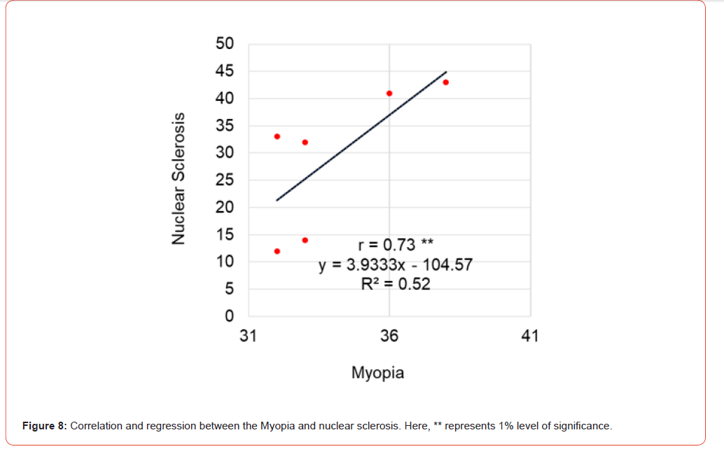
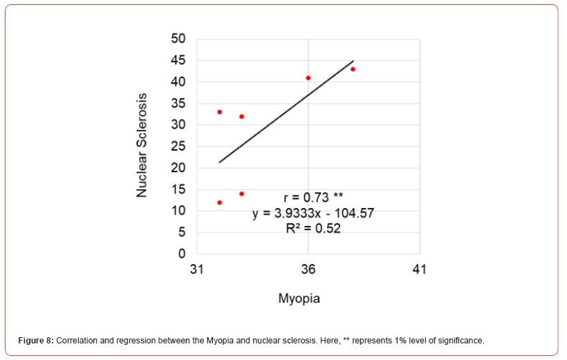
Correlation between Myopia and Nuclear Sclerosis: There is a stronger correlation between Myopia and Nuclear Sclerosis (r=0.73**). In the scatter plot, it was observed that myopia had a close relation with nuclear sclerosis (Figure 8). The gradient of this line for Myopia and Nuclear Sclerosis is 3.9333 which expressed that 3.9333 times of Myopia will be increased with the increase of 1 one times of Nuclear Sclerosis. The coefficient of determination (R2) was 0.52 suggesting that 52% of the nuclear cataract is accounted by the Myopia [1-5] (Figure 8).
Table 1: Baseline characteristics and clinical presentation of our patients.
Discussion
Hyperglycemia is the major cause of transient refractive changes in diabetic patients. In the 19th century it was recognised that the vision of diabetic patients is influenced by their changing blood glucose concentrations [6]. In 1925 Dyke, et al. refracted the eyes of two patients with diabetic ketoacidosis and again after their blood glucose had returned to the normal range. He concluded that hyperglycemia produced myopia and that lowering the blood sugar resulted in hyperopia. Since that time, no systematic studies to further define the relationship of serum glucose to changes in refraction have been performed. However, many differing opinions regarding this relationship are expressed [7]. Our study was the first to re-examine this phenomenon.
Grant, et al. suggested that the change in vision that accompanied chronic changes in serum glucose paralleled the degree of change in glucose concentration [6-8]. He also. suggested that the change in vision that accompanied chronic changes in serum glucose paralleled the degree of change in glucose concentration. These inferences agrees with our findings. Their studies suggest that an increase in serum glucose concentration, whether acute or chronic, is invariably associated with a more myopic or less hyperopic change in vision as long as the crystalline lens of the eye is intact. Additionally, they found that such changes do not occur in an eye from which the lens has been removed.
Kristian, et al. investigated the effect of intensive glycemic control on hyperglycemia-induced temporary changes in refractive error in Type 1 diabetic patients without retinopathy. Their study assessed the effect of variations in blood glucose level on the multifocal electroretinogram [9]. Their study design was similar to ours in regard to the presence of retinopathy, although it differed in terms of the type of diabetes. However, in their studies with our study is parallel to investigate changes in blood sugar levels [9]. That is; they compared hyperglycemia associated with an overall decrease in the implicit times and an increase in blood glucose levels.
Bozkurt, et al. also investigated the effect of glycaemic control on refractive changes in diabetic patients with hyperglycemia [10]. They measured blood glucose repeatedly over the same period and their results demonstrated the mechanisms by which elevated glucose affects cellular metabolism with a time course consistent with the transient nature of the effect observed. In our study, the fasting venous blood glucose was analysed in newly diagnosed diabetes patients.
In a study conducted by Elisabeth, et al. in patients with controlled diabetes, refractive and visual acuity test results were highly reproducible and stable in patients with variable blood glucose levels under routine care. They found that refraction was completely stable in 43 of the 53 patients. They also found no significant difference in blood glucose variability among the diabetic patients [11]. Their results suggest that refractive changes are minimal and assessments are highly reproducible provided that a standard method is used for testing, within a wide range of blood glucose levels in diabetic patients under routine care with different degrees of retinopathy [11]. However, in our study, refractive error was altered markedly by fasting plasma glucose levels. This difference could have been due to the fact that our patients had been newly diagnosed with diabetes.
Tatsuyuki, et al. studied blurred vision in diabetes mellitus patients [12]. Although the accurate incidence of transitory refractory change in diabetic patients is unknown, it ranges from 5 to 50% in untreated or uncontrolled patients. However, Bradley, et al. reported that working showed higher incidence of 90%, which agrees with the recent observation [13] Moreover, their results indicated that the magnitude of fpg decrement during the treatment was closely associated with the fluctuation of refraction of the eyes in diabetic patients, as reported by Holton [14, 15] Our study results showed blurred vision in 100% of patients.
Conclusion
Diabetes is a complex disease that can affect every ocular structure if left untreated. Many individuals remain unaware of their condition until they visit a hospital for other concerns. Blurred vision is a common symptom, often linked to refractive changes, with myopia being the most prevalent. Proper assessment and correction of refractive errors in diabetic patients not only enhance visual clarity but also improve overall eye comfort and quality of life. Regular ocular examinations play a crucial role in the early detection and management of diabetes-related vision changes, emphasizing the importance of comprehensive eye care.
Acknowledgement
None.
Conflict of Interest
None.
References
- Ali A, Iqbal F, Taj A, Iqbal Z, Amin MJ, et al. (2013) Prevalence of microvascular complications in newly diagnosed patients with type 2 diabetes. Pak J Med Sci 29(4): 899-902.
- Hayrullah Y, Mehmet B, Mustafa KB, Gocmen AY (2011) The effects of dialysers on some blood biochemical parameters in hemodialysis patients. A J P P Sci 5(22): 2513-2516.
- Daniel GV, Taylor A (1995) General Ophthalmology 4th Fourteenth edition A Lange medical book: 306-309.
- Robert KM, Daryl KG, Peter AM, Victor WR (2000) Harper’s Biochemistry Gluconeogenesis and control of the blood glucose. International Edition 25th Chapter 21 A Lange Medical Book: 216-217.
- Daniel G V, Taylor A (1995) General Ophthalmology 4th Fourteenth edition. Paul REA Lange medical book: 364-365.
- Grant G, Arnold V, Irvine C (1976) Relationship of Serum Glncose Concentration to Changes in Refraction. From the Division of Endocrinology University of California. Diabete 25(1): 29-31.
- Schneck ME, Fortune B, Switkes E, Crognale M, Adams AJ (1997) Acute effects of blood glucose on chromatic visually evoked potentials in persons with diabetes and in normal persons. Invest Ophthalmol Vis Sci 38(5): 800-810.
- Elschnig A (1923) Refraktionsanderungen bel Diabetes mellitus. Med Klin Berlin 79(17).
- Kristian K, Michael L, Birgit S, Allan V, Per BB, et al. (2004) Effect of Short-Term Hyperglycemia on Multifocal Electroretinogram in Diabetic Patients without Retinopathy. Invest Ophthalmol Vis Sci 45(10): 3812-3819.
- Bozkurt B, Atmaca A, Irkec M, Orhan M, Aslan U (2005) Effect of Glycemic Control on Refractive Changes in Diabetic Patients with Hypergly-cemia. Clin Sci Cornea 24(5): 531-537.
- Elisabeth A, Karl JH, Boel B (2011) Stable refraction and visual acuity in diabetic patients with variable glucose levels under routine care. Acta Ophthalmol 89(2): 107-110.
- Tatsuyuki I, Masaeumi M (1992) Refractory Changes of the Eyes in NIPPM During Treatment. Diabetes Care 15(7): 938-939.
- Bradley RF, Ramos E (1971) The eyes and diabetes. In: Marble A, White P, Bradley RF, Krall LP (). Josiin’s Diabetes Mellitus (11th edn.) Philadelphia, PA: Lea & Febiger, p. 478.
- Fledelius H, Fuchs J, Peck A (1990) Refraction in diabetics during metabolic dysreguration, acute or chronic: with special reference to the diabetic myopia concept. Acta Ophthalmol 68: 275-280.
- Holton IG (1982) Refractive changes in diabetes. Ophthalmic Optician 22: 734-736.
-

This work is licensed under a Creative Commons Attribution-NonCommercial 4.0 International License.