


 Review article
Postoperative refractive outcome of Senile Cataract
Received Date:March 13, 2026; Published Date:April 01, 2026
Abstract
Methods: This hospital-based prospective cross-sectional study included 100 patients aged 50-90 years with age-related cataract who attended the cataract clinic at Chevron Eye Hospital and Research Center. Demographic characteristics and ocular status were recorded. Postoperative refractive outcomes and visual acuity were evaluated after cataract surgery
Keywords: Manual small incision cataract surgery; Phacoemulsification; Refractive outcome; Visual acuity; Senile cataract
Abbreviations: VA: Visual acuity; ATG: astigmatism; WTR ASTG: With the rule astigmatism; AGNST ASTG: Against the rule astigmatism; ATG: Astigmatism; DM: Diabetes mellitus; HT: Hypertension; IHD: Ischemic heart disease; MSICS: Manual small incision cataract surgery
Introduction
Cataract is the leading cause of avoidable blindness worldwide, accounting for nearly half (47.8%) of all cases of blindness [1]. According to the World Health Organization, an estimated 20 million people worldwide are blind from bilateral cataracts, and this growing backlog poses one of the greatest public health challenges for the 21st century [8]. Because of the significant reduction in life expectancy and quality of life for the blind, sight-restoring cataract surgery is undoubtedly one of society’s most cost-effective medical interventions. The increase in economic productivity during the first postoperative year alone is estimated to exceed the cost of the surgery by a factor of 15.5 The cataract surgical rate (CSR) is an important public health metric, which represents the number of cataract operations annually performed per 1 million of population [1].
At present, there are a variety of common cataract surgery methods as such as the following (1) Phacoemulsification (PE)- the cataract is removed by an ultrasonic device (Phaco machine) [13] is the accepted. standard for cataract surgery in the developed world. Although phaco is often available in the developing world, particularly to those cataract patients who can privately afford it, there are numerous disadvantages to this method for the poorest societies. (2) Manual extracapsular cataract extraction (ECCE)- is a surgery requiring the creation of a large surgical wound (approximately 8-10 milimeters in size) and multiple stitches, which lead to a long time for recovery and more astigmatism after surgery [4].
Because of these problems associated with phaco in the developing world, alternative cataract surgical techniques such as sutureless manual small incision cataract surgery (MSICS) are gaining popularity. Manual small incision cataract surgery is able to achieve excellent outcomes with lower cost and average surgical time than phaco. This review covers the history, surgical variations, clinical outcomes, and cost-effectiveness of MSICS and its suitability for high- volume cataract surgery settings [1].
Performing phacoemulsification can be very challenging and is associated with increased risk of complications such as corneal decompensation and posterior capsular rupture (PCR) [7]. Manual small incision cataract surgery (MSICS) has been found to be a safe and effective technique for manual cataract extraction.7 The nucleus of the cataract is removed directly from the scleral tunnel wound without the need for sophisticated surgical technologies [2]. This enables a cost-effective treatment of cataract, which is particularly useful in healthcare settings that may be constrained by limited resources [2].
While performing phacoemulsification, the ophthalmic surgeon makes a clear corneal incision with a 2.8 to 3.2 mm keratome along with two paracenteses. Then after staining the anterior lens capsule, capsulorhexis of the desired size is performed, and the nucleus is emulsified and aspirated through an ultrasonic phaco probe inserted through the main wound. Following this, the remaining cortical matter is removed from the capsular bag, and a foldable IOL is implanted. The surgery is performed under topical or local anesthesia or as an elective procedure, and the patient can be discharged the same day. Postoperatively on day 1, uncomplicated cases usually have a perfect visual outcome, and the vision gradually improves until 4 to 6 weeks as globe remodeling takes place [9].
The purpose of study to evaluate postoperative refractive outcome of senile cataract at a tertiary eye hospital.
Methodology
Study Design and Ethical Approval:This study was a hospital based prospective cross sectional study with Cataract analysis approved by the Human Ethics Committee of Chevron Eye Hospital and Research Center (CEHRC). A thorough review of the medical records of patients who underwent cataract surgery at CEHRC from October 2024 to March 2025 was performed to investigate the clinical outcomes of the procedures. Data Collection The data were systematically extracted from the medical records, encompassing both demographic and clinical information. Demographic data included patient age and sex, while clinical data covered the history of systemic comorbidities, including diabetes mellitus (DM), hypertension (HT), ischemic heart disease (IHD), Ocular data were also collected, which included visual acuity, refractive status, lens opacity type, keratometry values, and results of fundus examination. Preoperative data, such as the exact date and time of surgery and the laterality of the operated eye, were meticulously recorded for each case. All patients underwent a comprehensive ophthalmic examination using a slit-lamp biomicroscope under the supervision of an experienced ophthalmologist to assess ocular health prior to surgery.
Visual acuity (VA) outcomes were classified following the guidelines outlined by the World Health Organization (WHO) for cataract surgery evaluation: Good Visual Acuity: 6/6 to 6/18, Borderline Visual Acuity: <6/18 to 6/60, Poor Visual Acuity: <6/60 [5]. This classification was used to assess the efficacy of cataract surgery, focusing on the postoperative visual outcome. Cataract hardness classification were classified based on the Lens Opacities Classification System III (LOCS III), which defines cataract hardness into two primary categories: Soft Cataracts: Nuclear sclerosis (≤ 3+), Cortical cataracts and posterior subcapsular cataracts (PSC), Hard Cataracts: Nuclear sclerosis (≥ 4+), Mature and hypermature cataracts [6]. The classification of cataract hardness provided insight into the surgical difficulty and potential impact on postoperative outcomes.
Two distinct cataract surgery methods were employed in this study: Manual Small Incision Cataract Surgery (MSICS), Phacoemulsification. These surgical approaches were selected based on the surgeon’s assessment and the patient’s individual needs, as well as cataract characteristics. Postoperative follow-up visits were scheduled at 1-month intervals to monitor visual acuity, refractive status, and ocular health. During these visits, the patients’ recovery process was evaluated, and any complications or changes in visual outcomes were documented. Inclusion Criteria: First, A diagnosis of cataracts confirmed preoperatively and underwent either Manual Small Incision Cataract Surgery (MSICS) or Phacoemulsification during the study period and second, those who had postoperative follow-up data at 1-month intervals. Exclusion Criteria: Patients were excluded from this study if they met any of the following conditions: Any cataract except senile, Underwent alternative cataract surgery techniques, such as Intracapsular Cataract Extraction (ICCE) or Extracapsular Cataract Extraction (ECCE). Lost to follow-up, with no available postoperative data. Presented with any ocular pathology other than cataract that could confound the surgical or visual outcomes.
Statistical analysis
Data analysis was performed with Microsoft Excel version 2020 and SPSS (26.0 for Windows, SPSS Ine, Chicago, IL, USA). All data were entered into a Microsoft Excel database (Microsoft, version 2019) and converted to SPSS for analysis. According to the normality test, all parameters were parametric and frequency data were used to evaluate the demographic, ocular and management characteristics for the study population. The mean and standard deviation were used for the descriptive study. Cross-tabulation was done to segregate the data according to age and gender range. The significant level was determined as P-value <0.05. Graphical structures formed by Microsoft Excel to represent data.
Results
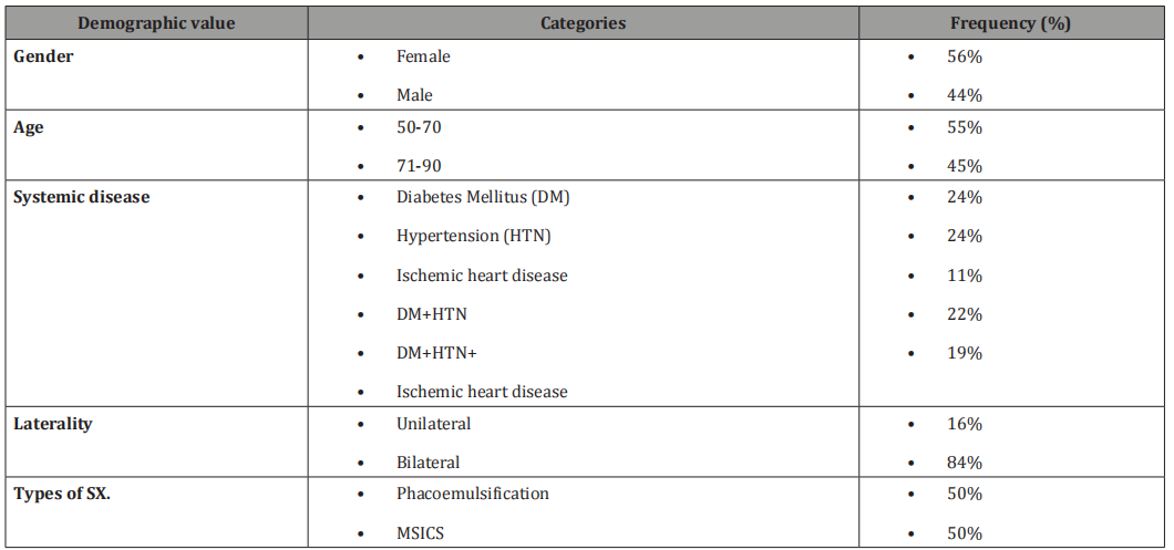
The men age of patients was 65.92 ± 11.8 years ranging from 40 to 90 years. Out of 100 patients, 56% were female. Most of the patients were over 40 years old, making up about 55% of the total group. Table 1 shows the basic demographic and clinical details of the patients with age-related cataract patients. A large number of patients also had cataracts, and 24% were found to have Diabetes Mellitus.
Table 1: Demographic and clinical characteristics of study population (n=100).
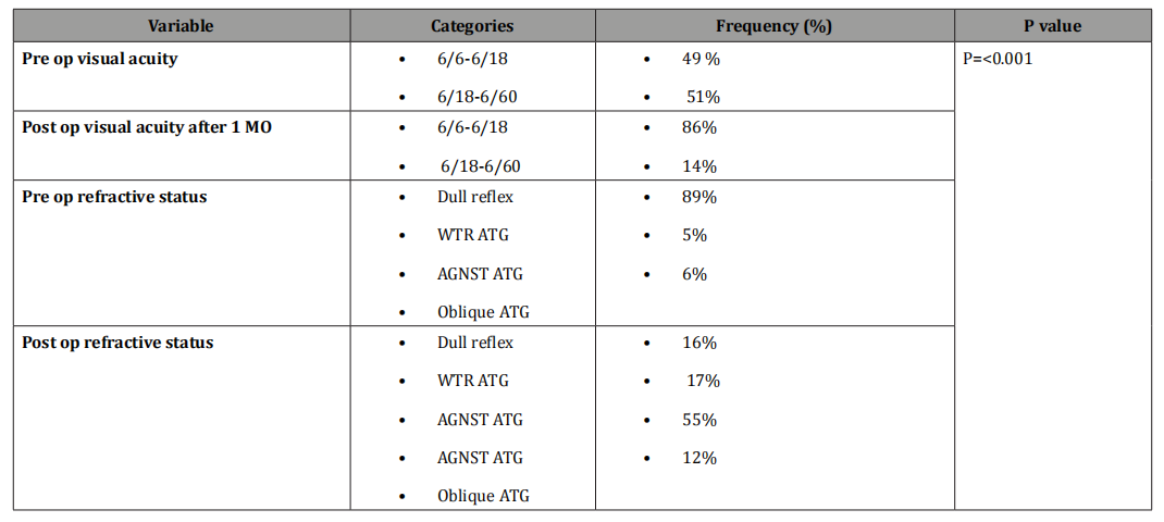
Before surgery, 51% of the patients had poor vision with a visual acuity of less than 6/18. One month after surgery, 86% of them showed improved vision, with visual acuity ranging between 6/6 and 6/18.Following surgery, 55% of the patients developed against-the-rule astigmatism, which was the most common type observed. This distribution significantly improved after surgery (P < 0.001).
In patients who had surgery in only one eye, 16% were found to have a dull red reflex in the other eye. These patients were informed about their condition and advised to consider surgery for the second eye.
Table 2:The clinical outcomes.
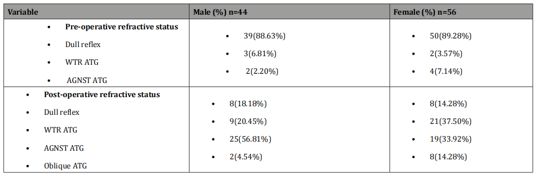
A total of 100 patients were included in the study, comprising 44 males and 56 females. Pre- operative refractive assessments showed that, among males, dull reflex was observed in 88.63% patients, with-the-rule astigmatism (WTR ATG) in 3 patients, and against-the-rule astigmatism (AGNST ATG) in 2 patients. In females, 50 patients had dull reflex, 3.57% had WTR ATG, and 4 had AGNST ATG. Post-operatively, 18.18% male showed dull reflex, while 9 patients had WTR ATG, 56.81% had AGNST ATG, and 2 patients developed oblique astigmatism. Among females, dull reflex was seen in 8 patients, WTR ATG in 21patients, AGNST ATG in 33.92% patients, and oblique astigmatism in 8 patients. After surgery majority of the male patients developed against the rule astigmatism, which is shown in Table 3.
Table 3:Refractive outcomes before and after surgery according to gender.
Discussion
Age-related cataract (ARC), also known as senile cataract, is a major cause of vision impairment and blindness among older adults. It occurs when the natural lens of the eye becomes cloudy due to the accumulation of protein, leading to blurred or decreased vision. This condition is most commonly linked with the aging process and continues to be a significant public health concern.
In this study, data were collected from 100 patients diagnosed with senile cataract. A greater number of cases were observed in individuals above 50 years of age, with females being more commonly affected than males. This finding is consistent with earlier research 10, which also reported a higher prevalence of cataracts in elderly females.
Many of the patients above 50 years were also found to have systemic diseases such as diabetes mellitus, hypertension, and cardiac conditions. These comorbidities are often associated with an increased risk of cataract formation, highlighting the importance of routine eye examinations, especially in patients with chronic illnesses.
Both Manual Small Incision Cataract Surgery (MSICS) and Phacoemulsification techniques were used, depending on the grade and density of the cataract. Among those who underwent bilateral cataract surgery, a majority developed against-the-rule astigmatism postoperatively. This distribution significantly improved from dull reflex to astigmatism after surgery (P= < 0.001). This type of astigmatism has also been commonly reported in other studies [13,14].
A significant improvement in visual acuity was observed after surgery. Most patients experienced an improvement from less than 6/18 to greater than 6/18, and this change was found to be statistically significant (P =< 0.001). These results are consistent with previous studies that show cataract surgery is highly effective in restoring vision [11,12].
This study also looked at changes in vision and astigmatism before and after eye surgery in 100 patients (44 males and 56 females). Before surgery, most patients had a dull reflex seen in 88.63% of males and 89.29% of females. Astigmatism was not common before surgery, with only a few patients showing with-therule (WTR) or against-the-rule (AGNST) astigmatism. After surgery, dull reflex greatly improved in both groups, showing better clarity of the eye’s optical system. However, the pattern of astigmatism changed noticeably. In males, AGNST astigmatism became the most common, found in 56.81%, possibly due to changes in corneal shape after surgery. Oblique astigmatism appeared in a few cases (2 males and 8 females). In females, there was a more even spread of WTR (21 patients) and AGNST (33.92%) astigmatism. After surgery the percentage of against the rule astigmatism more showed in male patients compared to female which reflects same thought like other study. 15 Despite some developing astigmatism post-surgery, the overall benefits underscore the importance of timely cataract surgery in restoring vision and enhancing quality of life.
Conclusion
Age-related cataract remains a predominant cause of vision loss in the aging population, with a marked preponderance in individuals over 50 years and a higher incidence among females. The frequent coexistence of systemic comorbidities such as diabetes mellitus, hypertension, and cardiac disease underscores the multifactorial nature of cataractogenesis. Surgical intervention, whether through MSICS or phacoemulsification, tailored to the severity of lens opacity, yielded substantial postoperative visual gains. Although a subset of patients developed against-the-rule astigmatism following bilateral surgery, the overall improvement in visual acuity statistically significant in most cases reinforces the critical role of timely cataract extraction in restoring functional vision and enhancing quality of life.
Acknowledgments
None.
Conflict of Interest
None.
References
- Venkatesh R, Chang DF, Muralikrishnan R, Hemal K, Gogate P, et al (2012) Manual small incision cataract surgery: a review. Asia-Pacific Journal of Ophthalmology 1(2): 113-119.
- Ting DS, Chua D, May KO, Aung M, Kumar A, et al. (2020) Modified manual small incision cataract surgery technique for phacoemulsification-trained surgeons. Therapeutic Advances in Ophthalmology 12: p.2515841420977372.
- Kaur K, Gurnani B (2022) Commentary: Tug of war between manual small-incision cataract surgery and phacoemulsification–A conflict or an agreement. Indian Journal of Ophthalmology 70(11): 3818-3819.
- Nampradit K Kongsap P (2021) The visual outcomes and complications of manual small incision cataract surgery and phacoemulsification: long term results. Romanian Journal of Ophthalmology 65(1): 31.
- Limburg H, Ramke J (2018) Cataract indicators: their development and use over the last 30 years. Community eye health 30(100): 82.
- Chylack LT, Wolfe JK, Singer DM, Leske MC, Bullimore MA, et al. (1993) The lens opacities classification system III. Archives of ophthalmology 111(6): 831-836.
- Foster GJ, Allen QB, Ayres BD, Devgan U, Hoffman RS, et al. (2018) ASCRS Cataract Clinical Committee 2018 hacoemulsification of the rock-hard dense nuclear cataract: options and recommendations. Journal of Cataract & Refractive Surgery 44(7): 905-991.
- Baltussen R, Sylla M, Mariotti SP (2004) Cost-effectiveness analysis of cataract surgery: a global and regional analysis. Bulletin of the World Health Organization 82(5): 338-345.
- Al Mahmood AM, Al Swailem SA, Behrens A, (2014) Clear corneal incision in cataract surgery. Middle East African journal of ophthalmology 21(1): 25-31.
- Hashemi H, Pakzad R, Yekta A, Aghamirsalim M, Pakbin M, et al. (2020) Global and regional prevalence of age-related cataract: a comprehensive systematic review and meta-analysis. Eye 34(8): 1357-1370.
- Forooghian F, Agrón E, Clemons TE, Ferris 3rd FL, Chew EY (2009) AREDS Research Group, 2009. Visual acuity outcomes after cataract surgery in patients with age-related macular degeneration: age-related eye disease study report no. 27. Ophthalmology 116(11): 2093- 2100.
- Koch CR, Neves GDF, Paredes RS, Siqueira ARAD, Kara-Junior N (2022) Impact of cataract surgery on visual acuity and quality of life assessed using the National Eye Institute Visual Function Questionnaire 25 in a public teaching hospital in Brazil. Revista Brasileira de Oftalmologia 81: e0011.
- Park RB, Aref AA (2024) Astigmatism management in modern cataract surgery. Vision 8(1): 9.
- Malakibungu SA, Makupa A, Makupa W (2024) Evaluation of Cataract Surgery as a Cause of Astigmatism among Patients Undergoing Phacoemulsification and Extracapsular Cataract Extraction at KCMC Hospital. medRxiv.
- Kim H, An Y, Joo CK (2019) Gender-differences in age-related changes of corneal astigmatism in Korean cataract patients. BMC ophthalmology 19: 1-8.
-

This work is licensed under a Creative Commons Attribution-NonCommercial 4.0 International License.