 Mini Review
Lens Induced Glaucoma
Dr Shams Mohammed Noman1*, Prof Dr Md Sharfuddin Ahmed2 and Dr Umme Salma Akbar3
1Assolciate professor, Bangabandhu Sheikh Mujib Medical University, Bangladesh
2Vice Chancellor Bangabandhu Sheikh Mujib Medical University, Bangladesh
3Associate consultant, Chittagong Eye Infirmary and Training Complex, Bangladesh
Dr Shams Mohammed Noman Associate Professor of Ophthalmology, Bangabondhu Shekh Mujib Medical University, Bangladesh
Received Date: February 03, 2022; Published Date: April 20, 2022
Abstract
Lens related elevation in Intraocular pressure results from a variety of mechanisms such as lens dislocation, lens swelling (intumescent cataract), inflammation due to phacoanaphylaxis and lens particle blocking the trabecular meshwork. This review was done with the aim of studying the types of lens-induced glaucoma, their clinical features and current management. Different published articles were discussed to see the result of management.
Keywords: Lens dislocation; Lens swelling; Lens-induced glaucoma
Introduction
Lens induced Glaucoma, one of the commonest cause of secondary Glaucoma due to senile cataract mandates an early recognition and management to prevent blindness [1]. Lensrelated elevation in intraocular pressure results from a variety of mechanisms such as lens dislocation, lens swelling, inflammation due to phacoanaphylaxis and lens particle blocking the trabecular meshwork. A study of these conditions is imperative since improper management leads to frequent complications and poor visual outcomes.
This review was done with the aim of studying the various types of lens-induced Glaucoma, their clinical features and current management. Apart from the textual literature, online search was conducted using search engines such as Google Scholar, PubMed, and Elsevier.
Phacolytic Glaucoma
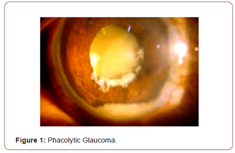
Phacolytic glaucoma is a condition characterized by leakage of lens contents through an intact capsule [2-4]. It is a principal complication of hypermature cataract. Hypermature cataract may cause leakage of lens protein from an intact capsule. The lens protein causes intense inflammation and blockage of trabecular meshwork, subsequently responsible for elevation of IOP [5]. The presentation is with an acute onset of pain and redness. The signs include congestion of the eye, corneal edema, deep AC and a hypermature cataract. The AC shows a significant amount of flare. Clumps of lens material may also be seen in the AC.
The majority of patients with phacolytic glaucoma can be managed through topical cycloplegia, topical steroids and aqueous suppressants. Manual small incision cataract surgery has proven successful in the management of phacolytic glaucoma.
Phacomorphic Glaucoma
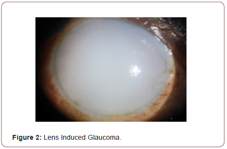
This condition is caused by a swollen, cataractous lens causing pupillary or angle block and elevating IOP [6]. In this type of Glaucoma, the swollen lens may block the anterior flow of the aqueous humour from the posterior chamber pushing the iris forward. Eventually, the trabecular meshwork gets blocked by the iris and leads to a sudden and extreme rise in IOP [7]. The diagnosis of phacomorphic glaucoma is made on the presence of pain and redness, shallow anterior chamber, corneal oedema and increased IOP with intumescent lens.
Initially, IOP-lowering medications are used. Topical beta blockers, carbonic anhydrase inhibitors and hyperosmotic agents are the mainstay of medical treatment. After IOP control and establishing intraocular inflammation, we can proceed for cataract extraction.
Lens Particle Glaucoma
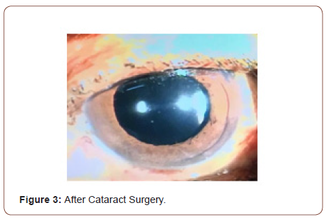
It is associated with a disrupted capsule and the presence of obvious fragments of lens material in the anterior chamber. It may occur after cataract surgery, trauma to the lens, or Nd:YAG posterior capsulotomy. The IOP increase is due to the obstruction of the aqueous outflow by the lens particles [8]. The patient reports with sudden pain and redness in the affected eye following trauma or surgery. On examination, the cornea is often oedematous, there are fluffy lens particles floating in the AC and/ a cataractous lens with ruptured capsule is present. Initially, a trial of medical antiglaucoma therapy maybe attempted. Mild to moderate steroid therapy can help to prevent synechiae, pupillary membrane, cystoid macular oedema, etc. If the glaucoma is severe and/ or there is a large amount of lens material in the anterior chamber, its surgical removal should be undertaken (Figures 1-3).


Phacoanaphylactic Glaucoma
It is a granulomatous, immune-mediated inflammation to lens proteins. The term phacoanaphylaxis is misleading since anaphylaxis requires mast cells, basophils, and immunoglobulin E, none of which are present during this process [9].
Capsule rupture leads to exposure of the eye to lens antigens which are antigenically privileged. When the eye is exposed to these antigens, a severe antibody response involving phacomorphonuclear leukocytes, lymphoid, epithelioid and giant cells occur. These inflammatory cells may block the trabecular macrophage and elevate IOP [10]. The clinical signs include lid oedema, chemosis, conjunctival injection, corneal oedema, AC reaction, posterior synechiae and mutton fat keratic precipitates. In the past, the differentiation between phacoanaphylaxis and sympathetic ophthalmia was confusing, but once it is understood that the former involves only the anterior segment of the eye, the diagnosis is easy [11,12]. The first step is to control IOP and inflammation medically. If unsuccessful, then the next step is surgical removal of the remaining lens material [13].
Epidemiology Prognostic Factors
There are 2 studies from the Indian Journal of ophthalmology and Nepal Journal of Ophthalmology that investigate lens-induced glaucoma, particularly phacolytic and phacomorphic glaucoma. The first study [14] deals with the frequency and types of lens-induced glaucoma, the reasons for late presentation, and the outcome of current management. The percentage of phacomorphic glaucomas was higher in this study (72%) than the phacolytic (28%). A similar result occurred in relation to gender since the ratio of females: males was 1.7:1. In this study, 38.6% achieved a visual acuity of 6/60 or better, 31.2% less than 6/60, and 30.2% less than 3/60. In the phacolytic group, 65.9% had a poor outcome compared with 59.8% of the phacomorphic group. This poor outcome was associated in the latter group with a longer distance from the hospital, longer duration of pain, and higher IOP at presentation. In the phacolytic group, only the distance was found to influence negatively the visual outcome. In this series, the final visual outcome is worse than in other studies, probably because the majority of the patients reported later than ten days after the onset of pain.
The second study [15] dealt with the age and sex distribution, causes for delayed presentation, immediate post-operative visual outcome and the reasons for poor visual outcome. The percentage of phacomorphic cases (57.5% ) was higher than the phacolytic glaucoma (42.5%). Female to male ratio was 2.1:1. The majority of patients (57%) presented after 2 weeks of symptoms and the reason for late presentation in more than half of the patients (52.5%) was financial constraints. Following surgery, 90% had an IOP less than 21 mm Hg at discharge. Visual acuity was either hand-movement or just perception of light in all eyes before surgery. At discharge, 65% of patients achieved a visual acuity of 6/60 or better, 5% had less than 6/60 and 30% less than 3/60. The reasons for poor VA in these patients were optic atrophy, uveitis, macular cause, and corneal edema.
Conclusion
This review shows that lens-induced glaucoma can have various modes of mechanism and presentation. It is imperative to identify the correct type of lens-induced glaucoma and manage it accordingly. There is a need to educate both the patient and the cataract surgeon of the dangers of lens-induced glaucoma and of the poor outcome if treatment is delayed.
Acknowledgement
None.
Conflict of Interest
The author reports no conflict of interest in this work.
References
- Gifford H (1900) The dangers of the spontaneous cure of senile cataract. Am J Ophthalmol 17: 289-293.
- Shields MB (1998) Glaucomas associated with disorders of the lens. In: Shields MB, (), Textbook of glaucoma. (4th edn.), Baltimore: Williams and Wilkins.
- Kothari R, Tathe S, Gogri P, Bhandari A (2013) Lens-induced glaucoma: the need to spread awareness about early management of cataract among rural population. ISRN Ophthalmol, pp. 581727.
- Mavrakanas N, Axmann S, Issum CV, Schutz JS, Shaarawy T (2012) Phacolytic glaucoma: are there 2 forms. J Glaucoma 21(4): 248-249.
- Johns KJ, Feder RS, Hamill MB (2003) Lens and cataract. American Academy of Ophthalmology.
- Rajkumari V, Kaminibabu KS, Bhabanisana RD, Victor R (2013) Manual small incision cataract surgery in phacomorphic glaucoma: Surgical technique and outcome in North-eastern India. J Curr Glaucoma Pract 7(2): 43-48.
- Jonathan PE, Ellant, Stephen A (1992) Lens induced glaucoma. Documenta Ophthalmia (81): 317-338.
- Yanoff M, Scheie HG (1968) Cytology of human lens aspirate. Its relationship to phacolytic glaucoma and phacoanaphylactic endophthalmitis. Arch Ophthalmol 80(2): 166-170.
- Skuta GL, Cantor LB, Weiss JS (2011) Basic and clinical science course. In: Intraocular inflammation and uveitis. San Francisco: American Academy of Ophthalmology.
- Allen JC (1967) Sympathetic uveitis and phacoanaphylaxis. Am J Ophthalmol 63: 280-283.
- Easom HA, Zimmerman LE (1964) Sympathetic ophthalmia and bilateral phacoanaphylaxis A Clinicopathologic Correlation of The Sympathogenic And Sympathizing Eyes Arch Ophthalmol 72: 9-15.
- Riise P (1965) Endophthalmitis phacoanaphylactica. Am J Ophthalmol 60(5): 911-915.
- Pradhan D, Hennig A, Kumar J, Foster A (2001) A prospective study of 413 cases of lens-induced glaucoma in Nepal. Indian J Ophthalmol 49(2): 103-107.
- Pant Sitoula (2016) Lens induced glaucoma in eastern Nepal. Nepal J Ophthalmol 8(16): 161-166.
-
Dr Shams Mohammed Noman, Prof Dr Md Sharfuddin Ahmed, Dr Umme Salma Akbar. Lens Induced Glaucoma. Clinical Case Report. W J Opthalmol & Vision Res. 4(1): 2022. WJOVR.MS.ID.000580. DOI: 10.33552/WJOVR.2022.04.000580.
-
Lens dislocation, Lens swelling, Lens-induced glaucoma, Senile cataract mandates, Textual literature, Topical cycloplegia, Topical steroids, Aqueous suppressants
-

This work is licensed under a Creative Commons Attribution-NonCommercial 4.0 International License.


