


 Research Article
An Overview of Pentacam® Indices
Received Date: February 26, 2025; Published Date: March 11, 2025
Summary
Background: Ethambutol and Isoniazid are major drugs in the management of pulmonary tuberculosis. This study aims to evaluate the incidence of optic neuropathy in pulmonary tuberculosis patients receiving ethambutol and isoniazid as part of the DOTS regimen at University of Port Harcourt Teaching Hospital, Nigeria.
Methods: This was prospective, descriptive study. Adult patients, enrolled for primary treatment of tuberculosis with the DOTS regimen, were recruited. They underwent an ocular examination before commencing treatment and 8 weeks after treatment. Best-corrected visual acuity, color vision, contrast sensitivity and central visual field were evaluated. Comparison between pre- and post-treatment measurements was done using McNemar test. Fisher’s exact test was employed to study the relationship between optic nerve dysfunction and known systemic risk factors. Statistical significance was set at p<0.05.
Results: A total of 150 subjects comprising 89 males and 61 females with age range of 18-64years participated in the study. The mean age was 37.3 ± 13.1years with male to female ratio of 1.5:1. A statistically significant reduction in color vision (p=0.03) and contrast sensitivity (p=0.016) from baseline was observed in 2% of subjects. Subjects who developed optic nerve dysfunction were older (mean age: 62 ± 2years) than those who did not (mean age: 36 ±12.8years) and it was statistically significant (p<0.001). Hypertension had significant association with optic nerve dysfunction (systolic: p=0.03; diastolic: p=0.007) on bivariate analysis. This association was not statistically significant on multivariate analysis (systolic: p=0.464; diastolic: p= 0.087).
Conclusion: The incidence of optic neuropathy in this study was 2%.
Keywords: Incidence; Pulmonary tuberculosis; DOTS therapy; Optic neuropathy
Introduction
Worldwide, tuberculosis (TB) is a chronic infectious disease of public health importance and has been known to mankind since ancient times [1] Epidemics of the disease were recorded in Europe in the 18th and 19th centuries with very high mortality rates [2, 3] There were an estimated 10.4 million new cases of active tuberculosis in the world in 2016 and 90% of these patients were adults, with HIV-positive patients constituting 10% of these cases [4]. Only 6.3million of the estimated 10.4million cases were detected, largely due to under-reporting and under-diagnosis. In the same period a total of 1.7 million tuberculosis-related deaths were recorded, representing a global decline of 22% from the pre-2000 era. The global estimate of new cases of the disease in 2019 was 10.0 million, representing a further 9% decline when compared with 2016 figures [5]. This decline has lifted tuberculosis out of the global list of top 10 causes of death though the disease has continued to be one of the major causes of mortality in low income regions of the world.
Most cases of tuberculosis occur in resource-limited countries of Africa and South-East Asia, where poverty, malnutrition, crowded living conditions, unavailability and lack of access to health care and dependence on traditional healers combine to facilitate disease transmission [6]. Although the disease shows no gender or age bias, the highest burden is seen in adult males, who accounted for 56% of cases globally in 2019, whereas females and children accounted for 32% and 12% respectively. Among these TB cases, 8.2% were HIVpositive [5]. At the beginning of 2020, Nigeria had the highest TB burden in Africa and ranked 6th among the 30 high-burden countries with an estimated incidence rate of 219 per 100,000 and mortality rate (excludes HIV+) of 64 per 100,000 population. This accounted for 4% of the global disease burden [5-7]. Combination therapy has been the mainstay of tuberculosis treatment for decades. It is said to exploit the differential properties of the drugs namely bacteriostatic or bactericidal, thereby enhancing treatment efficacy and reducing the risk of resistance [8, 9]. Treatment duration ranges from 6-9months depending on the clinical situation and is usually given in 2 phases-intensive and continuation.
The World Health Organization (WHO) Global Tuberculosis Program in 1993 declared tuberculosis a global emergency and introduced the Directly Observed Treatment Short-course (DOTS) strategy for managing the disease. It is recognized as a highly efficient and cost- effective TB treatment strategy [8-10]. DOTS strategy involves the direct monitoring of patients taking the medication by a health worker. The DOTS strategy specifies a 6-month treatment course using a combination of four drugsrifampicin, isoniazid, pyrazinamide and ethambutol for the first two months (intensive phase) followed by discontinuation of pyrazinamide and ethambutol (continuous phase). The W.H.O. recommends that the DOTS regimen be administered daily in the intensive phase. Where this is not possible, a three-times weekly dosing is allowed (intermittent regimen), in which patients receive doses of ethambutol and isoniazid that are higher than those of the daily regimen.
The most prominent ocular complication associated with isoniazid and ethambutol is optic neuropathy [11-14]. Optic nerve toxicity caused by ethambutol is typically bilateral, symmetrical and reversible upon discontinuation of the drug; however, several reports of permanent visual loss are documented [15-18]. Reversibility appears to depend on early detection of toxicity and prompt withdrawal of the drug. Isoniazid-induced optic nerve toxicity is also well recognized, and studies indicate that optic nerve toxicity from isoniazid is less severe and typically reversible [13, 14]. The onset of visual symptoms is often within ten days of commencement of therapy, but may occur later, up to 2-3 months into treatment. It is difficult to differentiate ethambutol-related toxicity from isoniazid-related toxicity in patients receiving both drugs in the DOTS regimen. Drug-induced pyridoxine deficiency is thought to underlie isoniazid-induced optic nerve toxicity but there are reports of neuropathy developing in patients receiving pyridoxine supplementation [19]. This study was undertaken to determine the incidence and early indicators of toxic optic neuropathy (TON) among patients on DOTS therapy at University of Port Harcourt Teaching Hospital. Data from this study will help the DOTS physician to identify patients with heightened risk of optic nerve toxicity thereby preventing potential severe visual impairment from TB treatment.
Materials and Method
This research was a hospital-based prospective observational study conducted at the Departments of Ophthalmology and DOTS centre of the Department of Community Medicine of the University of Port Harcourt Teaching Hospital. Patients 18 years and older who were diagnosed with pulmonary tuberculosis by PCR-based GeneXpert assay and enrolled for primary treatment with the DOTS regimen were included in the study. Primary TB treatment regimen consists of Rifampicin [R], Isoniazid [H], Pyrazinamide [Z] and Ethambutol [E] in a fixed-dose combination given by weight of the patient in two phases-intensive (RHZE x2months) and continuation (RH x4months) Subjects with pre-existing optic neuropathy or those taking medication (s) likely to cause optic neuropathy e.g., sildenafil, linezolid, HAART, anticancer drugs, digitalis; those with pre-existing retinal disease likely to interfere with measured parameters, e.g., diabetic retinopathy, retinal detachment, retinitis pigmentosa and those with pre-existing color vision, visual field or contrast sensitivity deficits were excluded. Subjects found to have narrow or potentially occludable anterior chamber angles on anterior segment examination, previous anti-TB treatment and subjects with best corrected VA < 6/9 at commencement of the study were also excluded. A minimum sample size of 150 was calculated and the study subjects were recruited by systematic sampling method from records from the DOTS center [20, 21].
Ethical Considerations
Ethical clearance to carry out this research was sought and obtained from the Research Ethics Committee of University of Port Harcourt Teaching Hospital. All procedures were in accordance with the standards specified by the Declaration of Helsinki for research involving human subjects. Signed or thumb printed informed consent was obtained from participants before inclusion in the study.
Data Collection Process and Examination Procedure
Upon identifying potential subjects at the DOTS clinic, the first author (O.I.F) counseled the participants regarding the study and obtained written informed consent as well as contact telephone numbers. Consenting subjects were led to the Eye clinic where questionnaires were administered by the researchers. Subsequent examination then followed. Subjects with unaided visual acuity < 6/9 were refracted and those with BCVA < 6/9 were excluded from the study but referred to the General Ophthalmology Clinics for further evaluation. Subjects with BCVA 6/9 or better were made to wear the optical correction for visual field, color vision and contrast sensitivity testing. Snellen acuity measurements obtained were converted to the LogMAR equivalent.
Assessment of color vision using the Ishihara plates and contrast sensitivity with the Pelli-Robson chart were performed at 75cm and 1meter respectively, under daylight illumination. Next, an anterior segment examination with pen torch and slit lamp was followed by intraocular pressure (IOP) measurement with Perkin’s handheld applanation tonometer. Perimetry (suprathreshold, 24-2) with Henson-9000 perimeter was then carried out. Blood samples for glucose and creatinine assay were collected by a medical laboratory scientist following which the pupils were dilated using tropicamide 1% for fundus examination (with +78D Volk lens) at the slit lamp.
At the end of examination, only subjects with BCVA ≥ 6/9, normal optic discs and having normal color vision (≥20/24 Ishihara plates), contrast sensitivity (≥1.50 log units) and visual fields were included in the study. Study participants were reexamined in the same order as stated above after 8 weeks of drug treatment. However, after the first examination, participants were counseled to present for immediate re-examination in the event of any visual symptoms occurring prior to 8weeks. All participants who developed impairment of visual function within the duration of the study were referred for follow up in the Eye clinic and the DOTS center physician was notified for proper adverse reaction reporting.
Data Analysis
The Data was entered into Microsoft Excel and exported to the Statistical Package for Social Sciences (SPSS) software (SPSS for windows version 25.0; SPSS Inc, Chicago, Illinois) for statistical analysis, under the guidance of a statistician. Continuous variables were summarized as means and standard deviations while categorical variables were summarized as frequencies and percentages. Summary statistics were presented using frequency tables, charts and mean. Paired t test was used to estimate the mean difference between pre- and post-treatment values of BCVA, color vision and contrast sensitivity. Related sample McNemar test was used to assess pre- and post-treatment results of perimetry and optic disc assessment. Fisher’s exact test was used to assess the association between systemic risk factors and the presence of optic nerve dysfunction. Risk factors that were significant (p<0.05) were subjected to logistic regression. Factors whose odds ratios were significant, were included in the multivariate model to assess the adjusted odds ratio. The level of statistical significance was set at p<0.05.
Results
Sociodemographic characteristics
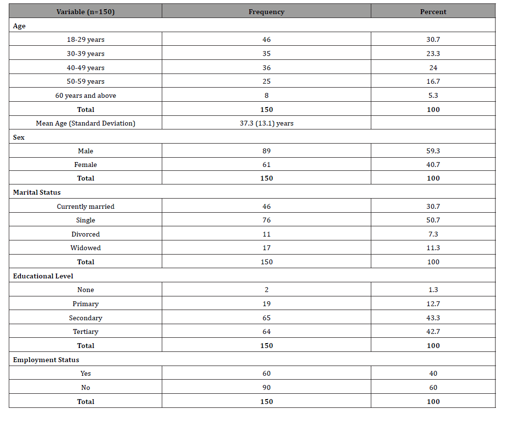
There were 89 males and 61 females with a male to female ratio of approximately 1.5:1. The mean age of the study group was 37.3 ± 13.1 years with a range of 18 to 64years. Of the 150 subjects, 76 (50.7%) were single, 90 (60%) were unemployed and 126 (86%) had obtained at least post primary education. In addition, 120 participants (80%) admitted to drinking alcohol, 41 (27.3%) were smokers of cigarette and 109 (72.7%) were not smokers. See table 1 Twenty-eight patients (18.7%) had a history of hypertension while 6 (4%) were known diabetic patients.
Table 1: Socio-demographic characteristics of study participants.
Visual function pre and post treatment
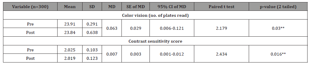
There was no change in mean best corrected visual acuity with the study period. See table 2 Colour vision impairment was noted in 6 eyes of three subjects within the study period. Table 3. All subjects had normal color vision at baseline (mean no. of plates read = 23.91 ± 0.29). After 8weeks of treatment, a reduction in color vision was observed (mean no. of plates read =23.84 ± 0.64) and this difference was statistically significant (p=0.03). See table 4 Similarly, 6 eyes of three subjects showed decreased contrast sensitivity.
Impairment in both functions were symmetrical. See table 3. Contrast sensitivity assessment showed mean pre-treatment score of the study group to be 2.03 ± 0.10. The mean post-treatment score was 2.02 ± 0.12. The difference between mean pre- and posttreatment scores was statistically significant (p=0.016). See table 4 Visual field defects were noted in three subjects. Out of these 3 subjects, 2 had bilateral and symmetric cecocentral scotoma whereas a unilateral marginal enlargement of blind spot was observed in the other.
See table 3. In summary, visual field defects were noted in 5 eyes of 3 subjects within the study period. Using McNemar’s test, there was no statistically significant difference between pre- and post-treatment results with respect to perimetry (p =0.063).
Table 2: Pre-and post-treatment best corrected visual acuity (LogMAR) of subjects

Table 3: Distribution of study subjects with and without impairment of color vision, contrast sensitivity and perimetry after 8 weeks of treatment.
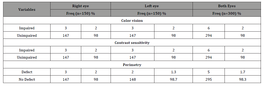
Table 4: Pre- and post-treatment results of color vision and contrast sensitivity.
Incidence of toxic optic neuropathy
After 8weeks of treatment, anterior segment examination findings with the slit lamp remained unchanged from pre-treatment status for all subjects. On posterior segment examination, bilateral optic disc hemorrhage was observed in 4 eyes of 2 (1.3%) subjects. Hemorrhages were flame-shaped and located at the disc margins. No subject had optic disc edema or pallor within the study period. Although optic disc hemorrhage was observed post-treatment, this finding was not statistically significant (p=0.125) using McNemar’s test. Three (2%) subjects developed optic nerve dysfunction within the study period, where optic nerve dysfunction is the presence of impairment in one or more of the following parameters in any individual: color vision, contrast sensitivity, perimetry, optic disc changes (pallor, hemorrhage and edema).
The subjects who developed optic nerve dysfunction were older (mean age 62 ± 2 years) than those who did not (mean age 36 ± 12.8 years) and were in the 55-70kg weight category. The difference (25.2 years) was found to be statistically significant (95% CI: 10.6- 39.9, t= 3.3982, df=148, p<0.001). They were also observed to be hypertensive patients, frequent alcohol consumers and had smoked tobacco for at least 3 years. See table 5 Among the risk factors reported in the literature, only hypertension (systolic: p=0.03; diastolic: p=0.007) was found to have significant association with optic nerve dysfunction on bivariate analysis in this study (Table XI). With reference to normal blood pressure (JNC classification), both systolic hypertension and diastolic hypertension showed slightly higher adjusted odds of optic nerve dysfunction (aOR=0.278, 95% CI=0.009-8.525 and aOR=0.051, 95%CI=0.002-1.542 respectively). However, this difference was not statistically significant (p>0.05) as shown in table 6.
Each subject in the 55-70kg weight group received 1100mg of ethambutol and 300mg of isoniazid daily. To determine if actual drug dose received in mg/kg within this weight category correlated with the incidence of optic nerve dysfunction (since individual weights vary within the group), the mean daily doses in mg/kg of isoniazid and ethambutol were calculated for subjects who showed optic nerve dysfunction and compared with those who did not. There was no statistically significant difference in mean dosage of both drugs between the two groups (p=0.927). See table 7
Table 5: Distribution of systemic risk factors for optic nerve dysfunction in study subjects
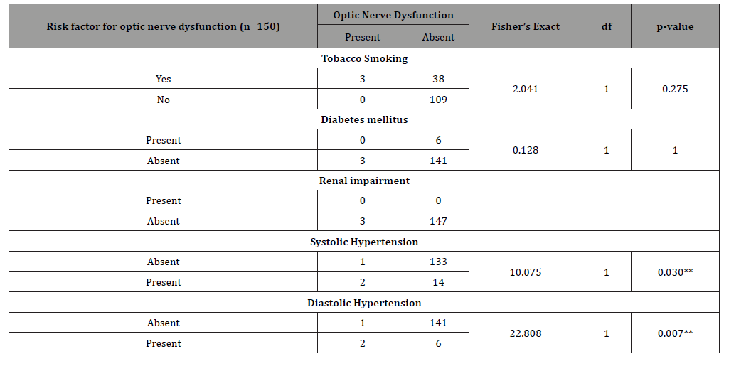
Table 6: Risk factors for optic nerve dysfunction in study subjects
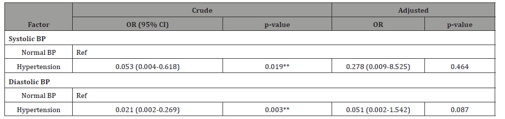
Table 7: Comparing Isoniazid and Ethambutol mean daily doses between

Discussion
The potential for optic nerve toxicity is a recognized complication of tuberculosis treatment. This study evaluated optic nerve function among patients receiving DOTS therapy at University of Port Harcourt Teaching Hospital. It assessed pre- and post-treatment measurements of best corrected visual acuity, color vision, contrast sensitivity and visual fields as well as optic disc changes. It also evaluated association of reported risk factors with optic nerve dysfunction among study subjects. All subjects in this study were adults, with a mean age of 37.3 ± 13.1years and there were more males. This is similar to the studies conducted among Indian populations by Garg, et al. and Mandal, et al. [22-23]. The male predominance is understandable since the burden of adult tuberculosis globally is greater in males [6]. However, Kandel, et al. [24] in Nepal, reported a lower mean age of 26 ± 9.5years with an equal male to female distribution. This was likely due to the inclusion of pediatric subjects in their study.
This study showed that the best corrected visual acuity was not affected by DOTS therapy. This is in agreement with most previous studies [23, 25-29]. In contrast, reduction in visual acuity within 2months of treatment was reported by Garg, et al. [22] This may be because subjects recruited for their study had a wide range of visual acuity (6/6-6/60) at baseline, implying possible pre-existing ocular disorder in some subjects. Their study also included both primary and secondary (re-treatment) TB cases which further increased the chances of having subjects with pre-existing optic nerve dysfunction from previous anti-TB treatment.
Dyschromatopsia is reported as one of the earliest detectable signs of toxic optic neuropathy. This study showed that there was significant impairment of color vision (p= 0.03) and contrast sensitivity (p=0.016) with DOTS therapy. Such impairment occurred without a reduction in the best corrected visual acuity. Similar trend was observed in studies by Cruz, et al. [25] and Kaimbo, et al. [30] whereas Garg et al in their study observed that impairment of color vision and visual acuity occurred simultaneously. However, the incidence of dyschromatopsia in the present study (2%) was by far lower than those recorded in the other studies by Cruz (47.88%), Kaimbo (15%) and Garg (12.6%) [25, 30, 22]. This difference is probably because these studies assessed subjects for both redgreen as well as blue-yellow defects whereas the present study tested for only red-green defect. Conversely, some other studies did not find any change in color vision or contrast sensitivity [23, 27, 28]. This may have been due to small sample size recruited for these studies.
The incidence of visual field abnormalities is highly variable among various studies and diverse visual field patterns have been reported [31, 32]. In this study, no significant change was observed with regards to visual fields. This finding correlated with studies in India by Mandal, et al. and in Nepal by Kandel, et al. [23, 24]. Similar trend was also observed in a study by Hong, et al. in Korea [26]. In contrast, Garg and Menon in their studies, found incidence of visual field abnormalities to be 6.3% and 7.6% respectively [22-33]. A large retrospective study in Taiwan by Chen, et al. found incidence of visual field defects to be as high as 33.9% [34]. This disparity was largely because many subjects had already developed visual symptoms prior to perimetry. This suggests that perimetry may not detect optic nerve dysfunction before the appearance of visual symptoms, a view first put forward by Citron [35]. In all study subjects, optic disc findings on fundoscopy remained unchanged after 8 weeks of DOTS therapy. This is in concurrence with most previous studies [23, 26]. On the other hand, Garg et al reported significant optic disc abnormalities of 4.7% in their study and these were in form of disc edema and pallor [22]. However, these subjects were category II (secondary treatment) patients who had previous exposure to anti-TB drugs and also received larger doses of ethambutol within the study period. It is possible that they had acquired some degree of unrecognized optic nerve dysfunction from previous TB treatment which may then be potentiated by higher drug dosage in the secondary treatment.
The dose-related incidence of ethambutol-induced optic nerve toxicity had long been described by Leibold and Citron, being 18% for doses >35mg/kg/day, 5-6% with 25mg/kg/day, 3% for dose of 20mg/kg/day and ≤1% for doses 15mg/kg/day or less [35]. In this study, the average daily doses of ethambutol and isoniazid received by subjects were well within the current standard recommendation of 15-20mg/kg/day. It was observed that optic nerve toxicity could still occur at this dose regimen This study showed that advanced age correlated with an increased risk of optic nerve dysfunction. Subjects that developed nerve dysfunction had a significantly higher mean age than those who did not (p<0.001). This agrees with other studies [16-17, 24]. The fact that optic nerve fiber count is known to decrease with advancing age could be reason for this observation. Again, the presence of diseases like hypertension and diabetes which are commoner in older individuals may increase vulnerability of the optic nerve to drug-induced toxicity. In the present study, all subjects that developed optic nerve dysfunction were hypertensive.
In this study, hypertension showed significant association with optic nerve dysfunction on bivariate analysis. Similar finding was reported in a population-based study conducted in Taiwan by Chen, et al. [34]. In contrast, hypertension was not associated with optic nerve dysfunction in studies by Kandel, et al. and Mandal, et al. [24, 23]. This difference is understandable since Kandel’s study recruited subjects within the pediatric age group alongside adult subjects; the mean age of subjects in their study (26.5 ± 9.5years) being lower than that of the present study (37 ± 13.1years). Although the mean age of subjects in the study by Mandal et al was like the present study, the study excluded patients with comorbidities like hypertension and diabetes.
Neither diabetes nor tobacco smoking showed significant association with optic nerve dysfunction in this study. This finding correlates with the studies by Cruz and Talbert-Estlin in United States of America [23, 36]. Similar findings were noted in other studies [29, 37]. Although smoking and diabetes are both mentioned in the literature as risk factors for optic nerve toxicity no study to the best of the authors’ knowledge directly implicated them [14, 38]. No subject in the present study had renal impairment. In evaluating subjects for color vision defects, this study employed the Ishihara color plates which detects only red-green color abnormalities; this means that subjects who may have had blue-yellow defect were not identified. However, testing for blue-yellow defects requires devices such as the Panel of Lanthony or the Farnsworth D-15 test panel, both of which are largely unavailable in general clinic use. Another limitation was the standard automated perimetry (SAP) in the visual field evaluation. More sensitive and techniques of visual field examination such as the short wave automated perimetry (SWAP) are more likely to detect field defects earlier than the SAP. Further studies with these more sensitive technologies will probably give a different outcome.
Conclusion
The incidence of optic nerve dysfunction from DOTS therapy in UPTH was 2% with subjects receiving the standard recommended dosage regimen. This study showed that color vision and contrast sensitivity testing are useful in detecting early optic nerve toxicity before the onset of visual symptoms. It was also observed from this study that older age (≥60years) is associated with optic nerve dysfunction in patients receiving the DOTS therapy Therefore elderly patients (60years and older) enrolled for DOTS therapy should be referred to the ophthalmologist for assessment of visual function before commencing treatment and ophthalmic evaluation should be repeated during treatment and where evidence of optic nerve toxicity is found, treatment should be reviewed.
Acknowledgements
Mr. B. Obazee, Miss C. Spiff and Mrs. P. Nwankwo, staff of the DOTS clinic University of Port Harcourt Teaching Hospital for their assistance in recruiting the participants
Competing interest
The authors do not have any competing interests.
Authors’ contributions
The article conceptualization, data collection and processing, and the writing of the initial draft were done by Idatonye F Ogolo. Godswill I. Nathaniel did the literature search and edited the manuscript; then Chinyere N Pedro-Egbe reviewed and wrote the final draft. The authors have read the manuscript and approved of it.
Data Availability
The data from which this study is based is available on request.
Disclaimer
The views expressed in this study are entirely those of the authors.
References
- Frith J (2014) History of tuberculosis Part 1-Pthisis, Consumption and the White plague. J Mil Veterans Heal 22: 29-35.
- Barberis I, Bragazzi NL, Galluzzo L, Martini M (2017) The history of tuberculosis: From the first historical records to the isolation of Koch’s bacillus. J Prev Med Hyg 58: 9-12.
- Daniel TM (2006) The history of tuberculosis. Respir Med 100: 1862-1870.
- World Health Organization (2017) Global Tuberculosis Report 2017. Geneva.
- World Health Organization (2020) Global Tuberculosis Report 2020. Geneva.
- World Health Organization Global Tuberculosis Report 2014. Geneva.
- Federal Ministry of Health (2019) National Tuberculosis and Leprosy Control Programme Annual TB Report.
- World Health Organization Global Tuberculosis Programme (1994) framework for effective tuberculosis control Geneva.
- Onozaki I, Raviglione M (2010) Stopping tuberculosis in the 21st century: Goals and strategies. Respirol 15: 32-43.
- Otu AA (2013) Is the directly observed therapy short course (DOTS) an effective strategy for tuberculosis control in a developing country?. Asian Pac J Trop Dis 3: 227-231.
- Grzybowski A, Zülsdorff M, Wilhelm H, Tonagel F (2015) Toxic optic neuropathies: An updated review. Acta Ophthalmol. 93: 402-410.
- Kumar J, Chaubey P, Singh VP (2018) Anti tubercular treatment (ATT) induced optic nerve changes: An observational study. J Dent Med Sci 17: 30-33.
- Sharma P, Sharma R (2011) Toxic optic neuropathy. Indian J Ophthalmol 59: 137-141.
- Makunyane P, Mathebula S (2016) Update on ocular toxicity of ethambutol. J Afr Vis Eye Heal 75: 1-4.
- Chuenkongkaew W, Samsen P, Thanasombatsakul N (2003) Ethambutol and optic neuropathy. J Med Assoc Thail 86: 622-625.
- Lim SA (2006) Ethambutol-associated optic neuropathy. Ann Acad Med Singapore 35: 274-278.
- Chen SC, Lin MC, Sheu SJ (2015) Incidence and prognostic factor of ethambutol-related optic neuropathy: 10-year experience in southern Taiwan. Kaohsiung J Med Sci 31: 358-362.
- Phillips KC, Clarke Farr PC (2018) Ethambutol toxic optic neuropathy: A case report and review. J Afr Vis Eye Heal 77: 419.
- Balaji O, Dipanjan B, Navin P (2017) Isoniazid, the culprit behind toxic optic neuropathy. Asian J Pharm Clin Res 10: 1-2.
- Abel N, Contino K, Jain N, Grewal N, Grand E, et al. (2015) Eighth joint national committee (JNC-8) guidelines and the outpatient management of hypertension in the African-American population. North Am J Med Sci 7: 438-445.
- Gavin JR, Alberti KG, Davidson MB, DeFronzo RA, Drash A, et al. (2003) Report of the Expert Committee on the diagnosis and classification of diabetes mellitus. Diabetes 26: 05-20.
- Garg P, Garg R, Prasad R, Mishra AK (2015) A prospective study of ocular toxicity in patients receiving ethambutol as a part of directly observed treatment strategy therapy. Lung India 32: 16-19.
- Mandal S, Saxena R, Dhiman R, Mohan A, Padhy SK et al. (2021) Prospective study to evaluate incidence and indicators for early detection of ethambutol toxicity. Br J Ophthalmol 105: 1024-1028.
- Kandel H, Adhikari P, Shrestha GS, Ruokonen EL, Shah DN (2012) Visual Function in Patients on Ethambutol Therapy for Tuberculosis. J Ocul Pharm Ther 28: 174-178.
- Cruz EM, Puentespina FG, Paolo Alejo KL, Santos Morabe ET, Lourdes Nañagas MR et al (2010) Color-vision abnormalities among patients undergoing tuberculosis treatment. Philipp J Ophthalmol 35: 3-9.
- Hong IH, Chung JK, Park SP (2012) Visual Function Test for Early Detection of Ethambutol Induced Ocular Toxicity. Invest Ophthalmol Vis Sci 53: 4884.
- Menon V, Jain D, Saxena R, Sood R (2009) Prospective evaluation of visual function for early detection of ethambutol toxicity. Br J Ophthalmol 93(9): 1251-1254.
- Kim KL, Park SP (2016) Visual function test for early detection of ethambutol induced ocular toxicity at the subclinical level. J Cutan Ocul Toxicol 35: 1-5.
- Jin KW, Lee JY, Rhiu S, Choi DG (2019) Longitudinal evaluation of visual function and structure for detection of subclinical Ethambutol-induced optic neuropathy. PLoS One 14(4): e0215297.
- Kaimbo WK, Bifuko ZA, Longo MB, Dralands L, Missotten L (2002) Color vision in 42 Congolese patients with tuberculosis receiving ethambutol treatment. Bull Soc Belge Ophtalmol 284: 57-61.
- Kho RC, Al-Obailan M, Arnold AC (2011) Bitemporal visual field defects in ethambutol-induced optic neuropathy. J NeuroOphthalmol 31: 121-126.
- Ashraf A, Bhat MA (2017) Visual Field Changes in Patients Receiving Antitubercular Drug Therapy at Tertiary Care Hospital: An Analytical Observational Study. Int J Contemp Med Res 4(2): 346-349.
- Addoor K, Raju V, Bhandary S, Menon S, Gupta A (2005) Optic neuropathy induced by low dose of ethambutol: A rare presentation. Asian J Pharm Clin Res 1 possible mechanism of injury J Pak Med Assoc 55: 10-11.
- Chen HY, Lai SW, Muo CH, Chen PC, Wang IJ (2012) Ethambutol-induced optic neuropathy: A nationwide population-based study from Taiwan. Br J Ophthalmol 96(11): 1368-1371.
- Citron KM (1969) Ethambutol: a review with special reference to ocular toxicity. Tubercle 50: 32-6.
- Talbert Estlin KA, Sadun AA (2010) Risk factors for ethambutol optic toxicity. Int Ophthalmol 30(1): 63-72.
- Ezer N, Benedetti A, Darvish Zargar M, Menzies D (2013) Incidence of ethambutol-related visual impairment during treatment of active tuberculosis. Int J Tuberc Lung Dis 17: 447-455.
- Melamud A, Kosmorsky GS, Lee MS (2003) Ocular Ethambutol Toxicity. Mayo Clin Proc 78: 1409-1411.
-

This work is licensed under a Creative Commons Attribution-NonCommercial 4.0 International License.