

 Research Article
Factors Influences the Effect of Anti-VEGF Drugs on Neovascular Age-Related Macular Degeneration- Complicated Multiple Interactions
Received Date: February 22, 2024; Published Date: April 15, 2024
Abstract
Neovascular age-related macular degeneration (nAMD), also known as wet age-related macular degeneration (wAMD), is a disease affecting the macular area of the retina. If not timely treatment, it will cause serious damage to vision. It is one of the main causes of severe vision loss in people over 60 years old. At present, intravitreal vitreous injection of anti- vascular endothelial growth factor (VEGF) drugs has become the firstline treatment of nAMD. Currently, the main anti-VEGF drugs include ranibizumab, aflibercept and conbercept. Although anti-VEGF drugs have made major breakthroughs in the treatment of nAMDs, therapeutic outcomes are not always the same, and patients show individualized different responses to therapeutic effects. The clinical factors, environmental influences and gene polymorphism affect the efficacy of anti-VEGF drugs. Understanding of the complex and influential factors during drug treatment are critical to achieve desirable outcomes from the patients. The efficacy of drugs, resistance to treatment, alternative approaches, and future directions are presented in this article.
Keywords: Neovascular age-related macular degeneration; Anti-VEGF drugs; Gene; Influencing factors; Single nucleotide polymorphisms
Abbreviations: AMD: Age-related macular degeneration; nAMD: Neovascular age-related macular degeneration; wAMD: Wet age-related macular degeneration; VEGF: Vascular endothelial growth factor; CNV: Choroid neovascularization; PED: pigment epithelial detachment; PIGF: Placental growth factor; RAAS: Renin angiotensin aldosterone system; PRA: Plasma renin activity; ALD: Aldosterone; AngII: Angiotensin II; ION: Ischemic optic neuropathy; RAMA: Retinal microaneurysm; VA: Visual acuity; CRT: Foveal retinal thickness; ELM: The outer membrane; CT: Choroidal thickness; PCV: Polypous choroidal vascular; ICGA: Indocyanine green angiography; VEGFR-2: Vascular endothelial growth factor receptor-2; SRF: Subretinal fluid; NSAIDs: Non-steroidal anti-inflammatory drugs
Introduction
Age-related macular degeneration (AMD) is the main cause of serious visual impairment and even visual acuity loss in the age of over 50, especially in developed countries such as Europe and North America. However, with the increasing number of patients with AMD in developing countries such as China, AMD has become the main disease that the visual acuity irreversible decline in the elderly [1]. According to fundus changes, AMD can be divided into dry AMD and wet AMD, among which wet AMD has the most serious visual impairment in patients, and it is difficult to treat and has a relatively poor prognosis [2,3]. In dry AMD geographic atrophy is common, while in wet AMD mainly is in the macular area choroid neovascularization (CNV) or retinal neovascularization for pathology, cause retinal exudation, hemorrhage, retinal or subretinal effusion, retinal pigment epithelial detachment (PED), late fibrovascular or atrophic scar changes, resulting in severe visual damage [4,5]. According to research findings, the formation of CNV is closely related to vascular endothelial growth factor (VEGF). VEGF is a dimer glycoprotein that stimulates the growth and leakage of blood vessels and is composed of VEGF-A, VEGF-B, VEGF-C, VEGF-D, VEGF-E, VEGF-F and placental growth factor (PIGF), where VEGF-A is an important promoter of CNV formation [6].
Currently, anti-VEGF drug therapy is the standard treatment for neovascular AMD (nAMD). These drugs include Ranibizumab, Aflibercept and Conbercept, etc. However, clinical follow-up results have shown that some patients did not improve or maintain vision after receiving anti-VEGF drug therapy, and the response to anti- VEGF drug therapy is inconsistent. Some patients fared better than average, while others became worse [7,8]. Environmental risk factors and genetic susceptibility have been shown to contribute the development of nAMD. Clinical and genetic factors have also been identified as important determinants of individual variability in response to anti-VEGF drug therapy. Therefore, it is important for clinical ophthalmologists to understand the factors that may affect the therapeutic effect, to guide the choice of treatment plan and determine the prognosis. By searching relevant literature, this paper summarizes the clinical factors and gene polymorphisms that affect the efficacy of anti-VEGF drugs in the treatment of nAMD.
Correlation Between Clinical Features and Efficacy of Anti-VEGF Drugs
Systemic disease effects
Hypertension, defined as systolic blood pressure >140mmHg or diastolic blood pressure >90mmHg, is a systemic disease that can have harmful effects on multiple organs such as the heart, kidneys, brain, and eyes [9]. Its pathogenesis mainly includes activation of renin angiotensin aldosterone system (RAAS), increased sympathetic nerve excitability, vascular endothelial cell damage, water sodium retention, etc. RAAS plays an important role in the formation of hypertension. Hypertension patients are often accompanied by excessive activation of RAAS Plasma renin activity (PRA) and the increased aldosterone (ALD) [10-13]. Among them, RAAS affects not only key components of the heart and cardiovascular system, but also blood vessels in the retina. Angiotensin II (AngII), a different important player in hypertension, not only causes apoptosis of retinal endothelial cells, but also upregulates VEGFR-2. The development of CNV is being promoted by crossing the blood-retinal barrier with apoptotic cells and upregulating VEGFR-2 which disrupts the balance between anti- VEGF and VEGF [14]. High blood pressure is considered a risk factor for retinal vascular occlusion (RVO), ischemic optic neuropathy (ION) and retinal microaneurysm (RAMA) [15,16].
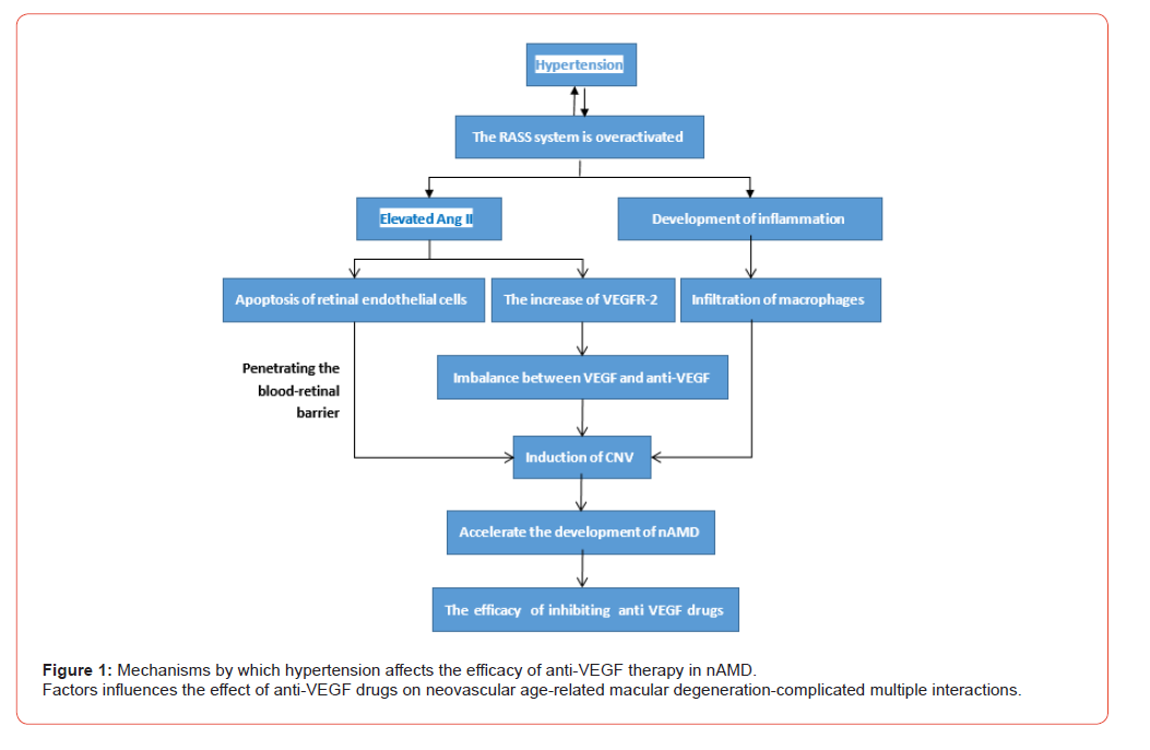
Pons, et al. [17] found that excessive activation of RAAS can promote inflammation, leading to macrophage infiltration and subsequently inducing retinal CNV. Therefore, RAAS may exacerbate the progression of nAMD, causing nAMD patients with hypertension requiring more anti VEGF therapy than those without hypertension. In addition, a study involving 3096 nAMD patients also reached the same conclusion, which may be related to abnormal activation of the RASS system [18]. However, other scholars have not found that hypertension has a significant impact on the response to VEGF therapy [19]. The impact of hypertension on the efficacy of VEGF drugs in treating nAMD still needs further study [Figure 1].
Baseline vision
Baseline visual acuity (VA) is one of the important predictors of final post-treatment visual outcome. Patients with worse baseline VA may be associated with better improvement after treatment, while patients with better baseline VA are less likely to gain as much improvement due to the upper limit effect [20]. The subgroup analysis of the MARINA and ANCHOR study showed that if baseline VA in one group was 5 ETDRS letters higher than in the other group (ETDRS eye chart), the mean VA change from began to 24 months of treatment would be 3.2 letters lower contrast the other group [21].
According to the study of Dai Hong, et al. [8] baseline VA has a significant impact on the prognosis of vision, which is usually related to lesion size, onset time, whether there is damage to photoreceptor cells and whether the fovea area is involved. Baseline VA is an important indicator reflecting the extent to which CNV lesions affect visual function. The better baseline VA is, the less impact the lesion has on visual function, and the better treatment prognosis. In a study of 97 subjects, Yan Fu, et al. [22] found that poor baseline VA was an independent factor influencing the efficacy of anti-VEGF therapy in nAMD patients. Asten, et al. [23] believed that baseline VA was a risk factor for ineffective treatment, and poor baseline VA predicted more serious structural and functional destruction of retinal photoreceptor cells. However, Hara, et al. [24] believed that baseline VA was not related to the response to anti-VEGF treatment. These findings suggest that the relationship between baseline VA and response to anti-VEGF therapy is controversial and requires further investigation. Based on the search and analysis of a large number of studies, we believed that the baseline VA is one of the important factors affecting the prognosis of anti-VEGF drugs in nAMD. Although the lower baseline VA showed more improvement, more satisfactory treatment effect remained in those patients with better baseline VA.
The age and course
Boyer, et al. [21] showed that younger age was associated with better clinical outcomes. In the subgroup analysis of the MARINA study, if the mean age of one group was 13.7 years younger than that of the other group at baseline. The change in VA in the younger group would be 5 letters higher than that in the older group. Similarly, the ANCHOR subgroup analysis also showed that patients in the younger group benefited more than those in the older group [25]. In addition, other studies found that patients younger than 70 gained 10.8 letters after treatment, while patients 70 or older gained only 5.6 letters after treatment [26]. At the same time, Lanzetta, et al. [27] also showed that the younger the nAMD patients, the greater the visual benefit after intravitreal injection of arbocept for 52 weeks.
The interval between symptom onset and treatment initiation was another important baseline predictor of final vision outcome. Shorter intervals from visit to treatment were associated with better VA outcomes [20]. The study showed that patients with nAMD with a treatment delay of 21w or longer had an odds ratio of 2.62 for visual deterioration after treatment compared with those with a treatment delay of 7w or less, suggesting that late treatment is an important predictor of poor treatment outcome [28]. The study of Gong Bin et al. [29] showed that the efficacy of anti-VEGF drugs in improving vision and macular edema in patients with nAMD with disease course less than 6 mo was better than those with NAMD with disease course greater than 6 mo. Early treatment could reduce the number of treatments, thus reducing the cost of treatment for patients.
Racial and sex differences
Among many factors that affect the efficacy of anti VEGF drugs in treating nAMD patients, although there are relatively few studies on racial differences, there are still some literatures documenting the relevant mechanisms of their impact on anti VEGF efficacy. Zhang [30]’s research found that in East Asian population, the nonresponse rate of nAMD patients with mutant allele A to anti VEGF treatment is higher than that of nAMD patients with wild genotype G. However, in Caucasian population research, this phenomenon has not been confirmed, which may be related to the lower mutation rate of mutant allele A in Caucasian population than that in East Asian population. In addition, gender was the largest factor affecting foveal retinal thickness (CRT), in which males reduced the effect of treatment on CRT.
This was supported by a study in which males were found to be a risk factor for poor vision prognosis in nAMD patients after anti- VEGF treatment for 60 months. The background of this finding is unclear, but it may be related to hormonal factors. This means that the information provided to patients about the expected treatment response should distinguish between male and female [31,32]. In a large prospective cohort study in a Danish population, Bek, et al. [31] found a statistically significant difference between gender and CRT changes, with a lower decline in men. However, some studies believe that there is no correlation between the two, which may be related to different population, sample size and other factors [33-35]. Although the different races and sex differ in genetics, the impact of these characteristics on the efficacy of anti-VEGF drugs in nAMD cannot be determined and should be confirmed by further studies.
Imaging features
Optical coherence tomography can produce noninvasive highresolution imaging of the retina [36,37]. Zhang ‘s research team [20] found that the integrity of the ellipsoid band (IS/OS) was highly correlated with the treatment effect. After anti-VEGF therapy in the vitreous body, the prognosis of patients with complete ellipsoid zone IS was better than those with partial ellipsoid zone. This study shows that interruption or absence of IS/OS layer is associated with poor VA prognosis. The length of destruction of ellipsoid zone will lead to different VA outcomes. In addition, the integrity of the outer membrane (ELM) is also directly related to VA. The interruption of ELM is a sign of severe damage to the inner segment or cell body of the photoreceptor. In a 1-year follow-up of nAMD patients treated with anti-VEGF drugs, it was found that the status of ELM is one of the important factors affecting VA outcomes in nAMD patients [38-40].
Whether CRT affects the outcome of VA is still controversial. It is generally accepted that CRT is a sensitive parameter and early sign for detecting baseline VA reduction [20]. In a hospital-based study, a total of 1105 participants of in all treated groups, age, larger CNV area and central foveal retinal thickness were negatively correlated with VA outcomes [28]. However, Gerding, et al. [41] studies show that CRT has nothing to do with the VA ending but was an early sensitive predictor of VA reduction. The study showed that the curative effect of the CRT affect anti-VEGF drugs still needs more research to confirm. In addition, in a 12-month follow-up study, it was found that the treatment effect was significantly better in cases without SRF compared to those with persistent SRF among patients still receiving on-demand injections after receiving three load doses of anti VEGF drugs [41,42].
It is widely believed that the pathogenesis of nAMD involves abnormalities in the choroidal vascular system. Baseline choroidal thickness (CT) may be another important factor in the prognosis of VA [43]. Studies have shown that after intravitreal injection of anti- VEGF drugs in nAMD patients, choroidal thickness is significantly reduced, which is associated with improved vision of patients [44]. However, in a retrospective sequential case series study, it was found that the greater the baseline choroid thickness of nAMD patients after intravitreal injection of anti-VEGF drugs, the better the clinical prognosis [45]. These studies suggest that the relationship between CT and the response to anti-VEGF therapy remains controversial and needs further study. Another OCT feature of nAMD-affected eyes is the bileval sign, which is also one of the OCT features of polypous choroidal vascular disease (PCV). Indocyanine green angiography (ICGA) is recommended in these cases to exclude PCV, as PCV patients may be less responsive to VEGF treatment [46].
Correlation Between Gene Polymorphism and Efficacy of Anti-VEGF Drugs
CFH gene
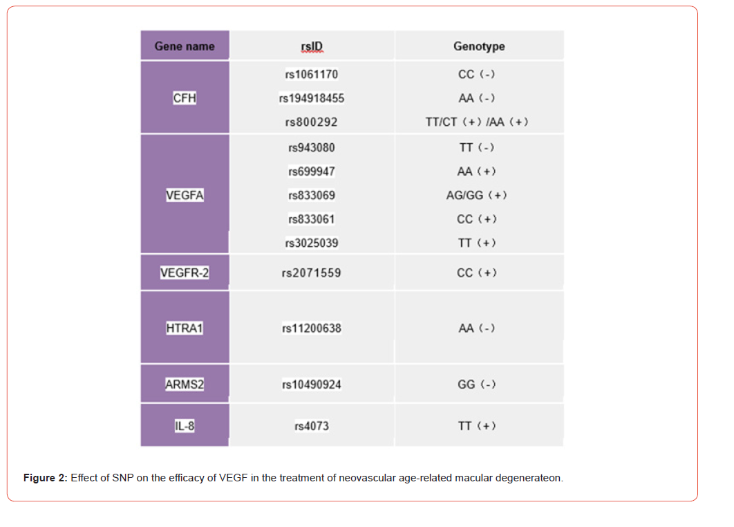
CFH gene, which is located in chromosome lq31.3 and contains 25 exons, has been confirmed as a risk factor for the occurrence of nAMD [47,48]. For nAMD patients, the study of Gourgouli K, et al. [49] showed a statistically significant correlation between CFH genotype and response to intravitreal injection of anti-VEGF drugs. Their analysis showed that patients with the CC genotype were less likely to respond to anti-VEGF drug therapy than those with the TC genotype. Another study showed that, after intravitreal injection of the anti-VEGF drug bevacizumab, patients with CC genotype had less visual improvement than patients with TC and TT genotype, meaning that patients with CFH CC genotype were more likely in the need of re-injection than those with TT genotype [50,51]. In addition, Kubicka-TrzaskaA’s group [52] and Kanoff’ s group [53] confirmed this idea by observing an association between the response to anti-VEGF drug therapy and the selected genotype of SNP rs1061170 in the CFH gene in nAMD patients. CC genotypes were found to have a higher risk of negative response after anti- VEGF therapy. At the same time, Dedania, et al. [54] also reached this conclusion by summarizing previous articles.
Moreover, Zhao, et al. [55] showed that CFH site rs1061170 CC genotype had a passive interaction in the anti-VEGF therapeutic effect of nAMD, suggesting that patients carrying CFH site rs1061170 CC genotype showed worse therapeutic effect after receiving anti-VEGF drug therapy [56]. However, TC genotype was not associated with the efficacy of anti-VEGF therapy. Other studies have shown that in nAMD patients treated with anti-VEGF drugs, patients carrying CFH rs194918455 AA genotype need more injections than those with AG genotype, that is, the fundus condition is more difficult to reach a stable state [57]. Tian, et al. [58] found that the therapeutic effect of CFH rs800292 TT and CT genotype carriers was significantly better than that of CC genotype carriers in their study on the correlation between genetic polymorphism and the response of the Chinese population to Bevacizumab treatment of nAMD. This suggests that CFH rs800292 may play an important role in the efficacy of Bevacizumab in the intervention of nAMD.
However, Cobos, et al. [59] pointed out that in a study based on Caucasian population patients with CFH rs800292 AA genotype had better prognosis than patients with other genotypes (AG/ GG). However, other studies have shown that CFH rs800292 has no significant correlation with nAMD response of anti-VEGF drug treatment [60]. Nevertheless, due to the short follow-up time of the study, the long-term efficacy of the above conclusion cannot be evaluated. In our opinion, that nAMD carrying the CFH CC genotype was generally less effective in patients treated with anti-VEGF than the other genotypes, which may be related to factors with higher VEGF levels in the vitreous fluid of patients.
VEGFA gene and VEGFR-2 gene
VEGFA gene is located on chromosome 6p21.1 and contains 9 exons. VEGF receptor-2 (VEGFR-2) gene is located on chromosome 4q12 and consists of 30 exons [47,61]. VEGFA is the strongest known angiogenic factor. Previous studies have described a significant association between response to anti-VEGF therapy in nAMD patients and polymorphism in VEGFA rs943080. Among them, the proportion of patients carrying risk T allele or TT genotype of VEGFA rs943080 gene polymorphism was higher in the poor efficacy group, which was 1.8 times of that of CC genotype [34]. These results suggest that the risk T allele or TT genotype of VEGFA rs943080 gene polymorphism may be one of the factors affecting the efficacy of anti-VEGF. Chang ‘s research team [62] found that there was a significant association between VEGFA rs833069 genotype variation and an anatomical response of Ranibizumab in the vitreous glass of nAMD patients, and that the central macular thickness of AA genotype patients was consistently lower than that of AG/GG genotype patients, indicating that AG/GG genotype patients may have a better therapeutic effect.
In 2017, Wu, et al. [63] conducted a meta-analysis of 9 variations of VEGFA and VEGFR-2. They are rs699947, rs699946, rs833069, rs833061, rs2146323, rs1413711, rs2010963, rs1570360 of VEGFA and one variant of VEGFR-2 rs2071559. The results of the meta-analysis concluded that only one SNP rs833061 was significantly associated with response to anti-VEGF therapy. For this polymorphism, anti-VEGF therapy was more effective in nAMD patients with CC genotypes, while allelic models (C vs T) and dominant models (CC+CT vs TT) were not associated with changes in treatment response. In addition, studies have shown that patients with the TT genotype of VEGFA rs3025039 require fewer injections when receiving anti-VEGF therapy in nAMD patients compared to other genotypes. This conclusion corresponded to their better results in terms of visual and tomography results [64]. Other studies concluded that the VEGFA gene locus rs3025018 variation had no significant effect on the efficacy of Ranibizumab in the treatment of nAMD [65].
In 2016, Lazzeri S, et al. [66] analyzed the inverse relationship of VEGFR-2 polymorphism affecting the efficacy of Ranibizumab and showed that there was a significant correlation between the efficacy of Ranibizumab and VEGFR-2 rs2071559 genotype. The mean central retinal sensitivity of VEGFR-2 rs2071559 TT and CT genotypes was significantly lower than that of CC genotypes. Other studies have shown that two SNPS identified in VEGFR-2 gene (rs4576072 and rs6828477), which are independently associated with better visual outcomes after 1 year of anti-VEGF treatment in nAMD patients, are predictors of anti-VEGF treatment response [67]. However, another study showed no pharmacogenetic association between VEGFR-2 rs2071559 and response to nAMD anti-VEGF therapy [68].
Similarly, a previous study found that VEGFR-2 rs4576072 and VEGFR-2 rs6828477 had no strong effect on visual response to anti-VEGF therapy in CATT and IVAN study patients with nAMD [69]. The difference in results may be related to factors such as the subjects coming from different races. Although the results were different, we prefer to believe this conclusion that patients carrying the VEGFA TT genotype have better effects on anti-VEGF treatment, because the study of Park et al had large sample size and relatively long follow-up time. The results are more acceptable.
ARMS2 gene and HTRA1 gene
ARMS2 gene and HTRA1 gene are located in chromosome 10q26.13. The ARMS2 gene consists of 2 exons and HTRA1 gene consists of 9 exons. The former is related to phagocytosis of retinal pigment epithelial cells, while the latter is a high-risk factor for AMD progression [47]. A study of 224 nAMD patients showed that patients with ARMS2 site rs10490924 GG genotype and patients with HTRA1 site rs11200638 AA genotype were significantly associated with poorer treatment outcomes after 12 months of anti-VEGF injection therapy [70]. It was also concluded in another study that patients with the rs11200638 GG/GA genotype of HTRA1 fared better than those with the AA genotype. The genotype of rs10490924 of ARMS2 gene was significantly correlated with the efficacy of Conbercept in the treatment of Chinese patients with nAMD. Patients with GG/GT genotype at rs10490924 of ARMS2 gene showed better response after 6 months of Conbercept, but no longer significantly associated after multiple corrections [71]. In addition, Yuan’s team [72] and McKibbin’ s team [73] both concluded that HTRA1 gene polymorphism may influence patients’ response to intravitreal Ranibizumab treatment for nAMD.
Other studies showed that the efficacy of Ranibizumab in the treatment of nAMD was independent of HTRA1 (rs11200638) and ARMS2 (rs10490924 and rs61544945) but varied according to the genotype of CFH. Although specific alleles of ARMS2 and HTRA1 predicted the development of nAMD, they did not predict response to anti-VEGF therapy [74,75]. In addition, some studies have found a potential pharmacogenetic association between rs10490924 genotype in ARMS2/HTRA1 and the therapeutic effect of Ranibizumab [76]. Due to the limitations of research methods, the conclusions need to be further confirmed. When Smailhodzic D, et al. [77] analyzed the cumulative effect of the risk alleles of CFH, ARMS2, and VEGFA on response to nAMD Ranibizumab treatment, it was found that carriers of high-risk CFH genotypes had less improvement in corrected vision after treatment. The nAMD patients with high-risk alleles in CFH, ARMS2, or VEGFA showed better improvement in VA than the nAMD patients with high risk alleles in CFH and ARMS2, suggesting that VEGFA genes play a greater role in treatment response to Ranibizumab than CFH and ARMS2 genes. Studies have shown that the additive effect of CFH, ARMS2, and VEGFA genotypes is partly responsible for the reduced response rate to Ranibizumab therapy. But the mechanism by which these genotypes interact with anti-VEGF therapy is currently unknown.
IL-8 gene
The IL-8 gene is located on chromosome q12-21 and contains four exons. In addition to being a powerful inflammatory cytokine, a chemokine for migratory immune cells and a activation factor for neutrophils, it is also a potent angiogenic factor. IL-8 is expressed not only by immune system cells, but also by vascular endothelial and RPE cells and plays a key role in angiogenesis under physiological and pathological conditions [78]. In related studies, fewer patients with nAMD and TT genotype carrying IL-8rs4073 had no significant change in macular fovea retinal thickness after receiving Conbercept. Yin Xinxuan [60] and his team speculated that TT genotype was a nAMD protective gene and was more sensitive to the treatment of Conbercept. In addition, Lazzeri, et al. [66] again found in the study that in nAMD patients treated with Ranibizumab, the therapeutic effect of patients carrying IL-8 rs4073 AA genotype was significantly lower than that of patients carrying AT and TT genotype, which was similar to the results of the above study. However, another study found that the AA genotype of IL-8 rs4073 was not significantly associated with the efficacy of bevacizumab in patients with nAMD, which may be related to the short followup time of this study and other factors [79]. Although there are relatively few studies on the IL-8 gene polymorphism affecting the efficacy of anti-VE GF drugs, it is still concluded that patients carrying the IL-8 TT genotype have better therapeutic effects than those carrying the AA genotype [Figure 2].
Drug Resistance and Non-Efficacy
Non-responsiveness among patients treated with anti- VEGF drugs
Although the treatment of nAMD in humans is gradually breaking through, there are still a small number of patients who have poor or no response to anti VEGF drugs used in standard vertebral therapy. After treatment with anti VEGF drugs, there are still some patients who do not show any therapeutic benefits. Instead, their vision continues to decline. The unresponsiveness of treatment is still widely observed in clinical work and scientific research. Niu’s research shows that low baseline VA is a strong predictor of unresponsiveness, meaning that patients with low baseline VA have poorer treatment response [80]. Piermarocchi, et al. [81] research also confirms this viewpoint.
In addition, Fu Yan, et al. [22] study found that elderly age and poor baseline VA are factors affecting the ineffective treatment of anti-VEGF drugs. However, Hara, et al. [24] study suggests that baseline VA does not affect the response to anti-VEGF drugs. Further research is needed to investigate the relationship between baseline VA and the response to anti-VEGF drugs. Furthermore, a study involving 179 nAMD patients emphasized the need for gene biomarkers to effectively distinguish responders from nonresponders. After treatment with anti VEGF drugs, the study cohort was divided into 128 responders and 51 non responders based on established treatment response criteria such as visual acuity and central retinal thickness, suggesting that certain genetic variations may affect the efficacy of anti VEGF drugs in treating Namd [82]. In summary, there are many factors that affect the unresponsiveness of nAMD patients treated with anti-VEGF drugs, including not only clinical factors but also genetic mutations. However, the relevant mechanisms of unresponsiveness in anti-VEGF drug treatment still need further research.
Drug resistance
At present, there are three main anti-VEGF drugs used in the treatment of nAMD in China: Ranibizumab, Aflibercept and Conbercept. Ranibizumab is a humanized monoclonal Fab fragment that binds to all VEGF-A isoforms [83]. Ranibizumab was shown to be effective in the MARINA and ANCHOR trials. Clinical follow-up results showed that approximately 90% of patients who received monthly intravitreal Ranibizumab treatment had visual loss of less than 15 letters after 2 years (ETDRS visual acuity chart) [84,14]. Aflibercept and Conbercept are recombinant fusion proteins that act as soluble decoy receptors for members of the VEGF family. Conbercept was tested in the AURORA study and most patients reported improvements in functional and morphological parameters. It has been shown that monthly or every 2mo intravitreal injections of Aflibercept after three months of initial monthly dose are similar to monthly intravitreal injections of Ranibizumab [85,86].
Although most nAMD patients have achieved good results after anti VEGF treatment, there are still a small number of patients who slowly lose the efficacy of anti VEGF drugs after repeated administration. After routine treatment, persistent subretinal fluid (SRF) still exists [85]. In addition, the CATT study showed evidence of persistent fluid leakage on optical coherence tomography in 51.5% of nAMD patients treated with intravitreal ranibizumab and 67.4% of nAMD patients treated with intravitreal bevacizumab, despite monthly anti-VEGF therapy and maintenance of 2y [87]. Active exudation on angiography or OCT was observed in some patients after 1y of conventional therapy with Aflibercept (q4w or q8w) [88]. Tranos, et al. [89] argued that despite continuous treatment with the current standard anti-VEGF methods, half of the patients showed no improvement, and about 10% of the patients showed no response at all and were resistant to anti-VEGF therapy. However, Bakall’s team [85] reported that some patients with initial response is good, and the subretinal fluid faded. But subsequent recurrent leakage produce resistance to further treatment. These studies suggest that drug resistance in patients may be a clinical factor affecting the efficacy of anti-VEGF drugs [Figure 3].
Drug Resistance and Non-Efficacy
Non-responsiveness among patients treated with anti- VEGF drugs
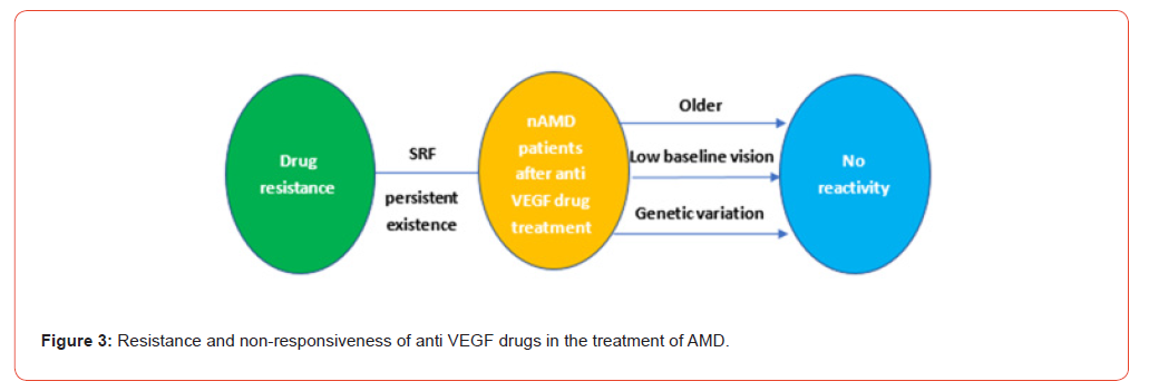
Although the treatment of nAMD in humans is gradually breaking through, there are still a small number of patients who have poor or no response to anti VEGF drugs used in standard vertebral therapy. After treatment with anti VEGF drugs, there are still some patients who do not show any therapeutic benefits. Instead, their vision continues to decline. The unresponsiveness of treatment is still widely observed in clinical work and scientific research. Niu’s research shows that low baseline VA is a strong predictor of unresponsiveness, meaning that patients with low baseline VA have poorer treatment response [80]. Piermarocchi, et al. [81] research also confirms this viewpoint.
In addition, Fu Yan, et al. [22] study found that elderly age and poor baseline VA are factors affecting the ineffective treatment of anti-VEGF drugs. However, Hara, et al. [24] study suggests that baseline VA does not affect the response to anti-VEGF drugs. Further research is needed to investigate the relationship between baseline VA and the response to anti-VEGF drugs. Furthermore, a study involving 179 nAMD patients emphasized the need for gene biomarkers to effectively distinguish responders from nonresponders. After treatment with anti VEGF drugs, the study cohort was divided into 128 responders and 51 non responders based on established treatment response criteria such as visual acuity and central retinal thickness, suggesting that certain genetic variations may affect the efficacy of anti VEGF drugs in treating Namd [82]. In summary, there are many factors that affect the unresponsiveness of nAMD patients treated with anti-VEGF drugs, including not only clinical factors but also genetic mutations. However, the relevant mechanisms of unresponsiveness in anti-VEGF drug treatment still need further research.
Drug resistance
At present, there are three main anti-VEGF drugs used in the treatment of nAMD in China: Ranibizumab, Aflibercept and Conbercept. Ranibizumab is a humanized monoclonal Fab fragment that binds to all VEGF-A isoforms [83]. Ranibizumab was shown to be effective in the MARINA and ANCHOR trials. Clinical follow-up results showed that approximately 90% of patients who received monthly intravitreal Ranibizumab treatment had visual loss of less than 15 letters after 2 years (ETDRS visual acuity chart) [84,14]. Aflibercept and Conbercept are recombinant fusion proteins that act as soluble decoy receptors for members of the VEGF family. Conbercept was tested in the AURORA study and most patients reported improvements in functional and morphological parameters. It has been shown that monthly or every 2mo intravitreal injections of Aflibercept after three months of initial monthly dose are similar to monthly intravitreal injections of Ranibizumab [85,86].
Although most nAMD patients have achieved good results after anti VEGF treatment, there are still a small number of patients who slowly lose the efficacy of anti VEGF drugs after repeated administration. After routine treatment, persistent subretinal fluid (SRF) still exists [85]. In addition, the CATT study showed evidence of persistent fluid leakage on optical coherence tomography in 51.5% of nAMD patients treated with intravitreal ranibizumab and 67.4% of nAMD patients treated with intravitreal bevacizumab, despite monthly anti-VEGF therapy and maintenance of 2y [87]. Active exudation on angiography or OCT was observed in some patients after 1y of conventional therapy with Aflibercept (q4w or q8w) [88]. Tranos, et al. [89] argued that despite continuous treatment with the current standard anti-VEGF methods, half of the patients showed no improvement, and about 10% of the patients showed no response at all and were resistant to anti-VEGF therapy. However, Bakall’s team [85] reported that some patients with initial response is good, and the subretinal fluid faded. But subsequent recurrent leakage produce resistance to further treatment. These studies suggest that drug resistance in patients may be a clinical factor affecting the efficacy of anti-VEGF drugs [Figure 3].
Conclusion and Future Direction
Neovascular AMD is a complex process involving and regulated by many factors. Among the clinically relevant influencing factors, most studies have confirmed that age, disease duration, BVA, some systemic diseases and gender have some effects on the efficacy of anti-VEGF drugs in neovascular AMD. The type of CNV, the morphological characteristics of the retina also made an important contribution to the efficacy, such as whether the lesion is type I CNV, the thickness of the retina, the structure of the macular IS/OS layer, the presence of macular traction and premacular membrane. These factors are implicated by causing disruption of nutrient and oxygen supply to the choroid, as well as affecting the diffusion of anti- VEGF drugs. In the genetic factors, some researchers believe that VEGFR is a key driving factor for the formation of CNV. However, the pathogenesis has not been fully understood. The understanding of how clinical factors and genetic variation affect the effect of anti- VEGF drugs on nAMD is still limited. Potential pharmacological effects need to be further investigated.
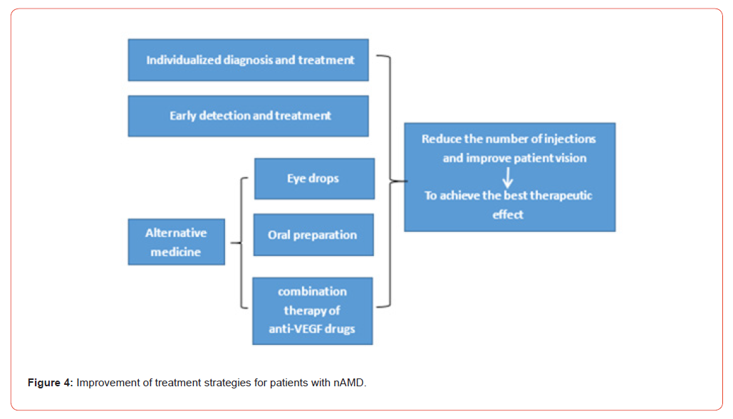
Anti-VEGF drugs are mainly targeted at CNV to improve patients’ vision and prognosis. However, there are still limitations of anti-VEGF drugs. After long-term use, the efficacy will not only decrease, but also lead to complications such as retinal hemorrhage and fibrosis, which will aggravate the condition and make it difficult to treat. Therefore, it is an important research direction in the future to formulate personalized treatment for individual genetic risk of patients, develop multi-target drugs simultaneously, improve the efficacy of anti-VEGF drugs and reduce the occurrence of complications. In addition, although individualized treatment regimens can reduce the frequency of anti-VEGF drug injections and visits, patient compliance with anti-VEGF drug therapy remains a challenge. On the one hand, increasing communication with patients so that patients can understand the occurrence, development and outcome of nAMD in more detail may be an effective plan to improve patient compliance.
On the other hand, improving the treatment system for nAMD patients and appropriately increasing the follow-up work of patients may reduce the frequency of re-visiting doctors for nAND patients. At present, there is still a delay in seeing patients with nAMD, and one of the main reasons is the lack of awareness of patients and their families about nAMD. By carrying out disease screening and publicity and strengthening the guidance to grassroots hospitals, we can improve the general population’s understanding of nAMD, so that nAMD patients can get timely and effective treatment, and finally achieve the purpose of early detection and early treatment. Although pharmacological studies have been carried out on the use of anti-VEGF drugs in the treatment of nAMDs, there are still some problems, such as racial differences in gene polymorphism, a large amount of sample evidence is needed for pharmacogenomics studies of Chinese nAMDs, and the study follow-up time is short. It is an important way to improve the prognosis of patients to find and study methods that can replace anti-VEGF drugs in the treatment of nAMD.
In most of the pharmacologic studies of anti-VEGF treatment of nAMDs discussed above, no control group was designed, and some studies had short follow-up periods. For these reasons, the correlation between SNP and the efficacy of anti-VEGF drugs for nAMDs was weakened. Therefore, the design of a large, prospective, double-blind, placebo-controlled, interventional study with longterm follow-up in patients with nAMD may provide stronger evidence to detect the correlation between clinical factors and genotypes anti-VEGF treatment response to nAMD. Understanding the relationship between various clinical and genetic factors and the effect of anti-VEGF drugs on nAMD may provide a new strategy for preventing the onset and progression of the disease.
Acknowledgment
None.
Conflict of Interest
Authors declare no conflict of interest.
References
- Zhang JY (2018) Anti-vascular endothelial growth factor for wet age-related macular degeneration. Chin J Ophthalmol and Otorhinolaryngol 18(2): 85.
- Ana IO, Á Pérez-Sala, A Pariente, Rodrigo O, Sara V, et al. (2021) Predictive Biomarkers of Age-Related Macular Degeneration Response to Anti-VEGF Treatment. J Pers Med 11(12): 1329.
- Gao L (2020) The Clinical Effect of Conbercept on Wet Age-related Macular Degeneration and Analysis of Its Prognostic Factor. Shandong University p: 1.
- Zhang JF, Zhao ZZ (2022) Pathogenesis and treatment of wet age-related macular degeneration. Recent Advances in Ophthalmology 42(02): 85-98.
- Ferris FL, Fine SL, Hyman L (1984) Age-related macular degeneration and blindness due to neovascular maculopathy. Arch Ophthalmol 102(11): 1640-1642.
- Ouyang LY, Xing YQ (2020) Current advance in the application of anti-vascular endothelial growth factor drugs in wet age-related macular degeneration. Guoji Yanke Zazhi 20(1): 74-78.
- I Habibi, F Kort, I Sfar, A Chebil, R Bouraoui (2016) Effect of Risk Alleles in CFH, C3, and VEGFA on the Response to Intravitreal Bevacizumab in Tunisian Patients with Neovascular Age-related Macular Degeneration. Klin Monbl Augenheilkd 233(4): 466-470.
- Dai H, Lu YY (2016) Influence factors and strategles of anti-VEGF therapy for wet AMD. Chin J Exp Ophthalmol 34(01): 1-4.
- Paul KW, Robert MC, Wilbert SA, Donald EC, Karen JC, et al. (2017) ACC/AHA/AAPA/ABC/ACPM/AGS/APhA/ASH/ASPC/NMA/PCNA Guideline for the Prevention, Detection, Evaluation, and Management of High Blood Pressure in Adults: A Report of the American College of Cardiology/American Heart Association Task Force on Clinical Practice Guidelines. Hypertension 71(6): 1269-1324.
- Du SG, Chen DJ, Chen ZF (2015) Change of vascular function state and RASS indexes of elderly patients with hypertension complicated with diabetes mellitus. Journal of Hainan Medical University 21(09): 1207-1209.
- Fan CN, Zhao HY, Wang H (2017) Changes and significances of plasma renin activity, angiotensin Ⅱ and aldosterone in patients with white-coat hypertension. Journal of Practical Diagnosis and Therapy 31(11): 1090-1092.
- Xue Q, Huang DZ, Zhou YC, et al. (2018) Association between raas-related microrna gene polymorphism and hypertension and diabetes. Shandong Medical Journal 58(16): 86-88.
- Xu MM, Deng HY, Li HH (2019) Mechanisms of MicroRNAs in Hypertension. Chinese Journal of Biochemistry and Molecular Biology 35(06): 606-613.
- Rosenfeld PJ, Brown DM, Heier JS, et al. (2006) MARINA Study Group Ranibizumab for neovascular age-related macular degeneration. N ENGL J Med 355(14): 1419-1431.
- Wong TY, Mitchell P (2004) Hypertensive retinopathy. The New England journal of medicine 351(22): 2310-2317.
- Wong T, Mitchell P (2007) The eye in hypertension. The Lancet 369(9559): 425-435.
- Marianne P, Scott WC, Oscar A, Gary ES, ME Marin-Castaño (2011) Angiotensin II-induced MMP-2 activity and MMP-14 and basigin protein expression are mediated via the angiotensin II receptor type 1-mitogen-activated protein kinase 1 pathway in retinal pigment epithelium: implications for age-related macular degeneration. Am J Pathol 178(6): 2665-2681.
- Ting W, Jun X, Meng Y, Xiaohang W, Yi Z, et al. (2021) Hypertension affects the treatment of wet age-related macular degeneration. Acta ophthalmologica 99(8): 871-876.
- Lee S, Song SJ, Yu HG, (2013) Current smoking is associated with a poor visual acuity improvement after intravitreal ranibizumab therapy in patients with exudative age-related macular degeneration. J Korean Med Sci 28(5): 769-774.
- Zhang X, Lai TYY, Lai TYY (2018) Baseline Predictors of Visual Acuity Outcome in Patients with Wet Age-Related Macular Degeneration. BioMed research international 2018: 9640131.
- Boyer DS, Antoszyk AN, Awh CC, (2007) Subgroup analysis of the MARINA study of ranibizumab in neovascular age-related macular degeneration. Ophthalmology 114(2): 246-252.
- Fu Y, Zhang YL, Wang LY (2021) Factors and outcomes associated with failure to anti-VEGF treatment in patients with wet age-related macular degeneration. Recent Advances in Ophthalmology 41(03): 254-258.
- Freekje van A, Maroeska MR, Yara TEL, Dzenita S, Philipp S M, et al. (2014) Predicting non-response to ranibizumab in patients with neovascular age-related macular degeneration. Ophthalmic epidemiology 21(6): 347-355.
- Chikako H, Taku W, Hiroshi T, Yoko F, Kaori S, et al. (2018) Characteristics of patients with neovascular age-related macular degeneration who are non-responders to intravitreal aflibercept. Br J Ophthalmol bjophthalmol 2018-312275.
- Peter KK, David MB, Kang Z, Henry LH, Frank GH, et al. (2007) Ranibizumab for predominantly classic neovascular age-related macular degeneration: subgroup analysis of first-year ANCHOR results. American journal of ophthalmology 144(6): 850-857.
- Jonathan HL, Sanjeewa SW, Jing X, Devinder SC, Paul NB, et al. (2012) Delay to treatment and visual outcomes in patients treated with anti-vascular endothelial growth factor for age-related macular degeneration. American journal of ophthalmology 153(4): 678-686.
- Paolo L, Alan FC, Salomon YC, Jason SS, Todd Katz, et al. (2018) Predictors of visual outcomes in patients with neovascular age-related macular degeneration treated with anti-vascular endothelial growth factor therapy: post hoc analysis of the VIEW studies. Acta ophthalmologica 96(8): e911-e918.
- Ying GS, Huang J, Maguire MG, et al. (2013) Baseline predictors for one-year visual outcomes with ranibizumab or bevacizumab for neovascular age-related macular degeneration. Ophthalmology 120(1): 122-129.
- Gong B, Fan CF, Wang Y (2014) Anti VEGF drugs (lucentis) to treat different courses of wet age-related maculardegeneration(62 cases report). Journal of Otolaryngology and Ophthalmology of Shandong University 28(05): 58-61.
- Zhang MH (2017) The Investigation of the Genetic Effect on the Non-response to the Anti-angiogenesis Treatment of Exclusive Age-related Macular Degeneration. Henan University of Science and Technology.
- Bek T, Klug SE (2018) Age, sex, and type of medication predict the effect of anti-VEGF treatment on central retinal thickness in wet age-related macular degeneration. Clinical ophthalmology 12: 473-479.
- Allen CH, Brandon GB, Carl DR, Mark RW, Sherri AVE, et al. (2014) Everen,Twenty-four-month efficacy and safety of 0.5 mg or 2.0mg ranibizumab in patients with subfoveal neovascular age-related macular degeneration. Ophthalmology 121(11): 2181-2192.
- Lee S, Song SJ, Yu HG (2013) Current smoking is associated with a poor visual acuity improvement after intravitreal ranibizumab therapy in patients with exudative age-related macular degeneration. Journal of Korean medical science 28(5): 769-774.
- Zhao L, Grob S, R Avery, Kimura A, Pieramici D, et al. Common variant in VEGFA and response to anti-VEGF therapy for neovascular age-related macular degeneration. Curr Mol Med 13(6): 929-934.
- Gwyn SW, Eulee S, Huw E, Muyiwa O, Sam E, et al. (2015) Factors affecting visual acuity after one year of follow up after repeated intravitreal ranibizumab for macular degeneration. Saudi journal of ophthalmology: official journal of the Saudi Ophthalmological Society 29(3): 187-191.
- Bora C, Jesse JJ, Sarah M, R Gallego-Pinazo, NA Yannuzzi, et al. (2015) Baseline Predictors for Good Versus Poor Visual Outcomes in the Treatment of Neovascular Age-Related Macular Degeneration with Intravitreal Anti-VEGF Therapy. Investigative ophthalmology & visual science 56(9): 5040-5047.
- AF Fercher, CK Hitzenberger, W Drexler, G Kamp, H Sattmann (1993) In vivo optical coherence tomography. American journal of ophthalmology. 116(1): 113-115.
- Florence C, Gabriel C, Marco L, Ali D, Mayer S, et al. (2015) Restoration of outer retinal layers after aflibercept therapy in exudative AMD: Prognostic value. Investigative Ophthalmology and Visual Science 56(6): 4129-4134.
- Akio O, Akitaka T, Kenji Y, Sotaro O, Hiroshi T, et al. (2015) One-year result of aflibercept treatment on age-related macular degeneration and predictive factors for visual outcome. American journal of ophthalmology 159(5): 853-860.
- Yoon HK, Dong KL, Hyung EK, Oh WK (2014) Predictive findings of visual outcome in spectral domain optical coherence tomography after ranibizumab treatment in age-related macular degeneration. Korean Jounal of Ophthalmology 28(5): 386-392.
- Heinrich G, Vlassios L, Juliane R, Lars H, Melanie T (2011) Results of flexible ranibizumab treatment in age-related macular degeneration and search for parameters with impact on outcome. Graefes’ archive for clinal and experimental ophthalmology 249(5): 653-662.
- Sanjeewa SW, Sukhpal SS, Lucy B, Jonathan L, Devinder SC, et al. (2012) Predictors of AMD treatment response. Ophthalmology 119(11): 2413-2414.
- Lilian HLK, Rupesh A, Neha K, Labishetty SC, Jay C (2017) Choroidal vascular changes in age-related macular degeneration. Acta ophthalmologica 95(7): 597-601.
- Hae MK, Hee JK, Jeong HY, Christopher SL, Sung CL, et al. (2014) Subfoveal choroidal thickness as a potential predictor of visual outcome and treatment response after intravitreal ranibizumab injections for typical exudative age-related macular degeneration. American journal of ophthalmology 157(5): 1013-1021.
- Magda G, Ludovico I, Francesco P, Umberto DV, Maria LM, et al. (2014) Visual and anatomical outcomes of intravitreal aflibercept for treatment-resistant neovascular age-related macular degeneration. BioMed research international Pp: 1-7.
- Thomas AC, Gui-Shuang Y, Maureen GM, Daniel FM, Glenn JJ, et al. (2015) Influence of the Vitreomacular Interface on Treatment Outcomes in the Comparison of Age-Related Macular Degeneration Treatments Trials. Ophthalmology 122(6): 1203-1211.
- M Gil-Martínez, P Santos-Ramos, M Fernández-Rodríguez, MJ Abraldes, MJ Rodríguez-Cid (2019) Research advance on pharmacogenomics of ranibizumab in the treatment of age-related macular degener. The Chinese Journal of Clinical Pharmacology 35(08): 816-820.
- Zhang H, Gao J, Bo DX, et al. (2021) Meta-analysis of risk factors for agerelated macular degeneration in China. International Eye Science 21(06): 1033-1039.
- Gourgouli K, Gourgouli I, Tsaousis G, Niskopoulou M, Efthimiopoulos S, et al. (2020) Investigation of genetic base in the treatment of age-related macular degeneration. Int Ophthalmol 40(4): 985-997.
- Lee AY, Raya AK, Kymes SM, Shiels A, Brantley MA Jr (2009) Pharmacogenetics of complement factor H (Y402H) and treatment of exudative age-related macular degeneration with ranibizumab. Br J Ophthalmol 93(5): 610-613.
- Dikmetas O, Kadayıfcılar S, Eldem B (2013) The effect of CFH polymorphisms on the response to the treatment of age-related macular degeneration (AMD) with intravitreal ranibizumab. Mol Vis. 19: 2571-2578.
- A Kubicka-Trząska, K Żuber-Łaskawiec, S Dziedzina, M Sanak, B Romanowska-Dixon, et al. (2022) Genetic Variants of Complement Factor H Y402H (rs1061170), C2 R102G (rs2230199), and C3 E318D (rs9332739) and Response to Intravitreal Anti-VEGF Treatment in Patients with Exudative Age-Related Macular Degeneration. Medicina (Kaunas) 58(5): 658.
- Kanoff J, Miller J (2013) Pharmacogenetics of the treatment response of age-related macular degeneration with ranibizumab and bevacizumab. Semin Ophthalmol 28(5-6): 355-360.
- Vaidehi S Dedania, Seanna Grob, Kang Zhang, Sophie J Bakri (2015) Pharmacogenomics of response to anti-VEGF therapy in exudative age-related macular degeneration[J]. Retina 35(3): 381-391.
- Zhao W, Zhao F, Li M, et al. (2022) Interaction between complement factor H gene polymorphism and vascular endothelial growth factor invitreous humor on anti-vascular endothelial growth factor efficacy in patients with age-related macular degeneration. Recent Advances in Ophthalmology 42(12): 962-966.
- Li MY, Xin XY (2021) Effect of CFH gene polymorphism on age-related macular degeneration and ranibizumab efficacy. Journal of Baotou Medical College 37(11): 128-132.
- Francis PJ (2011) The influence of genetics on response to treatment with ranibizumab (Lucentis) for age-related macular degeneration: the Lucentis Genotype Study (an American Ophthalmological Society thesis). Trans Am Ophthalmol Soc 109: 115-156.
- Jun T, Xueying Q, Kai F, Qing C, Jing H, et al. (2012) Association of genetic polymorphisms with response to bevacizumab for neovascular age-related macular degeneration in the Chinese population. Pharmacogenomics 13(7): 779-787.
- Estefania C, Sergio R, Jaouad A, M Hernandez-Sanchez, C Barreales, et al. (2018) Association between CFH, CFB, ARMS2, SERPINF1, VEGFR1 and VEGF polymorphisms and anatomical and functional response to ranibizumab treatment in neovascular age-related macular degeneration. Acta Ophthalmol 96(2): e201-e212.
- Yin XX, Xing DJ, Wang TL, et al. (2021) Correlation study of loading anti-vascular endothelial growth factor drug treatment response with single nucleotide polymorphisms in patients with exudative age-related macular degeneration[J]. Chinese Journal of Ocular Fundus Diseases 37(09): 693-701.
- Nana X, Qiyu B, Rong S, Jian L, Yuanqi Z (2019) Chitinase-3-like-1 Promotes M2 Macrophage Differentiation and Induce Choroidal Neovascularization in Neovascular Age-related Macular Degeneration. Invest Ophthalmol Vis Sci 60(14): 4596-4605.
- W Chang, DH Noh, M Sagong, ITKim (2013) Pharmacogenetic association with early response to intravitreal ranibizumab for age-related macular degeneration in a Korean population. Mol Vis 19: 702-709.
- Mingxing W, Haibo X, Yan X, Xiaojing X, Hongmi Z, et al. (2017) Association between VEGF-A and VEGFR-2 polymorphisms and response to treatment of neovascular AMD with anti-VEGF agents: a meta-analysis. Br J Ophthalmol 101(7): 976-984.
- UC Park, JY Shin, LC McCarthy, SJ Kim, JH Park, et al. (2014)Pharmacogenetic associations with long-term response to anti-vascular endothelial growth factor treatment in neovascular AMD patients. Mol Vis 20: 1680-1694.
- Song S, Yang F, Gu XY, et al. (2020) Effect of VEGFA-gene variation on neovascular age-related macular degeneration and efficacy of ranibizumab. The Chinese Journal of Experimental Ophthalmology 38(08): 680-685.
- Stefano L, Paola O, Paolo P, M Sole Sartini, G Casini, et al. (2016) IL-8 and VEGFR-2 polymorphisms modulate long-term functional response to intravitreal ranibizumab in exudative age-related macular degeneration. Pharmacogenomics 17(1): 35-39.
- Manuel MH, FV Asten, PS Muether, D Smailhodzic, P Lichtner, et al. (2014) Polymorphisms in vascular endothelial growth factor receptor 2 are associated with better response rates to ranibizumab treatment in age-related macular degeneration. Ophthalmology 121(4): 905-910.
- Hagstrom SA, Ying GS, Pauer GJ, et al. (2014) VEGFA and VEGFR2 gene polymorphisms and response to anti-vascular endothelial growth factor therapy: comparison of age-related macular degeneration treatments trials (CATT). JAMA Ophthalmol 132(5): 521-527.
- Stephanie AH, Gui-shuang Y, Maureen GM, Daniel FM, Jane G, et al. (2015) VEGFR2 Gene Polymorphisms and Response to Anti-Vascular Endothelial Growth Factor Therapy in Age-Related Macular Degeneration. Ophthalmology 122(8): 1563-1568.
- Farshad A, Sanjeewa W, Andrea JR, AFM Islam, RH Guymer, et al. (2013) Genetic influences on the outcome of anti-vascular endothelial growth factor treatment in neovascular age-related macular degeneration. Ophthalmology 120(8): 1641-168.
- Yan M (2018) Mechanism and Clinical Study of Age-related Macular Degeneration. Southern Medical University p:49-53.
- Dongqing Y, Donglan Y, Xiaoyi L, Songtao Y, Ping X, et al. (2013) Genetic association with response to intravitreal ranibizumab for neovascular age-related macular degeneration in the Han Chinese population. Ophthalmologica 230(4): 227-232.
- M McKibbin, M Ali, S Bansal, PD Baxter, Kumi W, G Williams, et al. (2012) CFH, VEGF and HTRA1 promoter genotype may influence the response to intravitreal ranibizumab therapy for neovascular age-related macular degeneration. Br J Ophthalmol 96(2): 208-212.
- FJ Rodríguez, HA Rios, María CA, SM Rosenstiehl, N Gelvez et al. (2019) Genetic association with intravitreal ranibizumab response for neovascular age-related macular degeneration in Hispanic population. Taiwan J Ophthalmol 9(4): 243-248.
- Stephanie AH, Gui-Shuang Y, Gayle JTP, GM Sturgill-Short, J Huang, et al. (2013) Pharmacogenetics for genes associated with age-related macular degeneration in the Comparison of AMD Treatments Trials (CATT). Ophthalmology 120(3): 593-599.
- Kenji Y, Keisuke M, Shigeru H, Mariko K, Yasuo Y, et al. (2017) A prospective multicenter study on genome wide associations to ranibizumab treatment outcome for age-related macular degeneration. Sci Rep 7(1): 9196.
- Dzenita S, PS Muether, J Chen, A Kwestro, AY Zhang, et al. (2012) Cumulative effect of risk alleles in CFH, ARMS2, and VEGFA on the response to ranibizumab treatment in age-related macular degeneration. Ophthalmology 119(11): 2306-2308.
- A Hautamäki, S Seitsonen, JM Holopainen, JA Moilanen, J Kivioja, et al. (2015) The genetic variant rs4073 A→T of the Interleukin-8 promoter region is associated with the earlier onset of exudative age-related macular degeneration. Acta Ophthalmol 93(8): 726-733.
- A Hautamäki, J Kivioja, S Vavuli, S Kakko, Eeva-Riitta S, et al. (2013) Interleukin 8 promoter polymorphism predicts the initial response to bevacizumab treatment for exudative age-related macular degeneration. Retina 33(9): 1815-1827.
- Niu Y (2021) Effect of Non-genetic Risk Factors on Anti-VEGF Therapy in Wet Age-related Macular Degeneration. Lanzhou University.
- Stefano P, Stefania M, Davide C, Alberta L, Tatiana S (2015) Combined effects of genetic and non-genetic risk factors affect response to ranibizumab in exudative age-related macular degeneration. Acta Ophthalmol 93(6): e451-457.
- Tobias S, M Pöllmann, Maria-Andreea G, Svenja T, Bernhard H F Weber (2022) Genetic Association Analysis of Anti-VEGF Treatment Response in Neovascular Age-Related Macular Degeneration. Int J Mol Sci 23(11): 6094.
- Sophie J Bakri, Melissa R Snyder, Joel M Reid, Jose S Pulido, Mohamed K Ezzat, et al. (2007) Pharmacokinetics of intravitreal ranibizumab (Lucentis). Ophthalmology 114(12): 2179-2182.
- David MB, Peter KK, Mark M, Gisele S, Jeffrey SH, et al. Ranibizumab versus verteporfin for neovascular age-related macular degeneration. The New England journal of medicine 355(14): 1432-144.
- Yang S, Zhao J, Sun X (2016) Resistance to anti-VEGF therapy in neovascular age-related macular degeneration: a comprehensive review. Drug design, development and therapy 10: 1857-1867.
- Xiaoxin L, Gezhi X, Yusheng W, Xun X, Xiaoling L, et al. (2014) Safety and efficacy of conbercept in neovascular age-related macular degeneration: results from a 12-month randomized phase 2 study: AURORA study. Ophthalmology 121(9): 1740-1747.
- Daniel FM, Maureen GM, Stuart LF, Gui-shuang Y, Glenn JJ, et al. (2012) Ranibizumab and bevacizumab for treatment of neovascular age-related macular degeneration: two-year results. Ophthalmology 119(7): 1388-1398.
- Jeffrey SH, David MB, Victor C, Jean-Francois K, Peter KK, et al. (2012) Intravitreal aflibercept (VEGF trap-eye) in wet age-related macular degeneration. Ophthalmology 119(12): 2537-2548.
- Paris T, Athanasios V, Solon A, Stavrenia K, Athanasios V, et al. (2013) Resistance to antivascular endothelial growth factor treatment in age-related macular degeneration. Drug design, development and therapy 7: 485-490.
- Wang YF, Liu Y, Luo XT (2021) Review and prospect of anti-vascular endothelial growth factor treatment for wet age-related macular. Journal of Shanghai Jiao Tong University 41(04): 530-534.
- Glenn JJ, Dean E, John AW, Jonathan LP, Andras P, et al. (2016) A Phase 1 Study of Intravitreous E10030 in Combination with Ranibizumab in Neovascular Age-Related Macular Degeneration. Ophthalmology 123(1): 78-85.
- Glenn JJ, Thomas AC, Antonio PC, Francois D, Pravin UD, et al. (2017) Dual Antagonism of PDGF and VEGF in Neovascular Age-Related Macular Degeneration: A Phase IIb, Multicenter, Randomized Controlled Trial. Ophthalmology 24(2): 224-234.
- Zhou QY, Peng H (2022) Overview of treatment of neovascular age-related macular degeneration by applying treat and extend regimen. Journal of Chongqing Medical University 47(04): 494-498.
- Stewart MW (2015) Individualized Treatment of Neovascular Age-Related Macular Degeneration: What are Patients Gaining? Or Losing? J Clin Med 4(5):1079-1011.
- Li H, Liu XQ, Fu YX (2022) Medical delay status and influencing factors in patients with wet age-related macular degeneration. Journal of Nurses Training 37(14): 1338-1343.
- Tolentino MJ, Dennrick A, John E (2015) Drugs in Phase II clinical trials for the treatment of age-related macular degeneration. Expert Opin Investig Drugs 24(2): 183-199.
- D Wyględowska-Promieńska, A Piotrowska-Gwóźdź, A Piotrowska-Seweryn, G Mazur-Piotrowska, W Rokicki (2014) Combination of bevacizumab and bromfenac therapy in age-related macular degeneration: a pilot study. Med Sci Monit 20: 1168-1175.
- Wyględowska-Promieńska D, Piotrowska-Gwóźdź A, Piotrowska-Seweryn A, G Mazur-Piotrowska (2015) Combination of Aflibercept and Bromfenac Therapy in Age-Related Macular Degeneration: A Pilot Study Aflibercept and Bromfenac in AMD. Med Sci Monit 21: 3906-3912.
-

This work is licensed under a Creative Commons Attribution-NonCommercial 4.0 International License.