

 Case Report
A Rare Case of Bilateral Double Primary Pterygium
Received Date:September 04, 2026; Published Date:October 13, 2026
Abstract
Background: Pterygium is a common ocular surface disease characterized by fibrovascular conjunctival tissue invading the cornea. Lesions
typically present as unilateral or bilateral single pterygia. Double pterygia within the same eye are uncommon, and bilateral double primary pterygia
are extremely rare.
Case presentation: We report a case of bilateral double primary pterygia obstructing the visual axis and causing severe visual impairment in
an elderly male patient. A 93-year-old man presented with progressive severe visual loss over 5 years. Ocular examination revealed mild bilateral
conjunctival congestion, with fibrovascular proliferations originating from both nasal and temporal conjunctiva. The nasal and temporal pterygial
heads converged on the corneal surface. The right pterygium encroached upon the pupil, while the left lesion completely covered the visual axis,
leading to marked visual impairment. The patient underwent simultaneous bilateral bare-sclera pterygium excision, with measurable improvement
in visual function after surgery.
Discussion: Bilateral double primary pterygium is a rare clinical entity. Excision combined with conjunctival autograft remains the gold standard
for pterygium surgery with the lowest recurrence rate. Nevertheless, single-session resection of multiple lesions represents a reasonable option for
elderly patients with bilateral double pterygia. This strategy reduces the total number of surgical procedures, alleviates patient discomfort, shortens
recovery time, and lowers medical costs.
Conclusion: Bilateral double primary pterygium can be safely managed with single-session bilateral bare-sclera excision without conjunctival
autograft in elderly patients, enabling effective visual rehabilitation.
Keywords: Nasal and Temporal pterygium; Pterygium excision; Bare-sclera pterygium surgery
Introduction
Pterygia are triangular fibrovascular degenerative overgrowths of the bulbar conjunctiva that progressively invade the cornea [1]. Major risk factors include chronic ultraviolet (UV) exposure, advanced age, and male sex [1-4]. Pterygia most frequently develop within the interpalpebral fissure and predominantly arise from the nasal conjunctiva. Double pterygia (coexisting nasal and temporal lesions) are infrequent, and bilateral double primary pterygia are exceptionally rare [5].
Common symptoms of pterygium include intermittent conjunctival redness, ocular irritation, foreign body sensation, dryness, and tearing, as well as progressive visual deterioration. Visual loss arises primarily from three mechanisms: encroachment on the visual axis, pterygium-induced astigmatism, and tear film instability. Surgical intervention is indicated for patients requiring symptomatic relief, visual improvement, or cosmetic correction. Multiple surgical techniques are available with varying recurrence rates, including primary conjunctival closure, conjunctival rotational grafts, limbal-conjunctival autograft, free conjunctival autograft, conjunctival flaps, and amniotic membrane transplantation.
Young age, fleshy thick pterygia, and severe preoperative inflammation are well-established risk factors for postoperative recurrence. Various adjuvant therapies have been adopted to reduce recurrence risk. Isolated bare-sclera excision without adjuvant treatment carries a relatively high recurrence risk [2,6,7], whereas conjunctival autograft combined with topical 0.05% cyclosporine eye drops achieves optimal recurrence control [1,8]. However, few controlled trials specifically compare surgical strategies among patients older than 70-80 years. One previous study even demonstrated that different pterygium surgeries did not significantly improve symptoms or quality of life in patients complicated with conjunctivochalasis [9]. Herein, we describe an elderly male farmer with bilateral double primary pterygia who received simultaneous bare-sclera excision with satisfactory postoperative outcomes.
Case Presentation
A 93-year-old male presented to our ophthalmic clinic with chief complaints of progressive, painless bilateral visual decline lasting five years. The patient had a long history of outdoor occupational labor without regular ultraviolet-protective eyewear. No prior ophthalmic medical records were available. There was no family history of pterygium. He denied ocular trauma, prior eye surgery, systemic hypertension, diabetes mellitus, or chronic medication use.
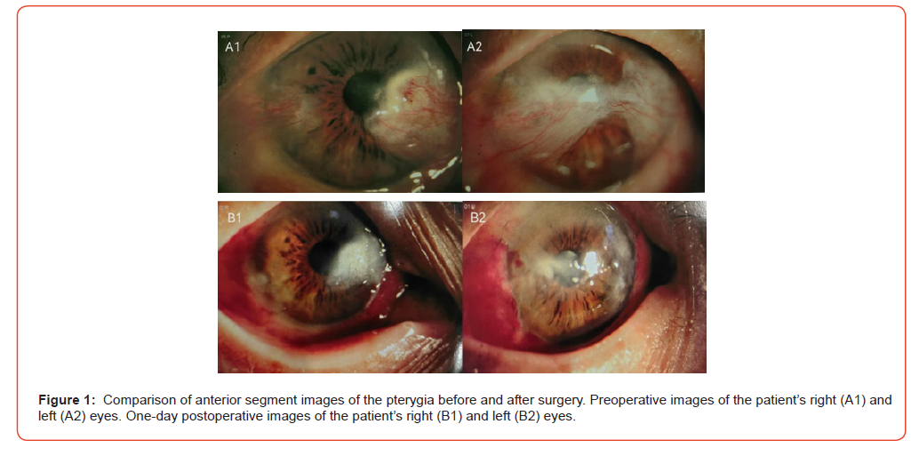
His best-corrected visual acuity (BCVA) was counting fingers at 30 cm (CF/30 cm) in the right eye (OD) and light perception (LP) in the left eye (OS). Slit-lamp examination of the right eye identified a fleshy fibrovascular nasal pterygium extending 5.5 mm from the limbus onto the cornea. A smaller temporal pterygium extended 2.5 mm from the temporal limbus toward the visual axis (Fig. 1A1). The left eye exhibited more advanced lesions: the nasal pterygium extended 6.5 mm from the limbus, and the temporal pterygium extended 4.5 mm. The two pterygial heads converged centrally on the cornea and obscured the visual axis (Fig. 1A2). Bilateral agerelated cataracts were also noted. The remaining anterior and posterior segment examinations were unremarkable.
After written informed consent was obtained, the patient underwent simultaneous bilateral pterygium excision under monitored local anesthesia; surgery was performed on the right eye first, followed immediately by the left eye.
For each lesion, standard bare-sclera excision was performed. The pterygial head was carefully dissected from the corneal epithelium using a crescent blade and toothed forceps. Fibrovascular tissue and subconjunctival Tenon’s capsule were meticulously removed. Hemostasis was achieved without cautery. The corneal surface was gently polished with a scalpel blade. Conjunctival margins were trimmed and anchored to bare sclera using interrupted 10-0 nylon sutures. A bandage contact lens was placed at the end of surgery.
On postoperative day 1, the patient only reported bilateral ocular redness and foreign body sensation. BCVA improved to 20/200 OD and 20/400 OS (Fig. 1B1, Fig. 1B2).
The postoperative regimen included topical 0.5% moxifloxacin and 1% prednisolone acetate four times daily for four weeks, followed by gradual tapering. The three-month follow-up revealed no evidence of recurrence, infectious complications, or corneal thinning. The patient reported substantial subjective improvement in visual function.
Discussion
Pterygium is one of the most prevalent ocular surface disorders worldwide. Nevertheless, bilateral double primary pterygia remain extremely rare [5]. Our patient harbored both nasal and temporal pterygia bilaterally. This atypical phenotype indicates widespread conjunctival actinic damage and chronic inflammation, consistent with his lifelong unprotected outdoor labor.
Double pterygia likely share the same pathogenic triggers as conventional single pterygium but involve more extensive conjunctival tissue [3,4]. It is hypothesized that chronic intense UV radiation induces diffuse conjunctival elastotic degeneration and persistent inflammation in susceptible individuals, permitting multifocal fibrovascular proliferation [10-12]. This case demonstrates that pterygium may manifest as multifocal disease in certain patients, requiring individualized surgical planning.
Surgical management of double pterygia poses unique challenges. Excision combined with conjunctival autograft (CAG) is regarded as the gold standard for primary pterygium, with superior recurrence control compared with amniotic membrane transplantation or primary closure [1,2,13,14]. Vertical split conjunctival autograft has been successfully applied for doublehead pterygium in previous reports [5]. In our patient, we performed simultaneous excision of nasal and temporal lesions without conjunctival autograft. This approach simplifies the surgical procedure, shortens operative time, and avoids graftrelated complications such as overlapping graft tissue or secondary conjunctival defects.
Young age is an established risk factor for postoperative recurrence [14], which supported our decision to omit conjunctival autograft in this nonagenarian patient. Adjuvant therapies may further lower recurrence risk [15-17]. Prospective comparative studies are required to evaluate recurrence rates among elderly patients older than 70-80 years treated with different surgical approaches and adjuvant pharmacotherapy.
A staged surgical strategy, prioritizing resection of the larger vision-threatening pterygium first, represents an alternative option. However, with careful surgical planning and meticulous dissection, single-session resection of all lesions is clinically feasible and beneficial. This approach minimizes repeated hospital visits, reduces patient discomfort, shortens overall recovery, and lowers financial burden. The absence of recurrence at the 3-month followup indicates that bare-sclera excision provides a simple, effective therapeutic option for elderly patients. Longer surveillance remains mandatory, given that pterygium recurrence can develop months to years postoperatively.
Conclusion
Bilateral double primary pterygium is a rare clinical presentation. For elderly patients, single-session bilateral baresclera pterygium excision without conjunctival autograft is an effective treatment strategy. This technique allows simultaneous resection of multifocal lesions to improve visual function and cosmetic outcomes. Long-term follow-up is essential to monitor disease recurrence. This case expands the existing literature and may guide management strategies for similar complex pterygium variants.
Acknowledgments
The authors thank Cheng-Zhi Yuan for technical assistance in capturing the anterior segment photographs used in this case report.
Statement of Ethics
This study was performed in accordance with the Declaration of Helsinki. This study protocol was reviewed and the need for approval was waived by Taizhou Zhangqin Eye Hospital, Bright Eye Hospital Group. Written informed consent was obtained from the patient for publication of this case report and all accompanying anterior segment clinical photographs. The patient understood that the published materials would not contain any identifiable personal information, and all data and images have been de-identified to protect privacy.
Conflict of Interest Statement
None.
Funding Sources
None.
Author Contributions
Yun-Zheng Cui: Data curation, Investigation, Writing – original
draft.
Gui-Yong Shi: Investigation, Writing – review & editing.
Qin Zhang: Investigation, Writing – review & editing.
Ji-Ping Cai: Conceptualization, Methodology, Supervision,
Writing – review & editing.
All authors approved the final manuscript and agreed to be
accountable for all aspects of the work.
Data Availability Statement
All data generated or analyzed during this study are included in this article and its supplementary material files. Further enquiries can be directed to the corresponding author.
References
- Fonseca EC, Rocha EM, Arruda GV (2018) Comparison among adjuvant treatments for primary pterygium: a network meta-analysis. Br J Ophthalmol 102(6): 748756.
- Janson BJ, Sikder S (2014) Surgical management of pterygium. Ocul Surf 12(2): 112-119.
- Zhao L, You QS, Xu L, et al. (2013) 10-year incidence and associations of pterygium in adult Chinese: The Beijing Eye Study. Invest Ophthalmol Vis Sci 54(2): 1509-1514.
- Marmamula S, Khanna RC, Rao GN (2013) Population-based assessment of prevalence and risk factors for pterygium in the South Indian State of Andhra Pradesh: The Andhra Pradesh Eye Disease Study. Invest Ophthalmol Vis Sci 54(8): 5359-5366.
- Kodavoor SK, Soundarya B, Dandapani R (2020) Comparison of vertical split conjunctival autograft with and without limbus to limbus orientation in cases of double-head pterygium-A retrospective analysis. Indian J Ophthalmol 68(4): 573-576.
- Noguera SI, Nicanor KSA, Ang RET, Cruz EM (2025) Clinical outcomes of pterygium surgery over a ten-year period: a review of recurrence and complication rates. BMC Ophthalmol. 2025;25(1): 380.
- Zheng K, Cai J, Jhanji V, Chen H (2012) Comparison of pterygium recurrence rates after limbal conjunctival autograft transplantation and other techniques: meta-analysis. Cornea. 2012;31(12): 1422-1427.
- Ryu S, Chung HY, Kim K, Tchah H, Koh K (2026) Comparison of two different topical 0.05% cyclosporine A formulations on ocular surface outcomes following pterygium excision with conjunctival autograft. Medicine (Baltimore) 105(11): e47941.
- Zhang N, Hao YN, Meng JK, et al. (2024) The Effect of Conjunctival Flap Transplantation, Pterygium Excision, and Scleral Fixation Surgery in Treating Pterygium Combined with Conjunctival Laxity and Its Impact on Postoperative Complications. Altern Ther Health Med 30(6): 188-195.
- Chen T, Ding L, Shan G, et al. (2015) Prevalence and racial differences in pterygium: a cross-sectional study in Han and Uygur adults in Xinjiang, China. Invest Ophthalmol Vis Sci 56(2): 1109-1117.
- Zhong H, Cha X, Wei T, et al. (2012) Prevalence of and risk factors for pterygium in rural adult Chinese populations of the Bai nationality in Dali: The Yunnan Minority Eye Study. Invest Ophthalmol Vis Sci 53(10): 6617-6621.
- Wang Y, Shan G, Gan L, et al. (2020) Prevalence and associated factors for pterygium in Han and Mongolian adults: A cross-sectional study in Inner Mongolia, China. BMC Ophthalmol 20(1):4 5.
- Kodavoor SK, Joseph AJ, Ramamurthy S, Dandapani R (2024) Management of pterygium: our experience and a simplified treatment algorithm. Rom J Ophthalmol 68(2): 108-113.
- Pardo Bayona M, Méndez Rodríguez LM, Benavides Cruz J, et al. (2026) Recurrence rate, time to recurrence, and associated factors after primary pterygium excision with conjunctival-limbal autograft: a prospective cohort study. BMC Ophthalmol 26(1): 263.
- Yap JY, Tai ELM, Kueh YC, et al. (2025) Effects of ranibizumab on microvasculature, oxidative stress and recurrence in primary pterygium surgery. Int Ophthalmol 45(1): 29.
- Richard S, Obied B, Abu Dbai J, et al. (2025) Teprotumumab reduces the proliferation and viability of pterygium fibroblasts. Exp Eye Res260: 110589.
- Rajagopal J, Das GK, Sharma S, Sahu PK (2024) Pterygium recurrence following preoperative topical Mitomycin C and 5-Fluorouracil eye drops. J Fr Ophtalmol 47(9): 104272.
-

This work is licensed under a Creative Commons Attribution-NonCommercial 4.0 International License.