 Case Report
Necrosant Fasciitis with Histoplasma Duboisii
Lenga loumingou IA1*, Ondima I², Soussa R1, Djouboue O1, Mouamba F3 and Peko JF3
1Department of Dermatology CHU Brazzaville
2Pediatric Surgery Department CHU Brazzaville
3Department of anatomy and pathology CHU Brazzaville
Ida Aurelie Lenga loumingou, Department of Dermatology CHU Brazzaville, Congo.
Received Date: April 09, 2024; Published Date: June 07, 2024
Abstract
Summary
Context and rationale: African Histoplasmosis has been described in the African tropical belt. Its prevalence is unknown in Congo. Histoplasmosis can be localized or systemic. Its skin manifestations vary. Necrotizing fasciitis is a serious bacterial condition. It is exceptionally fungal.
Observation
The authors report the case of a 12-year-old child, living in a rural area in Congo Brazzaville. The patient has no particular history. HIV serology is negative. He has a deterioration in general condition with anemia and asthenia, an infectious syndrome, hepatosplenomegaly. The diagnosis of histoplasmosis is histological. Improvement is obtained in a few days with Itraconazole.
Conclusion
Necrotizing fasciitis is classically due to common germs. It is useful to look for Histoplasmosis in certain skin necroses and ulcers.
Keywords: Necrotizing fasciitis; Histoplasmosis; child
Introduction
Necrotizing fasciitis is a deep skin infection that affects fascia. They are deadly. These are medical-surgical emergencies [1]. They are classically mono or poly microbial bacteria, notably streptococcal, associated or not with staphylococci, enterobacteria and rarely attributed to species such as Shewanella Alga [2] Antibiotic therapy is probabilistic during initial treatment [3] Mortality is high. Exceptional cases of fungal necrotizing fasciitis have been reported [4] We report necrotizing Histoplasma fasciitis in a 12-year-old child.
Observation
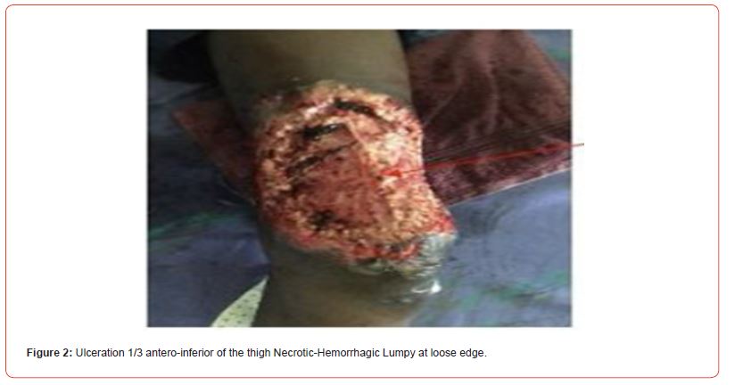
This was a male subject, aged 12, living in a rural area, with a low socio-economic level. He was admitted to the pediatric surgery department for ulceration of the left thigh. He had no particular background. The onset of symptoms dated back 2 months before admission, with pigmented popular lesions and umbilicated papules (Figure 1) diffuse but predominant in the pelvic limbs. One of the lesions had a gummy development leading to ulceration of the lower antero third of the left thigh (Figure 2). There was an infectious syndrome, anemia, hepatosplenomegaly, inguinal lymphadenopathy with an inflammatory appearance. HIV serology was negative. Histopathology revealed Histoplasma capsulatum Var Duboisii. ulcer cleaning and treatment with itraconazole at 100 mg per day were prescribed. An improvement was observed on the 5th day (Figure 3). Treatment was continued on an outpatient basis after 2 weeks of hospitalization.


Comments
It was a Histoplasma Duboisii necrotizing fasciitis in a 12-yearold child. Necrotizing fasciitis is rare. They are exceptional in children [5] their cause is classically bacterial. The probabilistic treatment codified by the French Society of Infectiology and Tropical Pathology includes a synergistic combination of antibiotics [3] But necrotizing fasciitis can be fungal. A form resistant to antibiotics was murcomycoses [4] immunocompromise is not obligatory in cutaneous and or systemic histoplasmosis [5] Mycobacteria must be discussed in particular buruli ulcer and multifocal tuberculosis. Itraconazole is effective and well tolerated [1,6].
Conclusion
This was an exceptional clinical form due to the age of the patient, the fungal etiology and the effectiveness and tolerance to itraconazole.
Acknowledgments
None.
Conflict of Interest
of Interest
References
- Damisa J, Ahmed S, Harrisson S Necrotisîg fasciitis: A narrative Review of the literature. British journal of Hospital Medicine 82 (1):1-9.
- Ramampisédrahova JB, Rafimahatratra R, Duval Solomalala G (2020) Monomicrobial necrotizing fasciitis of the leg due to multiresistant Acinobacter in a healthy adult: Case report. Pan Afr Med J 36: 344.
- SPILF / SFD (2019) Recommendations for the management of bacterial dermohypodermatitis.
- Dugourd PM, Hubiche T, Courjon J, Boissy C, Masseloer S, et al. (2017) Resistant necrotizing cellulitis. Ann Dermatol Venerol 144(12): S285-S286.
- Pfeifle VA, Gros SJ, Holland Cunz S, Kampfen A (2017) Necrotizîng fasciitis in children due to minot lesions. Journal of Pediatric Surgery case report 25: 52-55.
- Ekeng BE, Edem K, Amamilo I, Ranos Z, Denning D, et al. (2021) Histoplasma in children; HIV/AIDS not a major. J fungi (Basel) 7: 530.
-
Lenga loumingou IA*, Ondima I, Soussa R, Djouboue O, Mouamba F and Peko JF. Necrosant Fasciitis with Histoplasma Duboisii. World Journal of Dermatology & Cosmetics. 1(2): 2024. WJDC.MS.ID.000509.
-
Histoplasma, skin infection, necrotizing fasciitis, microbial bacteria
-

This work is licensed under a Creative Commons Attribution-NonCommercial 4.0 International License.


