


 Research Article
Experimental Infection of Immature Stages of Sarcophagid Flies with the Parasitoids Dirhinus Himalayanus Westwood, 1836 and Brachymeria Podagrica (Fabricius 1787) (Hymenoptera:Chalcididae) Under Laboratory Conditions
1Central Veterinary Research Laboratory, Dubai, United Arab Emirates
1Central Veterinary Research Laboratory, United Arab Emirates
Received Date: January 27, 2023; Published Date: February 21, 2023
Abstract
The development of two species of parasitoid wasps, Dirhinus himalayanus and Brachymeria podagrica, was studied under laboratory conditions at 26oC using Parasarcophaga dux and Wohlfahrtia nuba as hosts. The breeding success of B. podagrica in P. dux and W. nuba was 78.0 and 72.0 %, respectively and offspring emerged 22-28 d after 3rd stage larvae of flies were exposed to the wasps. D. himalayanus emerged 29-37 d after exposure of fly pupae. The breeding success of this wasp species in P. dux and W. nuba amounted to 90.0 and 98.0%, respectively. No fly imagines eclosed in the experimental groups. Out of 50 third stage larvae of P. dux and W. nuba in the uninfected control groups, 46 and 50 fly imagines eclosed, respectively. Their preimaginal development P. dux and W. nuba lasted 17-20 and 21-23 d, respectively.
Keywords: Parasitoid wasps, Dirhinus himalayanus, Brachymeria podagrica, Parasarcophaga dux, Wohlfahrtia nuba, United Arab Emirates
Introduction
Brachymeria podagrica and Dirhinus himalayanus are solitary parasitoid wasps of calyptrate flies. B. podagrica occurs in temperate, subtropical and tropical zones on all continents [1]. With regards to the Middle East, it has been registered in Iran [2], in the UAE [3] and recently also in Saudi Arabia (Al Galil et al. 2022). B. podagrica infects fly maggots and its larval stages grow inside the fly pupa. D. himalayanus is considered an indo-pacific species that has been introduced to countries of the Arabian Peninsula. Noyes [4] mentioned the occurrence of this wasp in the Central Asian Republics, Iran, Pakistan, India, Japan, Thailand, Malaysia and Philippines. Contrary to B. podagrica, D. himalayanus is an ectoparasitoid that infests fly pupariae. In an attempt to study the preimaginal devel-opment of facultative myiasis flies during the hot summer months in Dubai, both parasitoids were detected hatching from naturally exposed fly larvae and pupae of sarcophagid flies [5].
The aim of this study was to investigate details of the preimaginal development of these wasps and their efficacy to prevent the development of the two host flies, Parasarcophaga dux and Wohlfahrtia nuba under laboratory conditions.
Materials and methods
Establishing colonies of host flies
Colonies of Parasarcophaga dux and Wohlfahrtia nuba were established at the Central Veterinary Research Laboratory (CVRL) in late spring 2022. For this, 500 g of fresh camel meat deposited in a plastic container in shaded place was used as a bait. Already the same day, a large number of flies (Calli Phoridae, Sarcophagidae and smaller species) visited the meat and deposited their development stages. At daily visual examination, the development of fly maggots was observed. In order to avoid an overcrowding, but also since parasitoid wasps and ants were attracted, the container was covered with a layer of tissue paper. When the third stage fly larvae occurred the container was placed into a bucket with a 3 cm layer of fine grained sand at the bottom. To avoid the entrance of parasitoids and ants, the bucket was covered with a fine woven mesh. Creamy white fly larvae left the meat bait, fell into the sand and were sieved out four to five days after the bait was set. Harvested maggots were kept for pupation in plastic bowls on a layer of fine-grained sand in the laboratory at 26o C and pupae were transferred into Petri dishes until flies eclosed. By keeping the Petri dishes for 10 min at 4o C in the refrigerator fly imagines were temporarily immobilized in order to sort them by species under the stereoscopic microscope.
About 40 to 50 flies of P. dux and W. nuba were kept separately in wooden fly cages with a glass window in front, at 26 to 30o C on a diet of pasteurized milk and sugar. Camel meat was introduced as breeding substrate. With the appearance of 2nd stage maggots, the meat bait container was removed from the cage and kept in a small bucket on a layer of sand until the third stage larvae moved out.
Establishing colonies of parasitoids
When establishing the host fly colonies, the surplus of 3rd stage maggots was put in an open plastic bowl next to the decaying meat sample and left for pupation. Ten days later, fly pupae were collected and placed in large Petri dishes in the laboratory at 26o C. Emerging parasitoids were separated by species and kept at a diet of water diluted honey.
Infection trials
Metal flour sifters (Dayso, Japan), 165 mm in diaonter, 45 mm high, 0.8 mm mesh covered with a large Petri dish as lid were found ideal for the purpose of our infection trial because they allow sufficient ventilation and prevent smaller parasitoids (Nasonia spp.) to enter.
At the day of infection, parasitoids were five to ten days old. In the first trial, to each 50 third stage larvae of P. dux and W. nuba kept in separate containers five fertilized B. podagrica females were added and parasitoids were removed after three days when all maggots became pupae. In the second trial, each 50 freshly pupated P. dux and W. nuba in separate containers remained for 5 days in contact with five fertilized D. himalayanus. The observations were stopped five days after the last parasitoid wasp hatched. Remaining pupariae were dissected and their content was examined under a stereo microscope. The uninfected control group consisted of each 50 P. dux and W. nuba third stage larvae that were kept in separate containers under similar conditions as the experimental groups.
Statistical treatment
Confidence intervals for the wasps breeding success was generated using the software Quantitative Parasitology 3.0 [6].
Results
Table 1: Breeding success of parasitoid wasps Brachymeria podagrica and Dirhinus himalayanus using flesh flies Parasarcophaga dux and Wohlfahrtia nuba as experimental hosts.
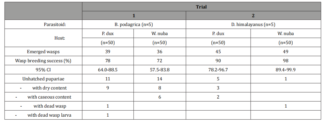
Both fly species deposited their larvae already at the day when the meat was placed in the fly cages. Four days later, creamy white maggots started to move out and completed pupation within the next three days. Immediately after female B. podagrica were added to fly maggots, parasitoids started to attack their hosts by piercing their ovipositor into the fly larvae. Maggots resisted by turning around their axis in order to dislodge the attacker. As a reaction of the attack, maggots released a small amount of a smelly grey liquid. The wasps had a maximum of three days to deposit their eggs and Brachymeria progeny hatched 22 – 28 d later in numbers of 39 and 36 from Parasarcophaga and Wohlfahrtia pupariae, respectively (Table 1). The dissection of the unhatched fly pupae revealed dry or caseous contents in 23 out of 25 puparie. Two more puparial shells contained a dead wasp larva and a dead B. podagrica imago that was unabale to hatch.
D. himalayanus observed the offered fly pupae carefully groping them with their antennae to find a suitable place to insert their ovipositor. This wasp species stayed in contact with the host puparium for up to 20 min. Dirhinus offspring emerged from fly pupariae 29 to 37 days after wasps were added to the fly pupae. The number of hatched progeny from Parasarcophaga and Wohlfahrtia pupariae amounted to 45 and 49, respectively. Five unhatched fly pupariae had a dry or caseous content and one D. himalayanus imago was unable to hatch.
When hatching, both wasp species gnaw the puparial shell all around at 2 mm from anterior end and hatch through this opening. In both experiments not a single fly eclosed. In the uninfected Parasarcophaga control group, 48 flies eclosed between day 17 and 20 after larval deposition, 11 to 14 days after pupation. Two 3rd stage larvae did not pupate and dried out. All flies hatched from 50 pupae in the Wohlfahrtia group, at day 21-23 after larval deposition, 15-17 days after pupation.
Discussion
Members of the Sarcophagidae family are necrophorous flies that can act as pathogens inducing myiasis in animals and humans. While W. magnifica causes obligatory myiasis and was recognized as a problem in Bactrian camels in Mongolia and China [7,8,9,10] W nuba was involved in cases of facultative myiasis in dromedaries in the UAE [11]. Regarding P. dux, there had been several human myiasis case described in the literature [12-14].
Flesh flies play an important role in criminal forensic investigations for the determination of the postmortem interval and the temperature of the environment plays a crucial role. In the current experiments under laboratory conditions at 26o C , the preimaginal period of P. dux and W. nuba in the unexposed control groups lasted 17-20 and 21-23 d, respectively. This time span agrees with reference literature data obtained at similar temperature conditions [15-19]. Contrary to the uninfected hosts, the development of the parasitoids took longer, 22 to 28 d and 29 to 37 d in the case of B. podagrica and D. himalayanus, respectively. And for this reason, these parasitoids can play a role in forensic investigations when flesh flies as the first colonizers of a carcass have eclosed leaving only empty puparial shells behind.
Both B. podagrica and D. himalayanus are solitary parasitoids meaning that only one progeny can be produced per host. While B. podagrica is an endo-parasitoid with larval development proceeding in the host tissues, the larval development of D. himalayanus takes place on the developing fly pupal stage within the puparial shell. For this reason, D. himalayanus is considered an ecto-parasitoid. Both species are attracted by the smell of decaying meat or carcasses.
Also the strategy of both wasp species differs. B. podagrica attacks crawling 3rd stage maggots and therefore has to act quickly to insert its eggs in heavily resisting hosts. Maggots were attacked several times by one and the same or by different wasps leading to multiple traumas and possibly to introduction of other pathogens as well. The time span for the infection is limited since fly larvae under conditions of this study pupate within three days after leaving the breeding substrate. It is our experience that B. podagrica observed the deposited meat baits for suitable hosts already when 3rd stage fly larvae were still in the breeding substrate but this will not substantially change the development time. D. himalayanus on the other hand infects immobile fly pupae and takes time to observe the host to find a suitable place to insert its ovipositor.
Since the larval stage of B. podagrica develops within the host, the impact of the infection compared to the ectoparasitoid D. himalayanus is more severe. This resulted in a higher premature death of the host when dry or caseous content or decaying remnants of fly development stages were found in uneclosed fly pupariae. D. himalayanus as an ectoparasitoid species in contrast drills a hole through the puparial wall and lays eggs on the developing fly pupa. Its larva develops between the puparial wall and the host, not entering the latter.
The life of the two flesh fly species in both experimental groups was terminated and not a single host eclosed while in the uninfected control groups all 50 (=100%) W. nuba and 46 (= 92%) out of 50 P. dux larvae completed their development.
Conclusion
Considering problems with insecticide resistance, more experiments have to be conducted to find out if these parasitoid species can be used for the control of house and stable flies in animal farms. Since B. podagrica and D. himalayanus emerge from their host pupariae later than the host flies from uninfected puparie, both parasitoids can become important in criminal forensic examinations in cases when corpses had started to decay. A precondition for the use of B. podagrica and D. himalayanus in forensic examination is the detailed study of the life cycle under different temperature conditions.
Acknowledgment
None.
Conflict of Interest
The authors declare that they have no conflict of interest.
References
- Iqbal T (2015) Taxonomic study of Chalcidididae (Chalcidoidea: Hymenoptera) of Khyber Pakhtunkhwa – Pakistan. PhD thesis University of Peshewar, pp: 132.
- Delvare G, Talaee L, Goldansaz SH (2011) New Chalcididae (Hymenoptera: Chalcidoidea) of economic importance from Iran. Ann Zool 61: 789-801.
- Delvare G (2017) Order Hymenoptera, family Chalcididae. Arthropod fauna of the UAE 6: 225-274.
- Noyes JS (2019) Universal Chalcidoidea Database. World Wide Web electronic publication.
- Schuster RK, Sivakumar S (2022) Natural infection of preimaginal stages of flies of the Sarcophagidae family with Brachymeria podagrica and Dirhinus himalayanus (Hymenoptera: Chalcididae) in the United Arab Emirates. Environ Anal Eco stud. 000734. 10(2).
- Reiczigel J, Marozzi M, Fabian I, Rozsa (2019) Biostatistics for parasitologists - a primer to Quantitative Parasitology. Trends Parasitol 35(4): 277-281.
- Schuhmann H, Ribbeck R, Beulig, W (1976) Wohlfahrtia magnifica (Schiner, 1862) Diptera: Sacrophagidae) causing a vaginal myiasis in domesticated two-humped camels in the Mongolian People's Republic. Arch Exp Vet 30(6): 799-806.
- Valentin A, Baumann M, Schein E, Bajanbileg S (1997) Genital myiasis (Wohlfahrtiosis) in camel herds of Mongolia. Vet Parasitol 73(3-4): 335-346.
- Xue J, Ai D, Xu X, Wang C, Jiang X, Han T, Er D (2022) Isolation and Identification of Volatile Substances with Attractive Effects on Wohlfahrtia magnifica from Vagina of Bactrian Camel. Vet Sci 9(11): 637.
- Zhi L, Ai D, Yong M, Bao H, Han B, et al. (2022) The effects of genital myiasis on the diversity of the vaginal microbiota in female Bactrian camels. BMC Vet Res 18(1): 87.
- Tsang CC, Tang JYM, Fong JYH, Kinne J, Lee HH, et al., (2018) Ignatzschineria cameli sp. nov., isolated from necrotic foot tissue of dromedaries (Camelus dromedarius) and associated maggots (Wohlfahrtia species) in Dubai. Int J Syst Evol Microbiol 68(11): 3627-3634.
- Chaiwong T, Tem-Eiam, N, Limpawithayakul M, Boongunha N, Poolphol W, et al. (2014) Aural myiasis caused by Parasarcophaga (Liosarcophaga) dux (Thomson) in Thailand. Trop Biomed 31(3): 496-498.
- Sukontason KL, Sanit S, Klong-Klaew T, Tomberlin JK, Sukontason K (2014) Sarcophaga (Liosarcophaga) dux (Diptera: Sarcophagidae): A flesh fly species of medical importance. Biol Res 47(1): 14.
- Chiewchanit S, Chaithong U, Sanit S, Samerjai C, Sukontason K (2017) Dermal myiasis caused by the flesh fly, Parasarcophaga (Liosarcophaga) dux (Thomson, 1869) (Diptera: Sarcophagidae) at the site of a malignant melanoma: a case report. Southeast Asian J Trop Med Public Health 48(1): 184-188.
- Amoudi MA (1993) Effect of temperature on the developmental stages of Wohlfahrtia nuba (Diptera:Sarcophagidae) J Egypt Soc Parasitol 23(3): 697-705.
- Shiravi AH, Mostafavi R, Akbarzadeh K, Oshaghi MA (2011) Temperature requirements of some common forensically important blow and flesh flies (Diptera) under laboratory conditions. Iran J Arthropoda Borne Dis 5(1): 54-62.
- Babu S, Sharma H, Upadhyay S (2017) Studies on the larval growth of forensically important flesh fly Sarcophaga dux Thomson 1869 (Diptera: Sarcophagidae) under outdor ambient temperatures from Central India. Int J Appl Res 3: 366-370.
- Zhang X, Li Y, Shang Y, Ren L, Chen W, et al. (2020) Development of Sarcophaga dux (Diptera: Sarcophagidae) at constant temperatures and differential gene expression for age estimation of the pupae. J Thermal Biol 93: 102735.
- Al Galil, FMA, Al-Keridis LA, Al-Mekhlafi FA, Al-Amri AM, Al-Khalifa MS (2022) First record of Brachymeria podagrica (Hymenoptera: Chalcididae) in Bisha city, Asir region, Saudi Arabia. J Med Entomol 59(5): 1556-1561.
-

This work is licensed under a Creative Commons Attribution-NonCommercial 4.0 International License.