 Research Article
Microscopic Appearance of Monolaurin-Treated Synthetic DNA (E. Coli) Crown Cells in Agar Medium Supplemented with Egg White
Shahed Uddin Ahmed Shazib, Department of Biological Sciences, Purdue University, West Lafayette, USA
Received Date: January 02, 2024; Published Date: February 15, 2024
Abstract
Synthesized DNA crown cells (artificial cells) can be prepared in vitro using sphingosine (Sph)-DNA-adenosine-monolaurin compounds. Previous studies have shown that assembly formation and cell proliferation of these synthetic DNA (E. coli) crown cells can observed on agar plates. In addition, these DNA crown cells can proliferate within egg whites in vivo, suggesting that synthetic DNA crown cells can become living cells when cultured in egg white. Therefore, the present experiments examined whether cell proliferation associated with egg white was observed when monolaurin-treated synthetic DNA (E. coli) crown cells were cultivated on agar plates supplemented with egg white. In addition, the microscopic characteristics of the proliferated cells are described.
Keywords:Synthetic DNA (E. coli) crown cells; agar plate cultures; Sphingosine-DNA; cell proliferation; monolaurin, egg white
Introduction
Artificial cells that are covered with DNA are referred to as DNA crown cells [1-3]. Synthetic DNA crown cells can be prepared by cultivating DNA crown cells in egg white supplemented with sphingosine (Sph)-DNA and adenosine-monolaurin (A-M). Such DNA crown cells have been shown to form assemblies or cells in cultures of synthetic DNA crown cells with monolaurin [4-6]. In a previous study, assembly formation and cell proliferation were observed after cultivation on agar plates [7]. Also, synthetic DNA (E. coli), human placenta, Akoya pearl oyster and HepG2) crown cells have been cultivated in test tubes containing egg white [8–11]. However, since these cultivation experiments were carried out using a liquid medium, it was not clear whether cell proliferation could be observed on agar plates. Hence, the present experiments examined whether cell proliferation associated with egg white was observed when monolaurin-treated synthetic DNA (E. coli) crown cells were further cultivated on agar plates supplemented with egg white. The findings were characterized microscopically.
Materials and Methods
Materials
The materials used were the same as those employed in previous studies [12,13], i.e., Sph (Tokyo Kasei, Japan), DNA (Sigma-Aldrich, Wako, Japan), adenosine (Sigma-Aldrich), and monolaurin (Tokyo Kasei). An adenosine-monolaurin (A-M) was prepared by mixing adenosine and monolaurin, as described previously [12,13]. Monolaurin solutions were prepared to a final concentration of 0.1 M in distilled water. Agar plates were prepared using standard agar medium (SMA) (AZS ONE Japan). Egg white was obtained from eggs procured at a local market.
Methods Preparation of synthetic DNA (E. coli) crown cells
Synthetic DNA (E. coli) crown cells were prepared as described previously [12,13]. Briefly, 180 μL of Sph (10 mM) and 90 μL of DNA (1.7μg/μL) were combined, and the mixture was heated and cooled twice. A-M solution (100 μL) was added to the mixture, which was then incubated at 37°C for 15 min. Next, 30 μL of monolaurin solution was added and the mixture was incubated at 37°C for a further 5 min. The resulting suspension was used as the synthetic DNA (E. coli) crown cells.
Culture of monolaurin with DNA (E. coli) crown cells and incubation
with egg white on agar plates was performed as follows:
a) 50.0 μL of cell sample was plated on the agar plate with a bacteria
spreader.
b) 1.5 mL of 0.1 M monolaurin (twice diluted) was poured onto
the agar plate.
c) After removing excess monolaurin, the plates were inverted
and incubated for 3.0 h at 37°C.
d) 1.5 ml of egg white was then poured onto the plate and excess
egg white was removed.
e) The plates were then inverted and incubated for 2.0 h and 2
days at 37°C.
Microscopic observations
The plates were observed under a light microscope.
Result and Discussion
a) Figure 1 shows an object associated with synthetic DNA (E. coli) crown cells on the plates 3.0 h after monolaurin addition and before the addition of egg white.
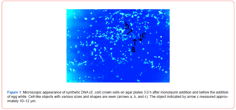
Numerous cell-like objects of various sizes and shapes (arrows
a, b, and c) were observed. The object indicated by arrow c measured
approximately 10–12 μm.
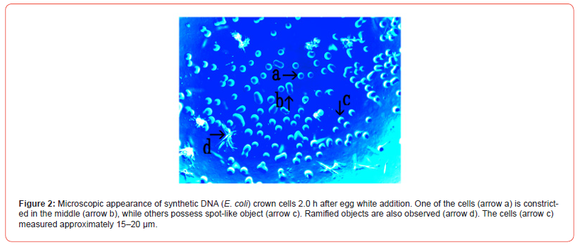
b) Figure 2 shows the microscopic appearance of synthetic DNA
(E. coli) crown cells 2.0 h after egg white addition.
Numerous round cell-like objects were observed (arrow a). One
of these was constricted in the middle (arrow b), while spot-like object
was observed (arrow c). Also, ramified objects were observed
(arrow d). The size of the cells (arrow c) was approximately 15–20
μm.
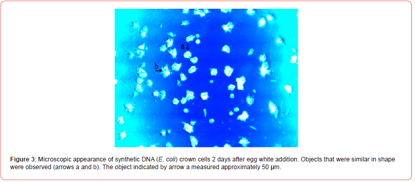
c) Figure 3 shows the microscopic appearance of synthetic DNA
(E. coli) crown cells 2 days after egg white addition.
Objects that were similar in shape were observed (arrows a and
b). These objects (arrow a) measured about 50 μm.
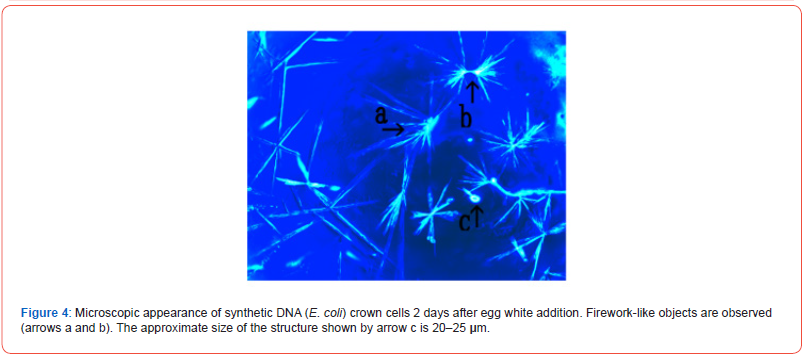
d) Figure 4 shows the microscopic appearance of synthetic DNA
(E. coli) crown cells 2 days after egg white addition.
Numerous firework-like objects (arrows a and b) measuring
approximately 20–25 μm (arrow c) were observed.
e) Figure 5 shows the microscopic appearance of synthetic DNA
(E. coli) crown cells 2 days after egg white addition.
Objects varying in shape and size were observed (arrows a, b, c, and d), most of which were enclosed by a membrane. An object enclosing a cell-like object was observed (arrows a and b), and some objects (arrow c) enclosed several cell-like objects.
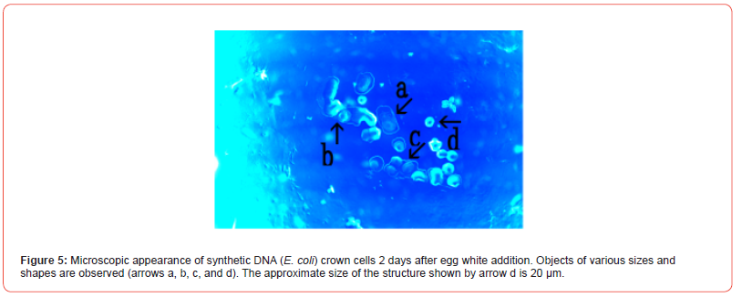
In addition, a cell-like object that was not enclosed was also observed
(arrow d). The approximate size of this cell-like object was
20 μm (arrow d).
f) Figure 6 shows the microscopic appearance of synthetic DNA
(E. coli) crown cells 2 days after egg white addition.
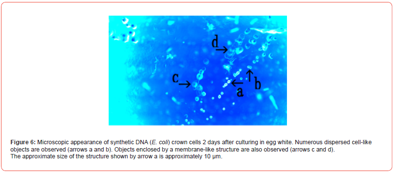
Numerous dispersed cell-like objects were observed (arrows a and b).
In addition, objects enclosed by a membrane-like structure were observed (arrows c and d); The size of these objects was approximately 10 μm (arrow a).
Previously, assembly formation and cell proliferation were observed in cultures of synthetic DNA (E. coli) crown cells cultured on agar plates supplemented with monolaurin [7]. In addition, synthetic DNA crown cells or monolaurin-treated synthetic DNA crown cells could be cultivated with the use of egg white in test tubes, suggesting that the change from non-living objects to living cells could be attributed to growth in media with egg white. Hence, the present experiments examined whether similar culture characteristics could be observed in agar and new proof of the transition from non-living to living cells could be obtained. Cell proliferation of various types was observed after 2 h and 2 days of cultivation in egg white. An interesting finding was that cell-like objects enclosed with a membrane-like structure were observed (Figures 5 and 6) However, because such objects were not observed in test tube cultures, the mechanism underlying their formation was unclear. These enclosed objects may be derived from monolaurin-treated synthesized DNA (E. coli) crown cells, and the membrane-like structures may be derived from components of egg white.
Taken together, the objects shown in Figures 5 and 6 may be composed of cell-like objects enclosed by egg components. On the other hand, similarly shaped objects (Figure 3) and firework-like objects (Figure 4) were observed. Such objects were also observed in test tube cultures [14]. In particular, the firework-like objects were considered to consist of Sph-DNA and its related components. Because both objects were frequently observed, they may play an important role in the change from non-living to living cells. On the other hand, various types of proliferated cells exist, suggesting that cells the proliferated through various mechanisms. It is considered that the peak in object formation in cultures, both in vivo and in vitro, may occur after 7 days of culture. As a result, the cultures were maintained for 7 days. Recently, it has been shown that new compounds, such as adenosine-DNA, were formed during cultivation, and that synthetic DNA crown cells may be successfully cultivated [14]. The present data were collected over only 2 days of culture. However, generation of new cells in agar cultures may occur over longer periods. Future studies will be conducted to test this idea.
References
- Inooka S (2012) Preparation and cultivation of artificial cells. App Cell Biol 25(1): 13-18.
- Inooka S (2016) Preparation of Artificial Cells Using Eggs with Sphingosine-DNA J Chem Eng Process Technol 7(1): 277-282.
- Inooka S (2016) Aggregation of sphingosine-DNA and cell construction using components from egg white. Integrative Molecular Medicine 3(6): 1-5.
- Inooka S (2021) Microscopic observation of assemblies formed in mixtures of DNA (Human placenta) crown cells with Bacilus subtilius. An archive of organic and inorganic science 5(2): 658-664.
- Inooka S (2020) Assembly Formation of Synthetic DNA (E.coli) crown cells with Salt “Reconstruction and Regeneration of Synthetic DNA Crown Cells. American Journal of Biomedical Science 11(2): 1-8.
- Inooka S (2022) Cell Proliferation from the Assembly of Synthetic DNA (E. coli) Crown Cells with Stimulation by Monolaurin Twice. Current Trends on Biotechnology & Microbiology 2(5): 493-498.
- Inooka S (2024) Microscopic appearance of Cell Assembly and Proliferation in Agar Cultures of Synthetic DNA ( coli) Crown Cells with Monolaurin Chemical & Pharmaceutical Research 6(1): 1-4.
- Inooka S (2022) Preparation of a DNA (E. coli) Crown Cell line in Vitro-Microscopic Appearance of Cells. Annals of Reviews and Research 8(1): 555728-555732.
- Inooka S (2023) Preparation and Microscopic Appearance of a DNA (Human Placenta) Crown Cell Line. Journal of Biotechnology & Bioresearch 5(1): 655-658.
- Inooka S (2023) Preparation of a DNA (Akoya pearl oyster) crown cell line. Applied Cell Biology Japan. 36(1): 33-42.
- Inooka S (2023) Preparation of a DNA (HepG2 is a hepatoblastorma-derived cell line) Crown Cell Line. Journal of Tumor Medicine & Prevention 4(2): 1-5.
- Inooka S (2017) Biotechnical and Systematic Preparation of Artificial Cells (DNA Crown Cells). The Global Journal of Researches in Engineering 17(1): 1-11.
- Inooka S (2017) Systematic Preparation of Artificial Cells (DNA Crown Cells). J Chem Eng Process Technol 8(1): 1000327-1000331.
- Inooka S (2023) Formation and Microscopic Appearance of Fireworks-like Objects Created from Synthetic DNA (E. coli) Crown Cells with Adenosine-DNA (E. coli). Annals of Review and Research 8(2): 555760-555766.
-
Shoshi Inooka*. Microscopic Appearance of Monolaurin-Treated Synthetic DNA (E. Coli) Crown Cells in Agar Medium Supplemented with Egg White. Sci J Biol & Life Sci. 3(3): 2024. SJBLS.MS.ID.000562.
-
Synthetic DNA (E. coli) crown cells; agar plate cultures; Sphingosine-DNA; cell proliferation; monolaurin, egg white
-

This work is licensed under a Creative Commons Attribution-NonCommercial 4.0 International License.


