




 Case Report
WOAKES’ Syndrome in an Adult: Advanced Nasal Polyposis with Nasal Pyramid Deformity
Service D’orl Et Chirurgie Cervico-Faciale, Hopital 20 Aout, Casablanca, Morroco
Received Date: April 02, 2024; Published Date: April 28, 2026
Abstract
Woakes’ syndrome is an uncommon and advanced manifestation of chronic rhinosinusitis with nasal polyposis, characterized by progressive widening of the nasal pyramid secondary to longstanding inflammatory remodeling. Although classically described in children, its occurrence in adults remains exceptional and is usually associated with delayed diagnosis or neglected disease. We report the case of a 58-year-old man who presented with long-standing bilateral nasal obstruction, anosmia, and progressive nasal deformity. Clinical examination revealed a broadened nasal dorsum, while nasal endoscopy demonstrated extensive bilateral nasal polyposis. Computed tomography confirmed diffuse pansinusitis associated with expansion of the nasal cavity and thinning of surrounding bony structures without evidence of destruction. The patient underwent functional endoscopic sinus surgery with satisfactory postoperative evolution. Histopathological findings were consistent with inflammatory polyposis. This case highlights the importance of early recognition and appropriate management of severe nasal polyposis in order to prevent irreversible structural changes.
Keywords: Woakes’ syndrome; nasal polyposis; chronic rhinosinusitis with nasal polyps; facial deformity; nasal pyramid widening; endoscopic sinus surgery; pansinusitis; sinonasal inflammation
Introduction
Woakes’ syndrome represents a rare and particularly severe form of chronic rhinosinusitis with nasal polyposis, in which prolonged inflammatory disease leads to progressive deformation of the nasal pyramid. Initially described by Edward Woakes in the late nineteenth century, this condition has become increasingly uncommon in contemporary clinical practice, largely due to advances in early diagnosis and therapeutic management of sinonasal inflammatory disorders [1].
The syndrome is traditionally associated with pediatric populations, where the inherent plasticity of facial bones allows progressive remodeling under chronic mechanical pressure. In contrast, its occurrence in adults is rare and often reflects a prolonged disease course with delayed or insufficient treatment [2, 3]. The pathophysiological mechanisms underlying Woakes’ syndrome are multifactorial and involve a combination of persistent mucosal inflammation, polypoid tissue expansion, and cytokine-mediated bone remodeling. These processes result in progressive thinningand outward expansion of bony structures rather than destructive changes, distinguishing this entity from malignant conditions [4].
Despite its rarity, recognizing Woakes’ syndrome remains clinically important, as early intervention may prevent permanent facial deformity and improve functional outcomes. Through this case, we aim to illustrate the clinical, endoscopic, and radiological features of this condition in an adult patient, while emphasizing the importance of timely diagnosis and multidisciplinary management.
Case Presentation
A 58-year-old male presented to our otorhinolaryngology department with a long-standing history of progressive bilateral nasal obstruction that had evolved over more than a decade. The patient described a gradual worsening of symptoms, particularly over the preceding two years, during which he also developed complete anosmia and persistent mucous rhinorrhea. He additionally reported a subjective sensation of facial heaviness but denied any episodes of epistaxis, visual disturbance, or headache suggestive of complications. His medical history was unremarkable, with no prior sinonasal surgery or known allergic disease.
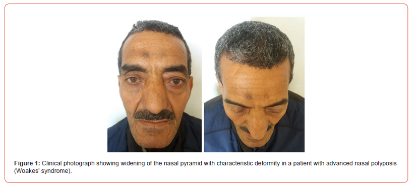
On external examination, a noticeable widening of the nasal pyramid was observed, predominantly affecting the nasal dorsum, which appeared broadened and flattened. This deformity was progressive according to the patient’s history and had become increasingly apparent over time. The overlying skin was intact, without erythema, ulceration, or other signs of inflammation. Palpation did not reveal tenderness or fluctuance.
The clinical appearance of the patient, characterized by a broadened nasal dorsum, was consistent with the typical deformity described in advanced cases of Woakes’ syndrome (Figure 1).
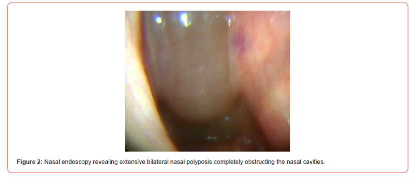
Anterior rhinoscopy demonstrated bilateral, pale, polypoid masses occupying the nasal cavities. These lesions appeared edematous and non-vascularized. Further evaluation by rigid nasal endoscopy confirmed the presence of extensive bilateral nasal polyposis completely obstructing the nasal cavities and extending posteriorly toward the choanae (Figure 2). The polyps exhibited a typical translucent and gelatinous appearance, without areas suggestive of necrosis or malignancy.
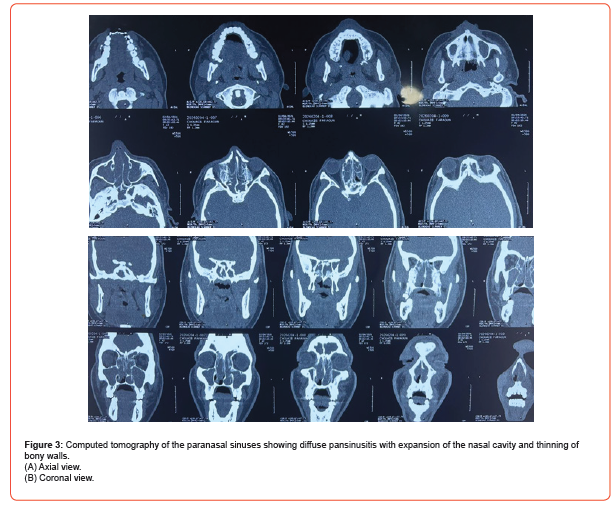
Computed tomography of the paranasal sinuses was performed to assess the extent of disease. Imaging revealed diffuse opacification of all paranasal sinuses, consistent with pansinusitis. Notably, there was marked expansion of the nasal cavity associated with thinning of the surrounding bony walls, particularly within the ethmoid labyrinth. The nasal bones appeared widened, contributing to the external deformity. Importantly, no evidence of bone destruction, orbital invasion, or intracranial extension was identified, supporting the diagnosis of a benign inflammatory process rather than a neoplastic lesion (Figure 3A and 3B) [5, 6].
Given the severity of symptoms and radiological findings, the patient was scheduled for functional endoscopic sinus surgery under general anesthesia. The procedure involved complete removal of polypoid tissue, anterior and posterior ethmoidectomy, maxillary antrostomy, and sphenoidotomy. Intraoperative findings confirmed extensive inflammatory polyposis filling the nasal cavities and sinus spaces without evidence of invasive behavior or abnormal vascularization.
Histopathological examination of the resected tissue revealed an edematous stroma with a dense inflammatory infiltrate predominantly composed of eosinophils. The respiratory epithelium showed areas of hyperplasia but no dysplasia or malignant transformation. These findings were consistent with benign inflammatory nasal polyposis [7].
The postoperative course was uneventful. The patient was treated with intranasal corticosteroids and saline irrigations, along with a short course of systemic corticosteroids. At six-month follow-up, the patient reported a marked improvement in nasal breathing and partial recovery of olfactory function. Endoscopic examination showed no evidence of recurrence, and the nasal deformity remained stable, with no further progression.
Discussion
Woakes’ syndrome is an uncommon clinical entity that represents the extreme end of the spectrum of chronic rhinosinusitis with nasal polyposis. Although it has become rare in modern clinical practice, it remains relevant in cases of neglected or severe disease, particularly when diagnosis and treatment are delayed.
The pathogenesis of Woakes’ syndrome involves a complex interplay between chronic inflammation and mechanical factors. Persistent mucosal inflammation leads to the formation and progressive enlargement of nasal polyps, which in turn exert continuous pressure on the surrounding bony structures. Over time, this pressure, combined with the action of inflammatory mediators such as cytokines and growth factors, induces bone remodeling characterized by thinning and expansion [4]. Unlike malignant tumors, which typically cause bone destruction, this process results in deformation without erosion, a feature that is crucial for differential diagnosis.
The rarity of this condition in adults may be explained by the reduced plasticity of facial bones compared to children. However, as illustrated in the present case, prolonged disease duration can still lead to significant structural changes even in older patients [2, 3].
Imaging plays a pivotal role in both diagnosis and management. Computed tomography is the preferred modality for evaluating sinonasal anatomy and disease extent. The characteristic findings of diffuse sinus opacification, expansion of sinus cavities, and thinning of bony walls are highly suggestive of advanced inflammatory disease [5, 6]. The absence of bone destruction helps differentiate Woakes’ syndrome from neoplastic conditions, which may present with similar clinical features but require different management strategies.
The treatment of Woakes’ syndrome is primarily surgical, with functional endoscopic sinus surgery representing the gold standard. The objective of surgery is to remove polypoid tissue, restore sinus ventilation, and prevent further progression of the disease [8]. However, surgery alone is insufficient, and longterm medical management is essential to control inflammation and reduce the risk of recurrence. Intranasal corticosteroids remain the cornerstone of postoperative therapy, while systemic corticosteroids may be used in selected cases. More recently, biologic therapies targeting specific inflammatory pathways have emerged as promising options for patients with severe or refractory disease [9].
The prognosis of Woakes’ syndrome depends largely on the timing of intervention. While surgical treatment effectively improves functional symptoms, established facial deformities may only partially regress, particularly in advanced cases. This underscores the importance of early diagnosis and appropriate management.
Conclusion
Woakes’ syndrome is a rare but significant complication of chronic nasal polyposis that may lead to progressive facial deformity if left untreated. Although predominantly described in pediatric populations, it can occur in adults in the setting of longstanding disease. This case highlights the importance of early recognition, appropriate imaging, and combined surgical and medical management in order to prevent irreversible structural changes. Long-term follow-up remains essential due to the risk of recurrence.
Acknowledgement
None.
Conflicts of Interest
No conflicts of interest.
References
- Woakes E (1885) On necrosing ethmoiditis and nasal polyps. Lancet.
- Lund VJ, Kennedy DW (1997) Staging for rhinosinusitis. Ann Otol Rhinol Laryngol.
- Fokkens WJ (2020) European Position Paper on Rhinosinusitis and Nasal Polyps 2020. Rhinology.
- Settipane GA (1996) Nasal polyps: epidemiology and pathogenesis. Allergy Asthma Proc.
- Stammberger H (1991) Functional Endoscopic Sinus Surgery.
- Kennedy DW (1992) Prognostic factors and outcomes in sinus surgery. Laryngoscope.
- Mygind N (1990) Nasal polyposis pathophysiology. J Allergy Clin Immunol.
- Meltzer EO (2004) Rhinosinusitis classification. Otolaryngol Head Neck Surg.
- Bachert C (2019) Biologic therapies in chronic rhinosinusitis. J Allergy Clin Immunol.
-

This work is licensed under a Creative Commons Attribution-NonCommercial 4.0 International License.




