



 Case Report
Rare Tooth-Like Sinolith in the Left Frontoethmoidal Region: Endoscopic Removal for Refractory Rhinosinusitis
1Department of Otorhinolaryngology, KMG Klinikum Güstrow, Germany
2Department of Radiology, KMG Klinikum Güstrow, Germany
Received Date: April 23, 2026; Published Date: May 05, 2026
Abstract
Background: Sinoliths are rare calcified masses found in paranasal sinuses, predominantly reported in the maxillary sinus, where they are referred to as antroliths.
Frontal and ethmoid sinoliths are distinctly uncommon, and research on ectopic or tooth-like structures in the ethmoid region remains limited.
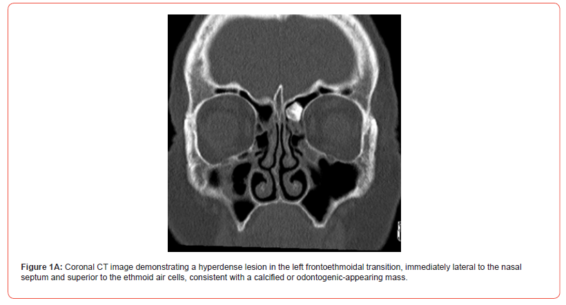
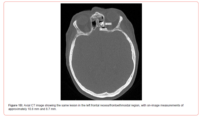
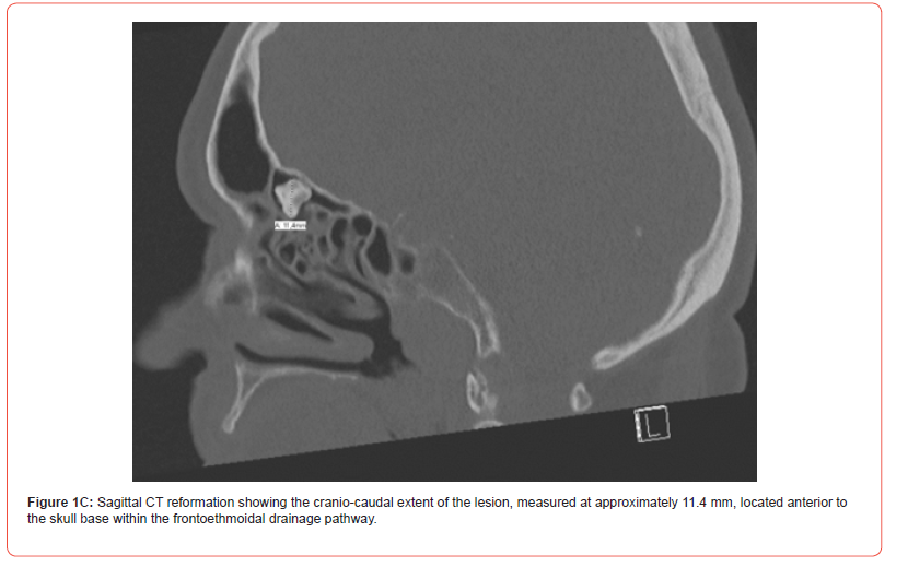
Case Presentation: A 74-year-old woman presented with a one-year history of nasal obstruction, more pronounced on the right side, accompanied by thick nasal secretions, rhinorrhea, a repeated urge to blow her nose, hyposmia, sneezing, and impaired sleep. Non-contrast computed tomography revealed a hyperdense lesion in the left frontoethmoidal transition, measuring approximately 10.8 mm in one plane and 8.7 mm in another, with a coronal image also showing a dimension of approximately 11.4 mm.
Endoscopic sinus surgery, including septoplasty and bilateral functional sinus surgery, was performed, resulting in the removal of a fixed, toothlike mass from the left frontoethmoidal/frontal recess region. Histopathology of the mucosa indicated chronic hyperplastic sinusitis with sinonasal polyps and eosinophilia, while the hard lesion was identified as tooth-like.
Conclusion: This case presents a combination of radiologic and intraoperative features of both frontal sinolithiasis and ectopic tooth-like sinonasal lesions, which differ from previously published reports in the frontoethmoidal location, loose appearance, and absence of known prior dental intervention.
High-resolution CT and endoscopic management are central to diagnosis and treatment in such unusual cases.
Introduction
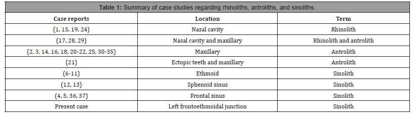
Calcareous masses in the nose or paranasal sinuses have been variously described as nasal dens [1], rhinoliths, maxillary sinus stones, antral calculi, antral rhinoliths, antroliths, and sinoliths [2]. In 1969, Bowerman introduced the term “maxillary antrolith” to differentiate stone formations in the paranasal sinuses from nasal stones [3]. Since then, stone formations within the nasal cavity have been termed rhinoliths, whereas similar pathologies within the maxillary sinus are referred to as antroliths. Rhinoliths and antroliths are common in the literature; however, the presence of a sinolith is exceedingly rare, particularly in the ethmoid sinus. A related but distinct entity is ectopic dentition within the sinonasal tract. Sinolith refers to the presence of calcareous collections of exogenous or endogenous origin within the frontal [4, 5], ethmoid [6-11], or sphenoid sinuses [12, 13].
Pathogenesis of stone formation remains unknown. A significant predisposing factor is long-standing infection and poor sinus drainage. The purulent fluid becomes concentrated, leading to the precipitation of mineral salts and resulting in the complete or partial encrustation of an antral foreign body [3-5, 14-17]. Anthroliths develop through the gradual deposition of mineral salts around an endogenous or exogenous nidus. The central core of an antrolith is typically of endogenous origin, less commonly of exogenous origin. If the central core arises around normal or abnormal tissue, an endogenous origin is assumed. Among these, tooth and bone fragments, blood, pus, and fungi are included [18-21]. In contrast, exogenous niduses can consist of various materials, such as cotton, cellulose [14], paper [22], wood [23], or other substances [24].
Antroliths may present with nasal obstruction, chronic rhinosinusitis, foul discharge, or facial symptoms. Most reported antroliths arise in the maxillary sinus, which makes non-maxillary presentations particularly notable.
There have been only a few reported cases of true sinoliths in the ethmoid sinus [6-11]. Diagnosis and management protocols vary depending on the etiopathogenesis of the sinolith. Frontal sinoliths have been reported but remain rare in the literature [4, 5].
Endoscopic removal of an ectopic tooth from the ethmoid sinus has been described, supporting the possibility that tooth-like lesions may be found outside their expected odontogenic location.
However, a loose tooth-like lesion in the frontoethmoidal recess without a history of trauma, dental implantation, or previous sinonasal surgery appears to be exceptionally uncommon based on currently available reports.
Case Presentation
A 74-year-old woman was referred for possible sinonasal surgery after persistent symptoms despite conservative treatment. She reported one year of nasal airway obstruction, which was more severe on the right than on the left, along with thick secretions, rhinorrhea, an urge to blow her nose, hyposmia, sneezing irritation, and difficulty initiating and maintaining sleep. She denied any prior nasal trauma, previous nasal surgery, allergies, headaches, or facial pressure.
Preoperative non-contrast CT of the paranasal sinuses showed a well-defined hyperdense lesion in the left frontoethmoidal region adjacent to the frontal recess.
The lesion appeared calcified and tooth-like on imaging, with measured dimensions of approximately 10.8 mm and 8.7 mm on one axial image and approximately 11.4 mm on a sagittal reformation. No definite radiologic signs of chronic rhinosinusitis were reported bilaterally aside from the focal lesion.
Surgical Procedure
Surgery was performed under general anesthesia. After positioning with neck extension, topical vasoconstriction was achieved, and the nasal vestibular hairs were shortened. Septoplasty was carried out through a right hemitransfixion incision with mucosal elevation, chondrotomy, resection of deviated septal components, and reduction of the bony floor crest while preserving bilateral mucosal integrity.
Functional endoscopic sinus surgery was then performed. On the left side, the middle turbinate was lateralized, the uncinate process was resected, the bulla ethmoidalis was opened, and the anterior and posterior ethmoid cells were cleared. The skull base was visualized and remained intact. The dissection continued to the frontal recess, and the maxillary ostium was widened with aspiration of retained secretions. With the help of a 45-degree endoscope, markedly thickened mucosa was removed, and a fixed tooth-like structure measuring approximately 2 × 1 cm was extracted from the left frontoethmoidal/frontal recess region (Figures 2 and 3). The structure was not embedded in bone and appeared intraoperatively similar to a small deciduous or molarlike tooth. The skull base remained intact.
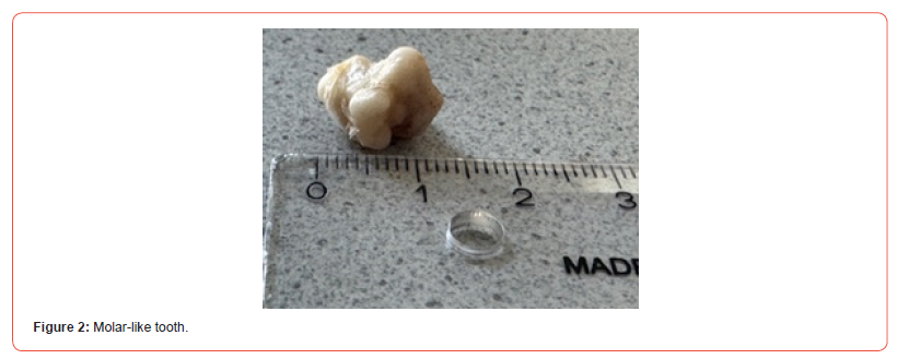
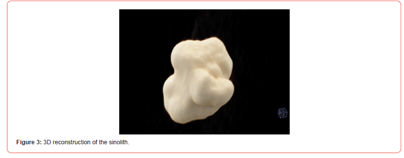
On the right side, uncinectomy, ethmoidectomy, frontal recess dissection, and widening of the maxillary ostium were performed in a similar manner, with thickened mucosa but no foreign body was found. The resected septal components were reimplanted, the hemitransfixion incision was closed, septal splints were fixed with mattress sutures, and nasal packing was inserted. All specimens were sent for histopathological analysis.
Outcome and Follow-Up
The patient was reviewed at two weeks and four weeks following the operative procedure for diagnostic nasal endoscopy and endoscopic cleaning of the operative cavity. It was observed that the patient’s symptoms had completely resolved.
Histopathology
Histopathological examination of the sinonasal tissue specimens showed chronic hyperplastic sinusitis with sinonasal polyps and eosinophilia. The extracted hard lesion was described by pathology as a tooth-like structure.
Taken together with the imaging appearance and intraoperative findings, this case supports the interpretation of a tooth-like calcified lesion in the left frontoethmoidal region.
Discussion
This case is notable because it overlaps with two rare categories of published sinonasal pathology: ethmoid/frontal sinoliths and ectopic tooth-like lesions. The formation of stone-like structures in the paranasal sinuses is rare, with the highest incidence observed in the maxillary sinus, followed by the frontal sinus [4, 5], the ethmoid sinus [6-11], and the sphenoid sinus [12, 13]. Only a few cases have reported ethmoid sinoliths in the global literature to date [6-11].
Rhinoliths, antroliths, and sinoliths may form around endogenous debris such as mucus, clot, fungal material [18], or bone fragments or around exogenous foreign bodies [23]. Ectopic teeth in the sinonasal tract [21] may arise through developmental disturbances, displacement, trauma, or odontogenic procedures.
The clinical presentation of stones in the paranasal sinuses varies. Common clinical manifestations of a sinolith include nasal obstruction, nasal discharge [occasionally foul-smelling], epistaxis, postnasal drip, and facial pain or headache [25, 26].
CT scans of the paranasal sinuses aid in diagnosis by demonstrating bony density. Osteoma, inverted papilloma, and osteoblastoma are the most common differential diagnoses of sinoliths [27]. However, the literature indicates that the presence of an isolated, distinct bony density without inflammation of the paranasal mucosa is usually caused by a sinolith. In contrast, longstanding infections and an increased number of bony densities are more likely to indicate tumors or infections than a sinolith. Nevertheless, multiple discrete densities are more likely to be associated with a tumor than with an inflammatory lesion [27]. Radiodensities may assist in refining a CT diagnosis, but just with that, one may not be able to determine whether the density is a calcification, ossification, or residual bone [27].
Table 1:
Biochemical analyses and infrared spectrography, along with histopathology, were performed to determine the composition of the sinolith [7, 8]. Infrared spectrography and chemical analyses revealed the presence of calcium salts, including phosphate [30], carbonate [30], and oxalate [2]; magnesium salts, including phosphate [16]; protein [5]; water [16]; organic matter [22]; and sometimes the presence of organic matter [33] and fungal elements [11]. Sinolith color varied depending on its composition, ranging from black or blackish-green to gray, brown, or white [2, 3, 7, 14, 16, 18, 24, 30]. The most common nidus is often of endogenous origin, typically tooth fragments [14, 17, 32] or bone, although some authors believe that tooth fragments may be of exogenous origin [18, 30, 38]. Histologically, sinoliths exhibit the same concentric rings as seen in stones found in other parts of the body [31]. Histopathology reveals the presence of osteocytes and bone marrow consistent with bone tissue.
The treatment of choice is endoscopic sinus surgery and removal of the stone [8, 11, 25].
In the present case, the combination of CT appearance, toothlike morphology, and lack of osseous fixation argues against a typical osteoma and favors either an odontogenic structure or a heavily calcified lesion with tooth-like architecture.
This case also emphasizes the diagnostic value of CT. Hyperdense lesions in the frontal recess or frontoethmoidal region should prompt consideration of osteoma, sinolith, fungal concretion, foreign body, and ectopic odontogenic structures.
Endoscopic sinus surgery offers both definitive diagnosis and minimally invasive treatment in such situations.
Conclusion
A tooth-like lesion in the left frontoethmoidal junction is an exceptionally unusual finding. In the literature, similar lesions have been reported either as frontal sinolithiasis or as ectopic tooth-related lesions of the ethmoid sinus, but not in the same configuration as in this case.
The case presented in this report broadens the differential diagnosis of hyperdense frontal recess lesions and underscores the importance of CT-based assessment and endoscopic surgical management.
Despite its rarity, frontal or ethmoid sinoliths should be considered in the differential diagnosis of radiodensities in the paranasal sinuses.
Conflict of Interest
None declared.
Funding
None.
Patient Consent
Obtained.
References
- Körner O, Steurer O (1994) Lehrbuch der Ohren-, Nasen-, Rachen- und Kehlkopf-Krankheiten. 14. Auflage 1944 ed: J. F. Bergmann-Verlag; 1944 01.01.1944. 552 p.
- Nass Duce M, Talas DU, Ozer C, Yildiz A, Apaydin FD, et al. (2003) Antrolithiasis: a retrospective study. J Laryngol Otol 117(8): 637-640.
- Bowerman JE (1969) The maxillary antrolith. J Laryngol Otol 83(9): 873-882.
- Grant DG, Hussain A, Burgel R (1998) Frontal sinolith. J Laryngol Otol 112(6): 570-572.
- Mori S, Lee K, Fujieda S, Kojima A, Saito H (2000) Antrolithiasis in the frontal sinus. ORL J Otorhinolaryngol Relat Spec 62(6): 335-337.
- Ogawa T, Nishioka K, Myoukai K, Nakagawa F, Uchida T (1992) Ethmodial Antrolith; A Report of Two Cases. Practica Oto-Rhino-Laryngologica 85: 1235-1243.
- Kanzaki S, Sakamoto M (2006) Sinolith in the ethmoid sinus. J Laryngol Otol 120: e11.
- Almasi M, Andrasovska M, Koval J (2010) Sinolith in the ethmoid sinus: report of two cases and review of the literature. Eur Arch Otorhinolaryngol 267(10): 1649-1652.
- da Costa ED, Verner FS, Peyneau PD, de Freitas DQ, de Almeida SM (2019) Diagnosis of ethmoid sinolith by cone-beam computed tomography: case report and literature review. Oral Radiol 35(1): 68-72.
- Nurrachman AS, Rahman FUA, Sarifah N, Ghazali AB, Epsilawati L (2024) Ostiomeatal complex inflammation with a rare ethmoid sinolith utilizing cone-beam computed tomography: A clinical and radiological approach to diagnosis. Radiol Case Rep 19(1): 268-276.
- Nayak DR, Bhandarkar AM, Valiathan M, K SV (2014) Incidental 'ethmoid sinolith'--an unusual cause of frontal recess obstruction. BMJ Case Rep 2014.
- Ozcan C, Vaysoglu Y, Gorur K (2013) Sinolith: a rare isolated sphenoid sinus lesion. J Craniofac Surg 24(2): e104-6.
- Sobczak A, Felber R, Leon L, Biglione A (2024) An Incidental Finding of a Sphenoid Sinolith: A Case Report. Cureus 16(5): e61386.
- Evans J (1975) Maxillary antrolith: a case report. Br J Oral Surg 13(1): 73-77.
- Cunningham AT, Lord OC (1945) Rhinoliths; the report of an antral and three nasal stones. J Laryngol Otol 60: 253-256.
- Sofat JR, Greval RS (1985) Maxillary antrolith around tooth root tip with oro-antral fistula--a case report. J Indian Dent Assoc 57(6): 227-229.
- Davis O, Wolff A (1985) Rhinolithiasis and maxillary antrolithiasis. Ear Nose Throat J 64(9): 421-426.
- Ogata Y, Okinaka Y, Takahashi M (1997) Antrolith associated with aspergillosis of the maxillary sinus: report of a case. Journal of Oral and Maxillofacial Surgery 55(11): 1339-1341.
- Stafne E (1962) Roentgeno-oddities. A dental root as a source of rhinolith. Oral Surg Oral Med Oral Pathol 15: 948-949.
- Wallace JR (1963) Chronically infected rhinolith of the maxillary sinus simulating osteomyelitis. Oral Surgery, Oral Medicine, Oral Pathology 16: 761-763.
- Brown RS, Coleman Bennett M, Abramovitch K (2002) Panoramic radiographic findings: case reports of ectopic teeth and antroliths within or adjacent to the maxillary sinus. Dent Today 21(1): 50-53.
- Lord OC (1944) Maxillary rhinoliths. J Laryngol 59: 218-222.
- Azoubel E, Carneiro MC, Azoubel MCF, Ribeiro NCR (2015) Removal of wood fragments from the sinus: a case report. Oral Surgery 8(4): 221-224.
- Dutta A (1973) Rhinolith. J Oral Surg 31(11): 876-867.
- Nair S, James E, Dutta A, Goyal S (2010) Antrolith in the maxillary sinus: an unusual complication of endoscopic sinus surgery. Indian J Otolaryngol Head Neck Surg 62(1): 81-83.
- Wu CW, Tai CF, Wang LF, Tsai KB, Kuo WR (2005) Aspergillosis: a nidus of maxillary antrolith. Am J Otolaryngol 26(6): 426-429.
- Som PM, Lidov M (1994) The significance of sinonasal radiodensities: ossification, calcification, or residual bone? AJNR Am J Neuroradiol 15(5): 917-922.
- Tsujimoto T, Sasaki R, Murata M, Sakai S (1982) Antrolithiasis, report of two cases. Practica Oto-Rhino-Lqaryngologica 75(8): 1635-1640.
- Swain SK, Sahoo RK, Sahu MC (2014) Two case reports of antrorhinoliths. Int J Otorhinolaryngol Clin 6(3): 118-120.
- Ishiyama T (1988) Maxillary antrolith: report of a case. Auris Nasus Larynx 15(3): 185-189.
- Cohen MA, Packota GV, Hall MJ, Steinberg J (1991) Large asymptomatic antrolith of the maxillary sinus. Report of a case. Oral Surg Oral Med Oral Pathol 71(2): 155-157.
- Blaschke DD, Brady FA (1979) The maxillary antrolith. Oral Surg Oral Med Oral Pathol 48(2): 187-189.
- Marr JT (1955) Stone in the maxillary sinus. Radiology 64(5): 718-720.
- Esser WJ (1965) Calculus Formation in the Maxillary Sinus. Oral Surg Oral Med Oral Pathol 19: 755.
- Allsobrook O, Rich AM (2013) Mycetoma of the maxillary sinus. Pathology 45: S9.
- Jagannath B, Praveen SSG, Bhardwaj S (2012) Frontal sinolith. Clinical Rhinology 5(1): 28-29.
- Lee DH, Yoon TM, Lee JK, C LS (2015) frontal sinolith. J Craniofac Surg 26(5): e385-6.
- Karges MA, Eversole LR, Poindexter BJ Jr (1971) Antrolith: report of case and review of literature. J Oral Surg 29(11): 811-814.
-

This work is licensed under a Creative Commons Attribution-NonCommercial 4.0 International License.




