

 Case Report
Intrathyroidal Thymic Tissue Masquerading as Carcinoma in a Child
1 University of Arkansas for Medical Sciences, College of Medicine
2 University of Arkansas for Medical Sciences, College of Medicine, Department of Pathology, Little Rock, AR, USA
3 University of Arkansas for Medical Sciences, College of Medicine, Department of Otolaryngology - Head & Neck Surgery, Division of Pediatric Otolaryngology, Arkansas Children’s Hospital, Little Rock, AR, USA
Received Date: September 11, 2026; Published Date: October 06, 2026
Abstract
Objective: To highlight a rare entity that has similar characteristics to pediatric thyroid carcinoma.
Methods: We report a single case of intrathyroidal thymic tissue that was evaluated by ultrasound, computed tomography, and fine needle
aspiration (FNA) prior to surgical excision. We discuss differences in radiographic findings and utilization of immunostains.
Results: A 21-month-old male with craniosynostosis was referred for further workup of bilateral TI-RADS 4 nodules and ultimately underwent
a hemithyroidectomy with pathology confirming intrathyroidal thymic tissue.
Conclusion: Similarities between papillary thyroid carcinoma and thymic lesions in the pediatric thyroid are subtle. Here we highlight the
differences to avoid additional comorbidities and review the literature on the natural progression of these lesions.
Introduction
Thyroid nodules in children are rare with 22-26% harboring malignancy. Papillary thyroid carcinoma is the most common, accounting for nearly 90% of thyroid carcinoma. Ultrasound findings of irregular margins, hypoechogenicity, taller-than-wide dimensions, microcalcifications, solid characteristics, and abnormal cervical lymph nodes are suspicious findings associated with malignancy [1]. Thyroid Imaging, Reporting and Data System (TIRADS) characterizes thyroid nodules by composition, echogenicity, shape, margin, and echogenic foci and stratifies them from TR1 (benign) to TR5 (highly suspicious) [2].
The primordial thymus, originating from the ventral wings of the third pharyngeal pouch, begins in the pharynx and finishes descending to the anterior mediastinum by the eighth to ninth week of gestation. Ectopic thymic (ET) tissue may thus be found anywhere along the tract between the pharynx and mediastinum. Intrathyroidal ectopic thymus (IET) is a rare entity with a reported prevalence ranging from 0.4% [3] to 11.7% [4], although these estimates vary by study population. More recently, a large 2025 screening study of children and adolescents analyzing approximately 320,000 thyroid ultrasound datasets reported an IET prevalence of 3.2% [5].
Ectopic intrathyroidal thymic nodules rarely cause symptoms [6] and are typically found incidentally. On ultrasound, IET is usually unilateral [6]. Reported lesion sizes vary across case reports and series, with lesions larger than 1 cm described in several instances [3, 6, 7]. A systematic review found that lesions are most commonly under 10 mm in maximum diameter [8]. IET can appear round, oval, or irregular [6]. Intrathyroidal thymic nodules can appear on either side and favor the middle or inferior aspect of the thyroid [7, 9] but bilateral IET are considered more rare [7]. Thymic tissue on ultrasound is hypoechoic with the internal septa and blood vessels producing linear and bright punctate echogenicities or “starry sky” appearance [6, 9]. This distinct appearance can often be confused with the diffuse sclerosing variant of papillary thyroid carcinoma, which can have a “snowstorm” appearance on ultrasound. Given the more aggressive nature of this disease, tissue sampling with fine needle aspiration (FNA) or surgical evaluation may be pursued even when the diagnosis remains uncertain [9-11].
With the family’s permission, we present a case report with incidental bilateral thyroid nodules that underwent radiographic, interventional radiology (IR), and finally surgical pathologic evaluation. The objective of this manuscript is to highlight a rare entity masquerading as a potential pediatric thyroid malignancy.
Case Presentation
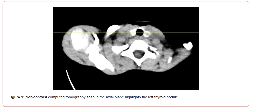
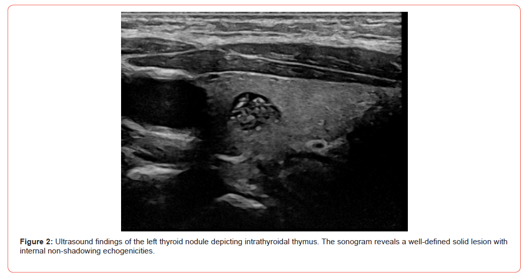
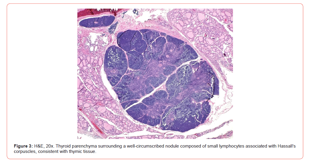
A 21-month-old male who underwent a non-contrast head CT (Figure 1) for the surgical planning of craniosynostosis repair. Thyroid nodules were incidentally noted, and he was referred to otolaryngology for further evaluation. On initial consultation, the family reported that his lower neck was fuller than normal and that he was having progressive dysphagia. His CEA and calcitonin laboratory values were negative. A follow-up ultrasound revealed bilateral TI-RADS 4 nodules (Figure 2). He subsequently underwent an FNA with IR, which revealed abundant small lymphocytes and occasional macrophages without Hassall’s corpuscles. No additional staining was reported. He subsequently underwent hemithyroidectomy, which confirmed the presence of IET (Figure 3). Surgery and recovery were uneventful.
Discussion
In a recently published article reviewing TI-RADS and Bethesda Classification in predicting pediatric papillary thyroid carcinoma (PTC), Amin et al. found that a high-risk TI-RADS score (3-5) was associated with a relative risk of 7.99 (95% CI 1.16-54.90) for PTC compared with a low-risk TI-RADS score (1 or 2). In the same cohort, patients who underwent FNA and received a high-risk Bethesda classification of 5-6 had a relative risk of 6.62 (95% CI 4.22-10.41) for PTC compared with a low-risk classification of 2, 3, or 4. From this analysis, the authors described a TI-RADS score ≥3 as “a potential cutoff for recommending FNA, independent of nodule size” [12]. However, smaller nodules are technically more difficult to sample. In a predominantly adult cohort, Moon et al. examined 1440 nodules ≤10 mm and found that 256 (17.8%) yielded inadequate specimens; a 6 mm cutoff significantly distinguished inadequate from adequate specimens [13].
ACR TI-RADS was developed and validated primarily in adults, and its application in pediatric patients remains less certain. A study evaluating ACR TI-RADS in a pediatric cohort found that applying the adult management criteria would have resulted in 17 of 77 malignancies (22.1%) being missed at the initial evaluation [14]. More recent pediatric data further suggest that the risk of malignancy associated with each ACR TI-RADS category may be higher in children than in adults. In a study incorporating data from 1,458 pediatric thyroid nodules, the observed risk of malignancy increased across TI-RADS categories and exceeded the corresponding adult risk estimates, leading the authors to suggest that the size cutoffs used to recommend FNA in adults may require modification in pediatric patients [15]. A 2023 commentary on the third edition of the Bethesda System for Reporting Thyroid Cytopathology notes that it includes pediatric thyroid disease, pediatric-specific estimates of risk of malignancy, and management algorithms, further emphasizing the differences between pediatric and adult thyroid disease [16]. Newer pediatric-specific analyses also suggest that clinical risk factors and modified biopsy cutoffs may improve the performance of ACR TI-RADS in children [17].
When managing diseases with a low prevalence, clinicians may face difficult management decisions when more than one reasonable treatment option exists. Shared decision making (SDM) is a collaborative process in which all involved parties develop a mutually agreed upon plan [18]. After discussion, the family elected to proceed with a hemithyroidectomy because concern about missing a thyroid malignancy outweighed the risk of surgery.
The presence of microcalcifications on ultrasound has been shown to be strongly correlated with thyroid cancer in pediatric thyroid nodules, with an odds ratio of [19].1 in the 2022 study by Fornwalt et al.19 Given that punctate echogenic foci can also be seen in IET,9 other features are likely more reliable for distinguishing between benign and malignant lesions.
IET Ultrasound Findings
Chang et al. examined 41 patients with IET (age 1 week to 3 years 10 months, average 8.7 months) and found the nodule diameter ranged from 3 to 25 mm, with the average being 7.3 mm [9]. IET are usually unilateral6 and often small, although larger lesions have been reported [8, 9]. They can appear round, oval, or irregular [6, 9]. Intrathyroidal thymic nodules can appear on either side and tend to favor the middle or inferior aspect of the thyroid [7, 9]. However, bilateral IET are rare [7]. Although uncommon, multifocal intrathyroidal thymic tissue has also been described [7]. Chen et al. reported an 8-year-old girl with two separate intrathyroidal ectopic thymic lesions, one in each thyroid lobe, measuring 0.9 and 1.1 cm in maximum diameter, respectively, that were both ultimately confirmed histologically following total thyroidectomy [20]. Our patient’s IET nodules were bilateral, measuring 0.7 cm in maximum diameter in the left mid-pole and 0.4 cm in maximum diameter in the right mid-pole. Table 1 compares TI-RADS scoring of IET and PTC ultrasound characteristics.
Table 1:TI-RADS Scoring for Ultrasound Findings of Intrathyroidal Ectopic Thymus versus Papillary Thyroid Carcinoma.
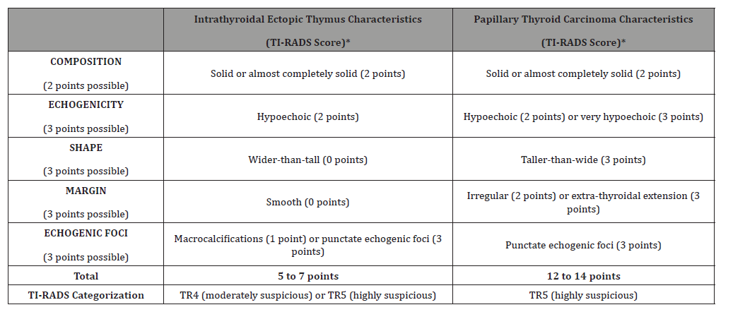
*Representative features; ACR TI-RADS scoring according to Tessler et al. [2].
Fine Needle Aspiration (FNA) & IET
Given suspicious findings on ultrasound, FNA is often incorporated as a diagnostic modality when evaluating thyroid nodules. In a systematic review of IET, four included studies reported FNA of concerning lesions for diagnostic confirmation, with cytology demonstrating abundant lymphoid stroma and lymphocytes of variable size; Hassall’s corpuscles were only rarely identified.8 If cellular material is adequate, tissue stains for specific thymic markers such as pancytokeratin or cytokeratin 5/6, terminal deoxynucleotidyl transferase (TdT), and CD3 can also confirm thymic origin [21, 22].
IET natural course
Ectopic intrathyroidal thymic nodules are generally asymptomatic [6], and longitudinal studies have demonstrated variable changes in size during observation [4, 6]. However, Segni et al. followed 9 patients with intrathyroidal thymic nodules for 34 months and found no progression in size, with regression in the two teenage patients [6]. In a larger study of 4,130 children, IET was identified in 51 patients and its prevalence decreased with age. Among 27 patients with serial imaging, 11 lesions decreased in size by at least 20%, 15 changed by less than 20%, and only one increased in size by at least 20% [23]. More recently, Jeffery et al. followed 21 children with sonographically detected intrathyroidal thymic tissue, all under 11 years of age. Only 3 of 21 (14%) underwent FNA, and follow-up scans demonstrated stable appearance, reduction in size, or resolution. The authors proposed an age-based classification that considered the expected size and appearance of thymic tissue at different ages to help distinguish IET from thyroid nodules and limit unnecessary biopsy and surgery [24]. A recent pediatric case series similarly reported stable lesions on serial ultrasound follow-up [25].
Role of surgery
Surgery is not indicated for treatment for IET. However, there are instances in which surgery can be considered and safely performed. For high-volume thyroid surgeons, risk of permanent hypocalcemia or recurrent laryngeal nerve injury is low [26]. In the series by Yildiz et al, one pediatric patient with congenital adrenal hyperplasia underwent thyroidectomy to exclude malignancy at her clinicians’ preference [21]. In another case reported by Chng et al., a 7-year-old boy with a PTEN gene mutation was found to have an intrathyroidal nodule with echogenic foci initially interpreted as microcalcifications, and total thyroidectomy was advised because of his predisposition for thyroid malignancy. However, FNA demonstrated numerous CD3-positive lymphocytes, and comparison of the nodule with the normal thymus on ultrasound supported a diagnosis of IET, allowing the patient to undergo continued observation rather than surgery [22].
Differential diagnoses beyond PTC to consider in a child with a thyroid mass include the nodular form of Hashimoto thyroiditis, lymphocytic thyroiditis, hyperplastic lymph nodes, and lymphoma. Other diagnoses to rule out include carcinoma showing thymus-like differentiation (CASTLE) and spindle epithelial tumor with thymuslike differentiation (SETTLE) [22].
Conclusion
As utilization of ultrasound and other imaging modalities for screenings in the pediatric population has expanded, incidental findings are likely to increase. Thus, the possibility of malignancy must be balanced with possible benign entities, especially when rare in occurrence, such as intrathyroidal ectopic thymic tissue. When the clinical and sonographic findings are strongly supportive of IET, observation with serial ultrasound can avoid unnecessary invasive evaluation or surgery [8, 24, 25]. In non-follicular based lesions on FNA, immunostains can define cell lineage and assist in identifying thymic tissue. Management of pediatric thyroid nodules highlights the role of shared decision making.
Acknowledgement
None.
Conflicts of Interest
No conflicts of interest.
References
- Paulson VA, Rudzinski ER, Hawkins DS (2019) Thyroid cancer in the pediatric population. Genes (Basel) 10(9): 723.
- Tessler FN, Middleton WD, Grant EG, Hoang JK, Berland LL, et al. (2017) ACR thyroid imaging, reporting and data system (TI-RADS): White paper of the ACR TI-RADS committee. J Am Coll Radiol 14(5): 587-595.
- Kim HG, Kim M, Lee M (2012) Sonographic appearance of intrathyroid ectopic thymus in children. J Clin Ultrasound 40(5): 266-271.
- Cimbek EA, Kaya S, Eyuboglu I, Dinc H, Karaguzel G (2022) Intrathyroidal ectopic thymus: An important entity in the differential diagnosis of thyroid nodules. Turk J Pediatr 64(5): 909-914.
- Suzuki S, Nagao M, Nagamine N, Kobashi Y, Iwadate M, et al. (2025) A comprehensive analysis of clinical factors interacting with ectopic intrathyroidal thymus in children and adolescents: The fukushima health management survey. Endocr J 72(12): 1317-1326.
- Segni M, di Nardo R, Pucarelli I, Biffoni M (2011) Ectopic intrathyroidal thymus in children: A long-term follow-up study. Horm Res Paediatr 75(4): 258-263.
- Bang MH, Shin J, Lee KS, Kang MJ (2018) Intrathyroidal ectopic thymus in children: A benign lesion. Medicine (Baltimore) 97(14): e0282.
- Gomez-Herrera J, Melo-Uribe MA, Parra-Medina R (2021) Echographic findings in ectopic intrathyroidal thymic tissue in children. A systematic review. Radiologia (Engl Ed) 63(6): 512-518.
- Chang YW, Kang HM, Lee EJ (2020) Long-term follow-up ultrasonographic findings of intrathyroidal thymus in children. Korean J Radiol 21(11): 1248-1255.
- Gimm O, Krause U, Wessel H, Finke R, Dralle H (1997) Ectopic intrathyroidal thymus diagnosed as a solid thyroid lesion: Case report and review of the literature. J Pediatr Surg 32(8): 1241-1243.
- Lignitz S, Musholt TJ, Kreft A, Engel R, Brzezinska R, et al. (2008) Intrathyroidal thymic tissue surrounding an intrathyroidal parathyroid gland, the cause of a solitary thyroid nodule in a 6-year-old boy. Thyroid 18(10):1125-1130.
- Amin SN, Branson M, Perkins JN (2025) TI-RADS and bethesda classification system correlate with predicting pediatric papillary thyroid carcinoma. Otolaryngol Head Neck Surg 173(4): 985-990.
- Moon HJ, Son E, Kim E, Yoon JH, Kwak JY (2012) The diagnostic values of ultrasound and ultrasound-guided fine needle aspiration in subcentimeter-sized thyroid nodules. Ann Surg Oncol 19(1): 52-59.
- Richman DM, Benson CB, Doubilet PM (2020) Assessment of american college of radiology thyroid imaging reporting and data system (TI-RADS) for pediatric thyroid nodules. Radiology 294(2): 415-420.
- Hess JR, Van Tassel DC, Runyan CE, Morrison Z, Walsh AM, et al. (2023) Performance of ACR TI-RADS and the bethesda system in predicting risk of malignancy in thyroid nodules at a large children’s hospital and a comprehensive review of the pediatric literature. Cancers (Basel) 15(15): 3975.
- Sengul I, Sengul D (2023) The 2023 bethesda system for reporting thyroid cytopathology: Novi sub sole, subdivision is no more debatable, in thyroidology. Rev Assoc Med Bras (1992). 69(12): e20231124.
- Lee DH, Cho YJ, Won J, et al. (2025) Pediatric thyroid nodules: K-TIRADS/ACR TI-RADS pediatric-specific biopsy cutoff incorporating clinical risk factors. Radiology 315(3): e241015.
- Ikeda AK, Hong P, Ishman SL, Joe SA, Randolph GW, Shin JJ (2018) Evidence-based medicine in otolaryngology part 7: Introduction to shared decision making. Otolaryngol Head Neck Surg 158(4): 586-593.
- Fornwalt B, Melachuri M, Kubina M, McDaniel J, Jeyakumar A (2022) Pediatric thyroid nodules: Ultrasound characteristics as indicators of malignancy. OTO Open 6(1): 2473974X211073702.
- Chen Y, Liu C, Cao Y (2020) Intrathyroidal ectopic thymus in 8-year-old girl: A case report. Transl Pediatr 9(1): 70-73.
- Yildiz AE, Ceyhan K, Siklar Z, et al. (2015) Intrathyroidal ectopic thymus in children: Retrospective analysis of grayscale and doppler sonographic features. J Ultrasound Med 34(9): 1651-1656.
- Chng CL, Kocjan G, Kurzawinski TR, Beale T (2014) Intrathyroidal ectopic thymic tissue mimicking thyroid cancer in children. Endocr Pract 20(12): e241-e245.
- Iwaku K, Noh JY, Watanabe N, Sugino K, Ito K (2024) Age-related reduction of the prevalence of ectopic intrathyroidal thymus in children and its size reduction with age. Thyroid Science 1(1): 100004.
- Jeffery H, Bosch KD, Brain C, et al. (2025) Significance of ectopic intrathyroidal thymic tissue detected on ultrasound in different paediatric age groups: A proposed classification to guide investigation and management. Eur J Pediatr 184(5).
- Pereira RS, Silva JA, Dias MI, Simoes AM, Pereira SM, Martins V (2026) Intrathyroidal thymic tissue: A pediatric case series from a secondary care center. Ann Pediatr Endocrinol Metab 31(3): 198-202.
- Francis GL, Waguespack SG, Bauer AJ, et al. (2015) Management guidelines for children with thyroid nodules and differentiated thyroid cancer. Thyroid 25(7): 716-759.
-

This work is licensed under a Creative Commons Attribution-NonCommercial 4.0 International License.