




 Case Report
Extramedullary Plasmacytoma of the Palatine Tonsil: A Case Report and Literature Review
ENT department, 20 Aout Hospital, Casablanca, Morocco
Received Date: January 19, 2026; Published Date: January 30, 2026
Abstract
Background: Extramedullary plasmacytoma (EMP) is a rare malignancy characterized by a monoclonal proliferation of plasma cells outside the bone marrow. Tonsillar involvement is exceedingly rare.
Case Presentation: We report the case of a 45-year-old man with a history of chronic smoking and anxiety-depression disorder, who presented with progressive dysphagia, odynophagia, muffled voice, and a sensation of a foreign body in the throat. Endoscopic examination revealed a bleeding, reddish-purple oropharyngeal mass involving the left tonsil and extending to the base of the tongue and supraglottic epiglottis. Imaging showed a well-limited contrast-enhancing oropharyngeal mass without systemic spread. Biopsy confirmed the diagnosis of EMP with CD138+ monoclonal plasma cell infiltration and lambda light chain restriction. The patient underwent prophylactic tracheotomy followed by curative-intent external radiotherapy (50 Gy in 25 fractions). The outcome was favorable with regression of obstructive symptoms. Work-up for multiple myeloma was negative.
Conclusion: EMP of the palatine tonsil is an exceptional entity requiring histopathological confirmation and exclusion of systemic disease. Radiotherapy remains the standard treatment, with close long-term follow-up due to the risk of progression to multiple myeloma.
Keywords: Extramedullary plasmacytoma; Palatine tonsil, Radiotherapy, Multiple myeloma
Introduction
Extramedullary plasmacytoma (EMP) is a rare, localized malignant plasma cell neoplasm without systemic bone marrow involvement. It accounts for less than 5% of all plasma cell dyscrasias. The head and neck region is the most commonly affected area, particularly the nasopharynx, paranasal sinuses, and larynx. Tonsillar involvement is extremely uncommon, warranting the presentation of this case.
Introduction
A 45-year-old man, a chronic smoker who had quit one year prior, with a medical history of anxiety-depression disorder under treatment, presented with a two-month history of progressive dysphagia, left-sided odynophagia, muffled voice, and a persistent pharyngeal foreign body sensation. He denied dyspnea or significant weight loss.
Nasofibroscopic examination revealed a reddish-purple, exophytic, bleeding mass in the left oropharynx, involving the homolateral tonsil, base of tongue, and supraglottic epiglottis, significantly narrowing the oropharyngeal lumen.
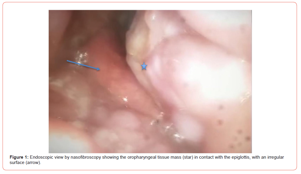
A targeted biopsy under local anesthesia confirmed an extramedullary plasmacytoma, characterized by monoclonal CD138+ plasma cell proliferation with lambda light chain restriction on immunohistochemistry.
Contrast-enhanced cervical CT scan revealed a well-defined oropharyngeal tissue mass with irregular contours, measuring 25 × 19 mm and extending 33mm in height. It was centered on the oropharynx and extended to the epiglottis. An additional 19 × 27 mm mass was observed at the base of the left tonsil. Bilateral subcentimetric cervical lymphadenopathy was present, the largest measuring 10 mm in level IIa. There was also polypoid thickening of the left maxillary sinus without bone invasion.
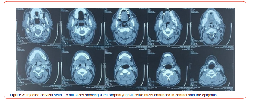
A full staging work-up, including thoracoabdominopelvic CT, serum and urinary protein electrophoresis, free light chain assay, and bone marrow biopsy, showed no evidence of multiple myeloma.
The patient received conformal external beam radiotherapy to a total dose of 50 Gy in 25 fractions (2 Gy per fraction, 5 days/ week). Due to the risk of post-radiation edema in the upper airway, a prophylactic tracheotomy was performed prior to treatment. The clinical course was favorable with progressive resolution of obstructive symptoms.
Discussion
EMP is a rare plasma cell neoplasm, representing approximately 3% of all plasma cell proliferations and a small subset of head and neck malignancies. In contrast to multiple myeloma, EMP is defined by the absence of bone marrow involvement, lytic bone lesions, hypercalcemia, or significant monoclonal proteinuria.
The most common sites of EMP include the nasopharynx, paranasal sinuses, oral cavity, and larynx. Tonsillar EMP is extremely rare, with few cases reported in the literature [1, 2].
Clinically, EMP in the upper aerodigestive tract typically presents with local obstructive symptoms such as dysphagia, odynophagia, hoarseness, and, in advanced cases, dyspnea. In this patient, the dominant symptom was dysphagia, necessitating a prophylactic tracheotomy before initiating radiotherapy due to the risk of airway compromise [3].
Diagnosis is based on histopathology and immunohistochemistry, with CD138 expression and light chain restriction confirming the monoclonal nature of the plasma cell infiltrate. A comprehensive work-up is essential to exclude multiple myeloma, including bone marrow biopsy, serum/urine protein electrophoresis, free light chain assay, and whole-body imaging [4].
Radiotherapy is the mainstay of treatment for localized EMP, with doses ranging from 40 to 50 Gy achieving local control rates above 85% [5, 6]. Surgery is generally reserved for diagnostic purposes or resection of accessible lesions. Combined surgery and radiotherapy may be considered for large, residual, or obstructive masses. Chemotherapy is not indicated in localized disease unless there is progression or transformation to multiple myeloma.
In this case, prophylactic tracheotomy helped prevent potential post-radiation airway obstruction in a critical anatomical region, enabling safe delivery of definitive radiotherapy.
The prognosis of EMP is favorable, with 10-year survival exceeding 70% [6]. However, progression to multiple myeloma occurs in 15-30% of cases, usually within the first 2-3 years postdiagnosis. Therefore, long-term clinical and biological monitoring is essential [7].
Conclusion
Extramedullary plasmacytoma of the palatine tonsil is an exceedingly rare entity. Diagnosis relies on biopsy and immunohistochemistry, and systemic disease must be ruled out. Radiotherapy is the treatment of choice for localized forms. Prognosis is generally good, but long-term follow-up is necessary due to the potential for progression to multiple myeloma.
Acknowledgement
None.
Conflict of Interest
No Conflict of Interest.
References
- Alexiou C (1999) Extramedullary plasmacytoma: tumor occurrence and therapeutic concepts. Cancer 85(11): 2305-2314.
- Bachar G (2008) Outcome of patients with extramedullary plasmacytoma of the head and neck. Ann Surg Oncol 15(6): 1754-1761.
- Raslan OA (2020) Primary extramedullary plasmacytoma of the tonsil: a rare case report. ENT J 99(6): NP100-NP104.
- Dimopoulos MA (2002) Solitary plasmacytoma of bone and extramedullary plasmacytoma. Curr Treat Options Oncol 3(3): 255-259.
- Soutar R (2004) Guidelines on the diagnosis and management of solitary plasmacytoma. Br J Haematol 124(6): 717-726.
- Weber DM (2005) Solitary bone and extramedullary plasmacytoma. Hematology Am Soc Hematol Educ Program 2005: 373-376.
- Dores GM (2009) Plasmacytoma of bone, extramedullary plasmacytoma, and multiple myeloma: incidence and survival in the United States, 1992–2004. Br J Haematol 144(1): 86-94.
-

This work is licensed under a Creative Commons Attribution-NonCommercial 4.0 International License.




