



 Research Article
Eustachian Tube Balloon Dilatation in the Treatment of Adhesive Otitis Media Before Tympanoplasty
1Department of Otorhinolaryngology, Head and Neck Surgery, KMG Klinikum, Güstrow, Germany
2Department of Audiology, KMG Klinikum, Güstrow, Germany
3Department of Otorhinolaryngology, Haugesund, Norway
Received Date: December 10, 2025; Published Date: December 17, 2025
Abstract
Pressure-less acoustic immittance (PLAI) is a novel technology that offers frequency-domain, pressure-less measurements in the open outer ear canal. It captures several parameters, including resonance frequency and peak admittance.
Objective: This study aimed to assess whether PLAI measurement is helpful in evaluating the effect of Eustachian tube (ET) dilatation in patients with adhesive otitis media (AdOM) and a negative Valsalva test before tympanoplasty.
Methods: A prospective study was conducted to compare subjective data (ETDQ-7) and objective data obtained through tympanometry, PLAI, and the objective Valsalva maneuver, before and 2 to 3 weeks after ET dilatation.
The study included two groups: (1) 15 AdOM patients (25 ears) with a mean age of 59.1 ± 11.1 years (5 men and 10 women) and (2) 57 healthy subjects with a mean age of 51.5 ± 17.3 years (35 men and 22 women). The healthy subjects had bilateral normal ears (114 ears), normal tympanic membranes and no history of inflammation or disease affecting the middle ear, recent hearing disabilities, or aural symptoms.
Results: Patients with AdOM demonstrated significantly higher PLAI measures of resonance frequency (RF) and peak admittance compared with healthy subjects (p < 0.001). Correlational analyses revealed a significant association between RF measurements and ETDQ-7 values in AdOM (r25 = 0.61, p = 0.016). Higher pre-dilatation symptom scores were associated with higher RF values. ET dilatation caused a significant decrease in RF in AdOM (p < 0.001). For all 25 ears presenting AdOM with a negative Valsalva test and intact tympanic membranes, the PLAI measurements after dilatation yielded an average RF of 457.2 Hz (ranging from 387.6 to 511.9 Hz) and an average peak admittance of 1.82*10-2 mmho (ranging from 1.10 to 2.07*10-2 mmho).
Conclusion:` PLAI revealed significantly elevated RF values in patients with AdOM and a negative Valsalva test. Therefore, improvement in ventilation function following ET dilatation can be effectively monitored using this method. These promising results, combined with the safe and quick applicability, qualify this technology as an additional diagnostic tool for measuring ET function in AdOM.
Introduction
Adhesive chronic otitis media (AdOM) is the end stage of a condition that originates from otitis media with effusion (OME) due to Eustachian tube dysfunction [1-3]. Tubal dysfunction can be caused by OME, recurrent infections, and atrophy of the tympanic membrane [1, 4], leading to atelectasis [4, 5], and ultimately AdOM. This condition is characterized by retraction and partial or complete adhesion of the tympanic membrane to the medial wall of the tympanic cavity, with no possibility of reversing the retraction through the Valsalva maneuver or pneumatic otoscopy. Adhesion and fibrosis may result in the destruction of the ossicular chain [4, 6]. In approximately 60-80% of patients with this disease, ossicular resorption can be observed [4], most commonly involving the erosion of the long process of the incus, which can lead to natural myringostapediopexy and the erosion of the stapes in about 25% of the cases [6]. Furthermore, AdOM can develop into cholesteatoma [7]. AdOM accounts for approximately 3-5% of chronic otitis media cases requiring surgery [8]. In most cases, a very poor or sclerotic mastoid pneumatization is found on CT scan. Since it appears easier to prevent AdOM than to treat it, the focus should be on improving tube function.
AdOM results in conductive hearing loss with an average air– bone gap of 20-50 dB. In advanced stages, a combined hearing loss may be apparent. Clinical assessment of the Eustachian tube (ET) function includes otoscopy/pneumatic otoscopy, Valsalva and Toynbee maneuvers, tympanometry, pure-tone audiometry, and nasopharyngoscopy. ET function tests include questionnaires (e.g., ETDQ-7), the ET Score (ETS) [9] and its extension ETS-7 [10], combined with subjective (e.g., clicking sound when swallowing, Valsalva maneuver) and objective measures of ET function (e.g., tubomanometry, tympanometry) [11].
Pressure-less acoustic immittance (PLAI) is an emerging technology that offers frequency-domain, pressure-less measurements in the open outer ear canal, measuring various parameters, including resonance frequency and peak admittance [12-15]. This prospective study aimed to determine whether PLAI measurement is useful in evaluating the effect of balloon dilatation in patients with AdOM and a negative Valsalva test before tympanoplasty. For this purpose, patients exhibiting an adhesive pathology, a negative Valsalva maneuver, and increased resonance frequency of admittance with proven increased stiffness were included.
Materials and Methods
Ethical considerations
The study design was approved by the Ethics Committee of the General Medical Council of Mecklenburg-West Pomerania (A2024- 0183). The study was conducted in strict adherence to the revised version of the Helsinki Declaration.
Participants
All patients with AdOM were admitted to the clinic for tympanoplasty. Due to a negative result in the Valsalva maneuver, balloon dilation was indicated prior to the planned tympanoplasty. The patients arrived with the results of a previously performed CT scan or underwent one during their initial presentation at the clinic. The healthy subjects (N = 57, Nears = 114) consisted of volunteer hospital employees or patients who presented at the clinic with non-otological and non-rhinological complaints. These subjects were examined between January and May 2025. All participants were informed about the aims of the study and written consent was obtained from them.
Inclusion criteria comprised patients aged 18-90 years with AdOM affecting one or both ears, meeting the following parameters: intact tympanic membrane, negative Valsalva maneuver, increased resonance frequency of admittance (RF), tympanometry types As, C, and B, and MedWave® dashboard diagnoses of “normal” or “stiff” (Figure 1).
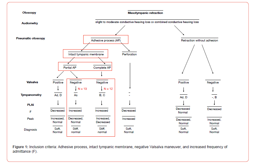
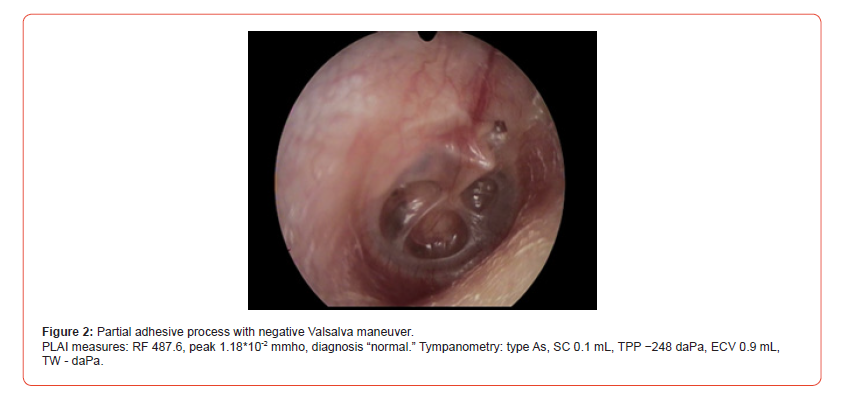
Exclusion criteria included patients younger than 18 years, those with perforation of the tympanic membrane, patients with AdOM and an open ET, and those with slight mesotympanic retractions without adhesion, resulting in a MedWave® dashboard diagnosis of “soft” and decreased RF.
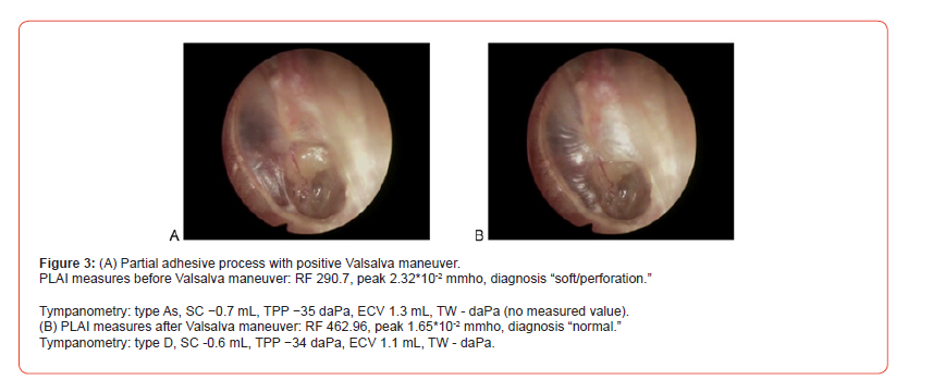
Figure 3 shows an example of a partial adhesive process before (A) and after the Valsalva maneuver (B). This case was excluded as the ET was open. The resonance frequency was significantly reduced before the maneuver.
The study included two groups: (1) 15 AdOM patients (25 ears) with a mean age of 59.1 ± 11.1 years (5 men and 10 women) and 57 healthy subjects with a mean age of 51.5 ± 17.3 years (35 men and 22 women). The healthy subjects had bilateral normal ears (114 ears), normal tympanic membranes, and no history of inflammation or disease affecting the middle ear, recent hearing disabilities, or aural symptoms.
Procedure
Medical evaluation
Ear, nose, and throat examinations were performed by experienced ENT surgeons. Only patients with a tympanic membrane partially or totally adhered to the medial wall of the middle ear, without a possibility of reversing the retraction through the Valsalva maneuver or pneumatic otoscopy, were categorized as “AdOM.” All patients underwent the Valsalva maneuver, video otoscopy, and pneumatic otoscopy, and the resulting images and videos were stored. Symptoms of ET dysfunction were scored using the ETDQ-7 [10] questionnaire.
Healthy subjects with acute or chronic otitis and other pathologies were excluded from the study. Any pathology of the tympanic membrane—including mesotympanic and/or epitympanic retraction, perforation, cholesteatoma, or myringitis— was documented in the otoscopic findings.
All patients with AdOM and healthy subjects older than 18 years underwent video otoscopy, pure-tone audiometry, PLAI using MedWave® (Neuranix, Italy), and conventional 226-Hz tympanometry.
PLAI (Pressure-Less Acoustic Immittance)
PLAI measures acoustic admittance via the outer ear canal, which remains at atmospheric pressure. The ear is stimulated using an acoustic signal ranging from 100 to 3000 Hz. The system estimates the proportion of acoustic energy transmitted versus reflected by the tympanic membrane and the middle ear. The resulting admittance curve displays a resonance peak, representing the maximum value of the admittance. MedWave® provides objective numerical values: the frequency at which the maximum value of the admittance curve is reached (Fr [Hz]) and the peak admittance value (P [*10-2 mmho]). Detailed information is presented elsewhere [12-16].
Conventional tympanometry
Tympanometry was performed in Güstrow (Germany) using the Madsen® Zodiac (type 1096; Otometrics) at 226 Hz or 1000 Hz, from −400 to +200 daPa. The following measures were used for calculation: tympanometric peak pressure (TPP), tympanic width (TW), equivalent ear canal volume (ECV), static peak compliance (SC), and type of tympanogram as per Jerger classification:
A (peak within normal limits for both pressure and height, with a normal admittance/compliance reading of 0.3-1.6 mL, a normal middle ear pressure reading of +50 to −50 daPa, and a normal ear canal volume reading of 0.5-2.5 mL,
Ad (peak between +100 and −100 daPa, with a compliance reading under 0.3 mL and ECV of up to 1.6 mL),
As (peak between +100 and −100 daPa, with a compliance reading over 1.5 mL and ECV of up to 0.4 mL),
B (flat with no significant peak), and
C (the height of the peak is or is not within normal limits and is more negative than normal).
Pure-tone audiometry
All audiological assessments were conducted in an audiometric sound-attenuated room, using calibrated signals and equipment in accordance with accepted ISO standards. For patient assignment, pure-tone audiometry was performed to measure air conduction (AC) and bone conduction (BC) thresholds. The air-bone gap was calculated across the frequencies of 0.5, 1, 2, and 4 kHz.
Statistical Analyses
Statistical analyses were performed using SPSS Version 30.0 (SPSS Inc., Chicago, IL, USA). Data were evaluated using descriptive statistical methods and comparative tests. Numerical tests were expressed as mean ± standard deviation, and categorical variables were presented as percentages. Statistical tests (Kolmogorov test and Shapiro-Wilk test) indicated a non-normal distribution for all parameters (p < 0.05). Nonparametric tests were performed for group comparisons whenever appropriate. Pearson statistics were used for correlational analyses. A p-value of < 0.05 was considered statistically significant in all analyses.
Results
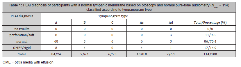
The bilateral normal ears (114 ears) of the 57 healthy subjects were classified as normal based on otoscopy and pure-tone audiometry.
Of these, 84 ears had a type A tympanogram (74%), 7 ears (6%) had a type B tympanogram, and 6 ears (5%) had a type C tympanogram, while 10 (9%) and 7 (6%) ears exhibited tympanogram types As and Ad, respectively (Table 1). Of the healthy subjects, 75% received a PLAI diagnosis of “normal,” while 15% were diagnosed with “OME/rigid” and 10% with “perforation/ soft” (Table 1).
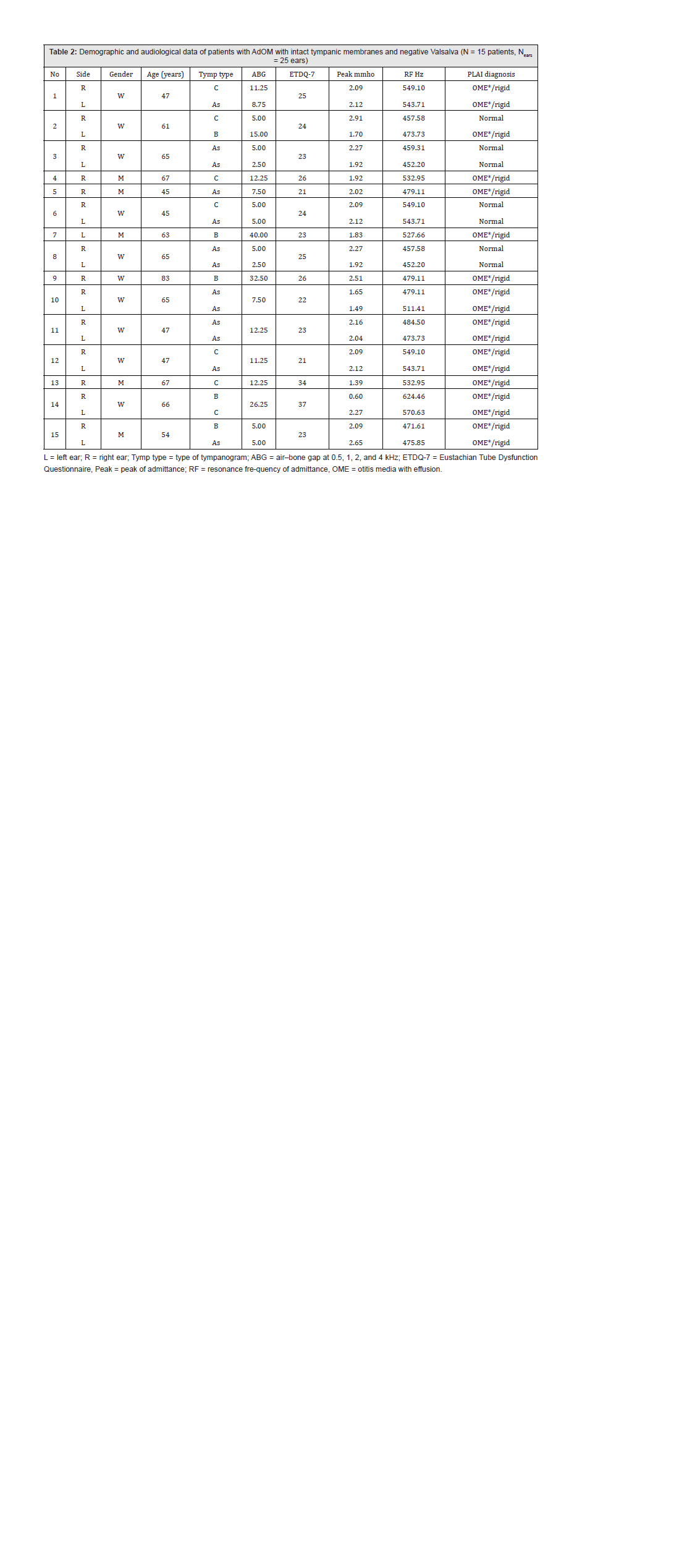
The demographic and audiometric data of patients with AdOM are presented in Table 2.
Using the PLAI measurements for all 114 normal ears assessed with otoscopy, an average RF of 403 ± 50.0 Hz (ranging from 258.4 to 516.8 Hz) and an average peak of admittance of 1.68 ± 0.39*10-2 mmho (ranging from 0.52 to 2.52*10-2 mmho) were determined (Figures 4A and 4B). Patients with AdOM demonstrated significantly higher PLAI values for RF and peak of admittance compared with healthy subjects (p < 0.001) (Figures 2A and 2B). Correlational analyses revealed a significant association between RF measurements and ETDQ7 values in AdOM (r25 = 0.61, p = 0.016). Higher symptom scores before ET dilatation were associated with higher RF values.


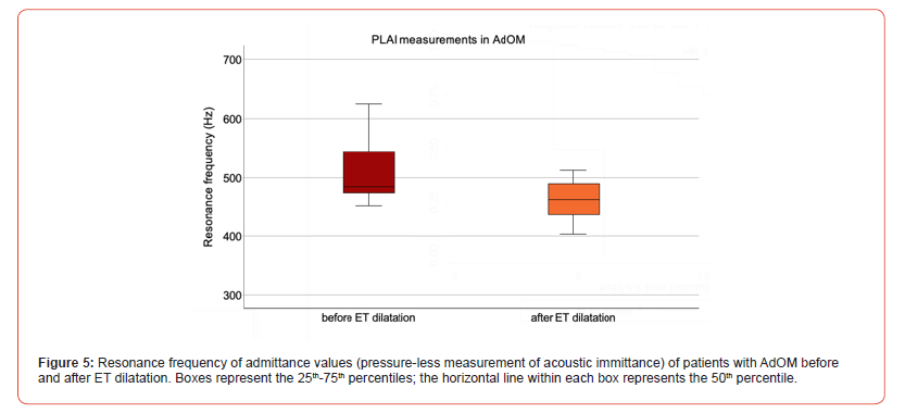
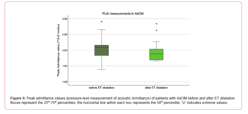
ET dilatation led to a significant decrease in RF in AdOM (p < 0.001). For all 25 ears presenting AdOM with a negative Valsalva test and intact tympanic membranes, the PLAI measurements after dilatation showed an average RF of 457.2 Hz (ranging from 387.6 to 511.9 Hz) and an average peak admittance of 1.82*10-2 mmho (ranging from 1.10 to 2.07*10-2 mmho) (Figures 5 and 6).
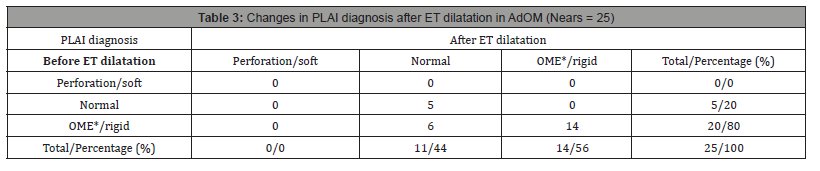
Overall, 44% of patients with AdOM received a PLAI diagnosis of “normal” after ET dilatation, 56% were diagnosed as “OME/ rigid,” and none were diagnosed as “perforation/soft” (Table 3). Six patients shifted in dashboard diagnosis from “OME/rigid” to “normal” after ET dilatation. A positive Valsalva test is regarded as the clinical correlate for this change in findings. In all six patients with partial adhesion at the promontory, ET dilation resulted in eardrum protrusion during the Valsalva maneuver and a corresponding decrease in resonance frequency during PLAI measurement.
Discussion
Considering the sequelae of otitis media with ET dysfunction— effusion, recurrent middle ear infections, atelectasis, and adhesive otitis media—the primary focus is to arrest and reverse the end stage of severe AdOM and the potential development of cholesteatoma.
While treatment of isolated ET dysfunction includes surgical Citation: Moayad Anbari, Rama Alslamah, Dörte Fischer, Blanda Kamin and Tino Just*. Eustachian Tube Balloon Dilatation in the Treatment of Adhesive Otitis Media Before Tympanoplasty. On J Otolaryngol & Rhinol. 8(2): 2025. OJOR.MS.ID.000684. DOI: 10.33552/OJOR.2025.08.000684. Online Journal of Otolaryngology and Rhinology Volume 8-Issue 2 Page 8 of 9 procedures such as adenoidectomy [17], balloon dilatation/ ET dilatation [18, 19], cleft repair [20], and septoplasty [21-23], treatment of atelectasis focuses on the insertion of ventilation tubes, which may reverse atelectasis and thereby prevent its progression to AdOM [24].
The treatment of severe AdOM remains challenging to date. In addition to moderate to severe conductive hearing loss, persistent otorrhea in AdOM is a significant indication for middle ear surgery with mastoidectomy. The timing and preparatory measures appear to be crucial for the success of middle ear surgery. It has been shown that the success rate of cartilage tympanoplasty in patients with AdOM and combined ET dysfunction increases after ET dilatation [19].
Simple practical tools such as tubomanometry [25], ETDQ-7 [10, 26], tympanometry, and the Valsalva maneuver are available for assessing ET function. Since tubomanometry is not commercially available at present [25], our study focused on evaluating ET function using ETDQ-7, audiometry, the Valsalva maneuver, otoscopy/ pneumatic otoscopy, and finally PLAI. The central question was whether PLAI could detect ET dysfunction in patients with AdOM and a negative Valsalva test, and whether changes in ET function following ET dilatation could be measured using PLAI. Preliminary examinations of patients with chronic ventilation disorders yielded varied results, which depended primarily on otoscopic findings/ middle ear pathology and middle ear ventilation [15]. Based on these early findings, inclusion and exclusion criteria were defined to address the study questions. Using the selection criteria applied in this study (AdOM, negative Valsalva), we demonstrated increased middle ear stiffness, reflected in elevated RF values, compared with healthy subjects and with AdOM patients who had a positive Valsalva test (exclusion criteria; results not shown). Using PLAI, we demonstrated a decrease in stiffness from 80% to 56% in AdOM patients, attributable to improved middle ear ventilation following ET dilatation. As expected, fixation of the stapes with total adhesion and ossicular chain interruption led to a minor reduction in the pretherapeutic elevated resonance frequency.
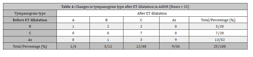
In contrast, improvement in middle ear ventilation after ET dilatation could not be demonstrated by tympanometry. Nevertheless, tympanometry remains the most frequently used technology for assessing ET function [18]. Its specificity for detecting ET dysfunction is about 93%, whereas its sensitivity is limited to about 61%. Therefore, tympanometry is unsuitable and insufficient for excluding obstructive ET dysfunction [19]. Regarding ETDQ-7, the American Academy of Otolaryngology-Head and Neck Surgery has published a clinical consensus statement indicating that ETDQ- 7 does not reliably diagnose obstructive ET dysfunction, as it can be influenced by several non-ET disorder-related factors [27]. Tubomanometry enables quantitative assessment of ET function with a high sensitivity of about 92%. According to Fontes Lima et al. [28], it is the most accurate method compared with tympanometry and ETDQ-7.
The correlation observed in our study between the resonance frequency values for bilateral adhesive processes and the ETDQ-7 should be interpreted with caution, as the questionnaire refers to both ears, whereas the extent of the adhesive process and the resonance frequencies differed between ears. Despite these limitations, future studies in bilateral cases should employ separate pre- and post-therapeutic, side-specific ETDQ-7 for each ear. Another limitation of this study is the small number of patients with AdOM included. As strengths of the present study, we highlight the carefully selected patients and the controlled postoperative followup. Future research should ideally conduct randomized and blinded controlled trials that evaluate the effectiveness of PLAI in assessing tube function in AdOM.
Conclusion
PLAI demonstrates significantly elevated RF values in patients with AdOM and a negative Valsalva test. Improvement in ventilation function following ET dilatation can be monitored using this method. These promising results, combined with the safe and quick applicability, make this technology an additional diagnostic tool for measuring ET function in AdOM.
Acknowledgment
None.
Funding
No conflict of interest.
Introduction
References
- Sade J (1993) Atelectatic tympanic membrane: histologic study. Ann Otol Rhinol Laryngol 102(9): 712-716.
- Tos M (1981) Experimental tubal obstruction. Changes in middle ear mucosa elucidated by quantitative histology. Acta Otolaryngol 92(1-2): 51-61.
- Zechner G (1983) Adhesive otitis media. J Laryngol Otol Suppl 8: 59-62.
- Sade J, Berco E (1976) Atelectasis and secretory otitis media. Ann Otol Rhinol Laryngol 85(2 Suppl 25 Pt 2): 66-72.
- Maw AR, Bawden R (1994) Tympanic membrane atrophy, scarring, atelectasis and attic retraction in persistent, untreated otitis media with effusion and following ventilation tube insertion. Int J Pediatr Otorhinolaryngol 30(3): 189-204.
- Sade J, Ar A (1997) Middle ear and auditory tube: middle ear clearance, gas exchange, and pressure regulation. Otolaryngol Head Neck Surg. 116(4): 499-524.
- Larem A, Haidar H, Alsaadi A, Abdulkarim H, Abdulraheem M, et al. (2016) Tympanoplasty in adhesive otitis media: A descriptive study. Laryngoscope. 126(12): 2804-2810.
- Dommerby H, Tos M (1986) Sensorineural hearing loss in chronic adhesive otitis. Arch Otolaryngol Head Neck Surg 112(6): 628-634.
- McCoul ED, Anand VK, Christos PJ (2012) Validating the clinical assessment of eustachian tube dysfunction: The Eustachian Tube Dysfunction Questionnaire (ETDQ-7). Laryngoscope 22(5): 1137-1141.
- Schroder S, Lehmann M, Sauzet O, Ebmeyer J, Sudhoff H (2015) A novel diagnostic tool for chronic obstructive eustachian tube dysfunction-the eustachian tube score. Laryngoscope 125(3): 703-708.
- Schilder AG, Bhutta MF, Butler CC, Holy C, Levine LH, et al. (2015) Eustachian tube dysfunction: consensus statement on definition, types, clinical presentation and diagnosis. Clin Otolaryngol 40(5): 407-411.
- Bassi F, Accardo A (eds.), Estimation of middle ear characteristics by an innovative pressure-less acoustic immittance (PLAI™) device. 9th European Mediacal and Biological Engineering Conference; 2024; Portoroz, Slovenia: EMBEC 2024, IFMBE Proceedings 113.
- Bassi F, Miladinovic A, Accardo A (eds.), Relations between age, tympanometric parameters and pressureless measurements. 9th National Bioengineering Group (GNB) Congress; 2025. Palermo, Italy
- Anbari M, Zhumabaeva S, Beishenova M, Fischer D, Sulaimanov S, Just T (2024) Comparison of conventional single-frequency tympanometry with pressure-less acoustic immittance measurements of pathological and normal middle ears. On J Otolaryngol & Rhinol 7(2): 1-11.
- Anbari M, Zhumabaeva S, Beishenova M, Fischer D, Sulaimanov S, et al. (2025) Pressure-less acoustic immittance measurements of patients with persistent ventilation disorders. On J Otolaryngol & Rhinol 7(3): 1-11.
- Polterauer-Neuling Née Polterauer D, Molenda C (2025) Early Results from a Pressureless Middle Ear Diagnostic and Its Relation to the Types of Tympanometry Results.
- Manno A, Iannella G, Savastano V, Vittori T, Bertin S, et al. (2021) Eustachian Tube Dysfunction in Children with Adenoid Hypertrophy: The Role of Adenoidectomy for Improving Ear Ventilation. Ear Nose Throat J 145561321989455.
- Poe DS, Hanna BM (2011) Balloon dilation of the cartilaginous portion of the eustachian tube: initial safety and feasibility analysis in a cadaver model. Am J Otolaryngol 32(2): 115-123.
- Azevedo C, Moreira F, Lima AF, Mar FM, Vilarinho S (2023) Prospective Evaluation of the Efficacy of Isolated Balloon Eustachian Tuboplasty: Short- and Medium-term Follow-up Results Based on Tubomanometry, ETDQ-7, Tympanometry, and Valsalva Maneuver. Int Arch Otorhinolaryngol 28(1): e76-e82.
- Smith TL, DiRuggiero DC, Jones KR (1994) Recovery of eustachian tube function and hearing outcome in patients with cleft palate. Otolaryngol Head Neck Surg 111(4): 423-429.
- Patil N, Jain S, Deshmukh PT, Gaurkar SS, Singh CV, et al. (2024) Quantifying the Domino Effect of Septoplasty on Eustachian Tube Function and Nasal Resistance in Patients with Chronic Otitis Media: A Cross-Sectional Study. Cureus 16(9): e69819.
- Low WK, Willatt DJ (1993) The relationship between middle ear pressure and deviated nasal septum. Clin Otolaryngol Allied Sci 18(4): 308-310.
- Fontes Lima A, Carvalho Moreira F, Esteves Costa I, Azevedo C, Mar F, et al. (2022) Nasal septum deviation and Eustachian tube function: A prospective case-control study based on tympanometry, tubomanometry, and ETDQ-7. Acta Otorrinolaringol Esp (Engl Ed) 73(1): 35-41.
- Valtonen H, Qvarnberg Y, Nuutinen J (1999) Tympanostomy in young children with recurrent otitis media. A long-term follow-up study. J Laryngol Otol 113(3): 207-211.
- Holm NH, Mirz A, Ovesen T, Pfeiffer CJ (2025) Tubomanometry-Score as Predictor of Outcome for Balloon Eustachian Tuboplasty in Patients with Eustachian Tube Dysfunction. Otolaryngol Head Neck Surg 172(4): 1387-1393.
- Holm NH, Ovesen T (2025) The Usefulness of ETDQ-7 Score in Assessing ETD. Clin Otolaryngol 50(5): 840-847.
- Tucci DL, McCoul ED, Rosenfeld RM, Tunkel DE, Batra PS, et al. (2019) Clinical Consensus Statement: Balloon Dilation of the Eustachian Tube. Otolaryngol Head Neck Surg 161(1): 6-17.
- Fontes Lima A, Carvalho Moreira F, Sousa Menezes A, Azevedo C, Costa I, et al. (2020) Eustachian Tube function tests: Prospective analysis of diagnosis accuracy. Rev Port Otorrinolar- ingol E Cir Cabeça E Pescoço 58(1): 31-66.
-

This work is licensed under a Creative Commons Attribution-NonCommercial 4.0 International License.




