



 Case Report
Cochlear Implantation for Single-sided Deafness Following a Transverse Temporal Bone Fracture
1Department of Otorhinolaryngology, Head and Neck Surgery, KMG Klinikum, Güstrow, Germany
2Department of Audiology, KMG Klinikum, Güstrow, Germany
3Department of Diagnostic and Interventional Radiology, KMG Klinikum, Güstrow, Germany
Received Date: June 25, 2026; Published Date: July 10, 2026
Introduction
Approximately 18–40% of skull base fractures involve the petrous bone [1]. Most petrous bone fractures are longitudinal, extending through the mastoid and the external auditory canal without involving the otic capsule. In cases involving otic capsule fractures, the fracture line extends through the cochlea, vestibular organ, and their connecting structures, resulting in severe hearing loss or deafness [1]. tinnitus, vertigo, facial nerve paralysis, and cerebrospinal fluid leaks, all of which may significantly reduce quality of life [2].
Transverse temporal bone fractures (TBFs) following head trauma may also occur bilaterally, leading to otological symptoms and complications on both sides [3]. Persistent hearing loss in one ear that cannot be corrected with a conventional hearing aid may be managed with a conventional contralateral routing of signal (CROS) hearing aid system. Alternatively, bone-anchored hearing aids (BAHAs) and other passive implantable or nonimplantable bone-conduction systems may be considered as CROS solutions [4]. In cases involving active middle-ear implants and active transcutaneous implantable bone-conduction systems [5]. adequate residual cochlear function is required. Severe hearing loss and deafness cannot be effectively treated with these systems.
Cochlear implants (CIs) represent a promising option for hearing rehabilitation in patients with severe sensorineural hearing loss following TBFs [4]. However, patients with isolated hearing loss and an intact cochlear nerve following transverse TBFs present a preoperative diagnostic challenge, as the likelihood of successful hearing rehabilitation must be carefully assessed and discussed before surgery. Additionally, the possibility of facial nerve co-stimulation following CI surgery must be considered during preoperative counseling.
This paper describes the diagnostic and therapeutic approach to isolated unilateral transverse TBFs and presents the associated hearing outcomes.
Materials and Methods
The preoperative neuro-otological evaluation included clinical examination, pure-tone audiometry, the Freiburger monosyllabic word recognition test at 65 dB SPL (Fb65), speech intelligibility testing, caloric testing, and the Head Impulse Test.
Brainstem audiometry was performed using a multi-stimulus chirp consisting of a 500-Hz chirp and a click. All audiological All audiological assessments were conducted in a sound-attenuated audiometric room using calibrated equipment and stimuli in accordance with accepted ISO standards. Pure-tone audiometry was performed to measure air-conduction (AC) and bone-conduction (BC) thresholds. The pure-tone average across four frequencies (PTA4) was calculated using thresholds at 0.5, 1, 2, and 4 kHz.
The Freiburger monosyllabic word recognition test was administered through headphones at presentation levels of 60, 80, and 100 dB SPL. Speech intelligibility testing was conducted in the free field using the Freiburger monosyllabic word test in both quiet and noise conditions. Loudspeakers were positioned 1 m from the participant’s head.
In the quiet condition (S0), speech intelligibility was measured at 65 dB SPL and 80 dB SPL. In the noise condition (S0N0), speech intelligibility was assessed using a fixed noise level of 60 dB SPL and speech presentation levels of 65 dB SPL and 80 dB SPL, resulting in signal-to-noise ratios (SNRs) of 5 dB and 20 dB, respectively.s
The taste testing was performed bilaterally on the tongue using four concentrations of each of four taste solutions: sweet (0.03, 0.1, 0.4, and 2 g/mL sucrose solution), sour (0.01, 0.05, 0.1, and 0.15 mL citric acid), salty (0.025, 0.075, 0.15, and 0.36 mL sodium chloride solution), and bitter (0.0002, 0.0005, 0.001, and 0.01 mL quinine hydrochloride) [5].
Tympanometry was performed using the Madsen® Zodiac (type 1096; Otometrics) at 226 Hz over a pressure range of −400 to +200 daPa. The resonance frequency of admittance (Fr [Hz]) and the peak admittance value (P [*10-2 mmho]) were measured using MedWave®. Detailed information regarding this procedure has been published elsewhere [6].
Caloric testing and head impulse testing were performed using the ICS® AirCal caloric test device (GN Otometrics) and ICS® Impulse system (Nautus Sensory), respectively.
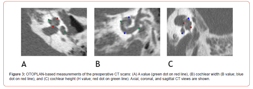
OTOPLAN software (CAScination, Bern, Switzerland) was used for anatomical measurements of the cochlea. For this purpose, the DICOM dataset obtained from the computed tomography (CT) scans was imported into the software (Version 3). After landmark placement, cochlear diameter, width, and height were measured. OTOPLAN uses the integrated “Elliptic Circular Approximation” algorithm to calculate cochlear duct length (CDL). A detailed description of the measurement procedure has been published elsewhere [7].
Case
A 70-year-old patient was referred to our clinic because of persistent hearing loss in the left ear after treatment for sudden hearing loss following head trauma caused by a fall from a ladder. Pure-tone audiometry revealed high-frequency sensorineural hearing loss in the right ear and severe pancochlear sensorineural hearing loss in the left ear. The patient also reported dizziness when standing up but denied lateropulsion. He did not report tinnitus, middle-ear infections, otorrhea, taste disturbances, or otalgia before or after the head trauma. Aside from the sudden hearing loss in the left ear and dizziness following the Trauma. He had no prior otological history.
Otoscopic examination revealed medium-sized external auditory canals bilaterally. The tympanic membranes were intact but non-translucent on both sides, without mesotympanic or epitympanic retraction or perforation.
In Weber tuning fork testing (a1, c3), sound was lateralized to the right ear. Rinne testing was positive on the right side and negative on the left side. Facial nerve function was normal bilaterally.
Single-frequency 226-Hz tympanometry demonstrated a type A tympanogram in the right ear and a type C tympanogram in the left ear. Acoustic immittance measurements obtained through the external auditory canal revealed admittance resonance frequencies of 474 Hz in the right ear and 517 Hz in the left ear. Video nystagmography demonstrated a low-frequency horizontal spontaneous nystagmus toward the right with a vertical component, as well as preserved bilateral thermal excitability of the vestibular organs.
A fistula test was performed by increasing pressure within the external auditory canal of the left ear and yielded a negative result, indicating the absence of pressure-induced nystagmus. Positional testing provoked nystagmus when the patient turned to either side and during head-shaking maneuvers. The clinical head impulse test demonstrated a corrective saccade involving the left anterior semicircular canal. However, the video head impulse test revealed reduced vestibulo-ocular reflex gain in the left posterior canal and increased gain in the left anterior canal. Taste testing demonstrated normogeusia for all tested taste qualities on both sides of the tongue.
The results of pure-tone and free-field audiometry confirmed left-sided hearing loss that could not be improved with either a conventional hearing aid or a BAHA in a CROS configuration (Table 1).
Vestibular testing revealed an almost completely compensated vestibular deficit on the left side. A CROS hearing aid system was recommended and trialed for four weeks; however, the patient did not perceive any improvement in everyday listening situations involving background noise. Consequently, he was referred for CI evaluation.
Brainstem audiometry confirmed profound hearing loss in the left ear. During promontory stimulation testing, the patient perceived slight vibration at frequencies of 50 Hz, 250 Hz, and 500 Hz, but no tonal perception was detected. The total Nijmegen Cochlear Implantation Questionnaire score was 57.
Table 1:
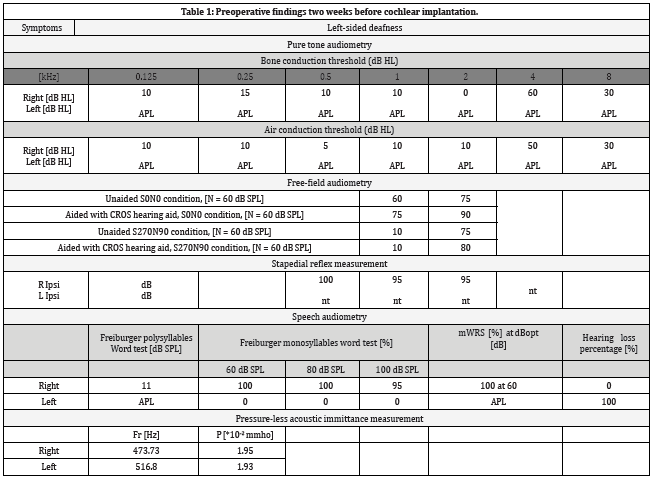
Unaided maximum word recognition score (mWRS); audiometer power limit (APL); resonance frequency of the admittance (Fr [Hz]); peak admittance value (P [*10-2 mmho]); not triggered (nt); and word recognition threshold (WRT).
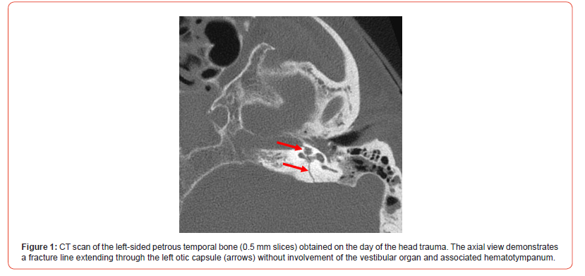
The patient underwent a preoperative high-resolution CT scan of the temporal bone three days after the trauma (Figure 1) and magnetic resonance imaging (MRI) at 1.5 Tesla (Figure 2). The CT scan demonstrated a transverse TBF.
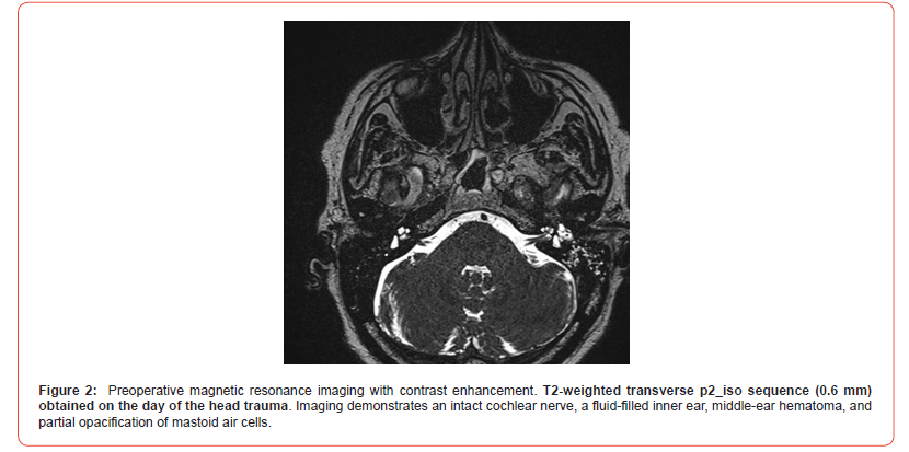
A second MRI examination was performed two weeks before cochlear implantation for surgical planning. Imaging continued to demonstrate a fluid-filled inner ear, an intact vestibulocochlear nerve, and aeration of both the middle ear and mastoid.
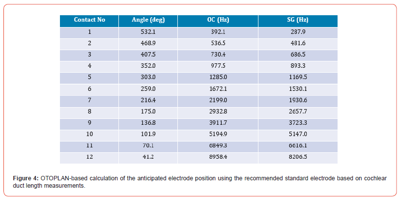
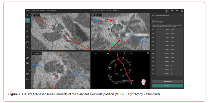
Morphological measurements of the cochlea were obtained from the preoperative CT scans using OTOPLAN (MED-EL®). The estimated CDLs were 38.8 mm for the Organ of Corti (OC) measured from 0° and 41.3 mm for the full OC (Figures. 3 and 4). The estimated electrode position is illustrated in Figure 4. Based on these measurements, OTOPLAN estimated electrode length options for an insertion angle for the first electrode contact up to 590° insertion angle (MED-EL FLEX34) and 532° (MED-EL Standard).
Cochlear parameters
• Diameter (A Value): 10.4mm
• Height (H Value): 4.4mm
• Width (B Value): 7.8mm
• Estimated CDL (full OC): 41.3mm
• Estimated CDL (OC from 0°): 38.8mm
• Number of turns:
Selected Electrode: STANDARD
The patient was informed about the need for intraoperative electrically evoked compound action potential testing, and cochlear implantation was recommended if a neural response could be detected. Following a standardized presentation by the implant manufacturers, the patient selected a MED-EL® CI system.
Results
Standardized cochlear implantation was performed using a posterior tympanotomy and an extended cochleostomy located anterior and inferior to the round window. An alternating rhythm phenomenon was observed during manipulation of the short process of the incus.
Upon opening the round window membrane, a fluid-filled scala tympani was identified. No fibrosis or ossification of the cochlea was observed. A test electrode was initially inserted to perform electrically evoked brainstem response audiometry (E-BERA). These measurements demonstrated neural responses by stimulating the first contact directing the tip of the test electrode.
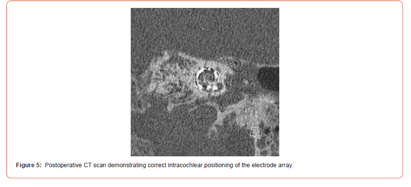
Based on the positive neural responses, cochlear implantation was subsequently performed. The standard electrode was successfully inserted into the cochlea via the scala vestibuli. Electrode impedances ranged from 3 to 7 kΩ. Neural responses were detected following stimulation of all electrodes. Postoperative CT imaging confirmed appropriate electrode placement (Figure 5).
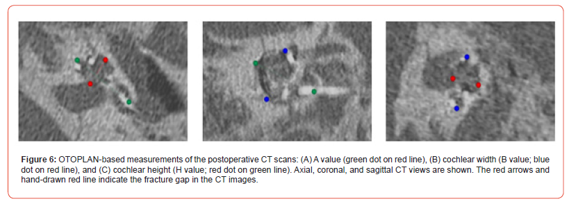
Postoperative measurements obtained with OTOPLAN demonstrated the final electrode positions. Electrodes 3, 6, and 12 were located at insertion angles of 302°, 218°, and 30°, respectively.
Cochlear parameters
• Diameter (A Value): 10.5mm
• Height (H Value): 4.3mm
• Width (B Value): 7.8mm
• Estimated CDL (full OC): 41.4mm
• Estimated CDL (OC from 0°): 38.9mm
• Number of turns:
Initial fitting of the speech processor was performed four weeks after cochlear implantation, and auditory rehabilitation was subsequently initiated.
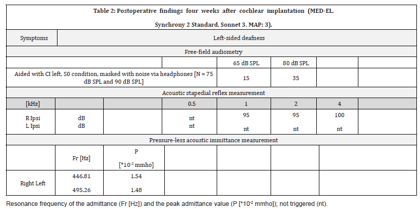
The first Freiburger monosyllabic word test demonstrated speech intelligibility scores in quiet of 15% at 65 dB SPL and 35% at 80 dB SPL (Table 2). The speech recognition threshold was 31 dB HL. The functional gain curve ranged between 10 and 30 dB HL. During left-sided speech perception testing, the normalhearing contralateral ear was masked using headphones with noise presented at +10 dB SNR.
Table 2:
All 12 electrodes were stimulated at maximum comfortable levels (MCLs) ranging from 31.0 to 75.24 qu, with effective pulse durations between 63 and 84 μs. Facial nerve co-stimulation occurred only during stimulation of electrode 6 at an MCL of 53 qu and above, with an effective pulse duration of 63 μs. Due to the high charge levels and the resulting increase in pulse width, a threshold for facial nerve co-stimulation was reached, thereby limiting the effective maximum charge that could be delivered.
Postoperative stapedius reflex thresholds were measured using the MedWave system. Although no stapedius reflexes could be elicited acoustically on the deaf ear, either ipsilaterally or contralaterally, ipsilateral reflexes were elicited on the right side. Ipsilateral and contralateral stapedius reflex measurements were obtained during stimulation of electrodes 1, 5, and 12.
During stimulation of electrode 1 (MCL 75 qu; effective pulse duration 84 μs), a contralateral stapedius reflex threshold of 85 dB HL was detected. During stimulation of electrode 12 (MCL 53 qu; effective pulse duration 63 μs), ipsilateral but not contralateral stapedius reflexes were observed. Neither ipsilateral nor contralateral stapedius reflexes were detected during stimulation of electrode 5.
Discussion
The predominant etiologies of post-traumatic hearing impairment include loss of hair-cell function caused by direct trauma to the cochlea, otic capsule, cochlear nerve, or auditory pathway; disruption of the membranous labyrinth; hemorrhage or circulatory disturbances within the inner ear; the development of a perilymphatic fistula; and endolymphatic hydrops. Long-term complications of temporal bone trauma may include labyrinthine contusion as well as fibrosis and/or ossification of the cochlea, which can subsequently lead to hearing loss [8, 9].
Cochlear implantation is a well-established treatment for restoring hearing in patients with profound unilateral or bilateral traumatic sensorineural hearing loss, provided that the auditory pathway remains intact [10-12]. Outcomes following cochlear implantation have been promising in various case series and are generally comparable to those achieved in conventional CI recipients [10, 13] However, cochlear implantation can be challenging in cases involving cochlear fibrosis or ossification. These conditions may complicate electrode placement or even result in incomplete insertion, ultimately leading to suboptimal hearing outcomes [14, 15].
Additional factors may influence postoperative hearing rehabilitation after cochlear implantation, including perilymphatic fistulae of the cochlea [16, 17] and facial nerve co-stimulation, which occurs when electrical current spreads from the electrode array to the spiral ganglion cells and subsequently to the facial nerve [18]. This phenomenon may be facilitated by reduced electrical resistance at the fracture site [18, 19].
In a retrospective study, the authors differentiated between otic capsule-sparing fractures and otic capsule-involving fractures [15] Within the latter group, they further distinguished between patients with and without evidence of cochlear ossification. Postoperative hearing outcomes were poorer in patients with otic capsuleinvolving fractures, particularly in those with ossification. It has been proposed that this fracture type may damage the first neural element of the central auditory pathway, namely the spiral ganglion cells. Subsequent degeneration of these cells may lead to significant alterations in central auditory processing and limitations in speech perception [22].
Because of the reduced number of functioning ganglion cells, central auditory processing may require targeted auditory training to enhance speech-processing capacity within the central nervous system [23]. Therefore, it is important to counsel patients regarding realistic expectations of the hearing outcomes that may be achieved. Patients with cochlear fractures and associated ossification generally achieve poorer hearing outcomes than typical CI recipients [15]. This finding underscores the significant impact of cochlear ossification on postoperative speech perception outcomes following cochlear implantation [20].
Cochlear ossification can be detected using either highresolution CT imaging or intraoperative assessment by the surgeon [15, 25]. However, ossification cannot always be visualized on imaging studies, particularly during the early stages when mineralization remains insufficient for radiographic detection [26]. Therefore, high-resolution contrast-enhanced MRI should also be performed preoperatively in all cases. If post-traumatic intracochlear fibrosis or ossification is not identified before surgery, it may significantly affect the intraoperative procedure because complete electrode insertion may no longer be possible.
In the present case, a measurement electrode was inserted for E-BERA testing without difficulty. However, if the promontory test had demonstrated a clearly positive auditory response, E-BERA testing would not have been necessary.
Specialized electrode arrays have been developed for ossified cochleae, and several surgical techniques have been described to address these challenging cases [27]. To access a partially ossified cochlea, the scala tympani may be drilled open, or alternatively, the scala vestibuli may be used if it remains patent. In cases of complete obliteration, a second cochleostomy may be required in addition to the drill-out technique. This approach permits the use of electrode arrays containing two electrode carriers [21, 22].
In some patients with complete cochlear ossification, incomplete electrode insertion remains unavoidable. In addition to ossification, the timing of surgery following trauma appears to play an important role in determining outcomes. Longer durations of hearing loss are associated with poorer CI performance [30]. Therefore, early implantation following a petrous bone fracture involving the cochlea is important to minimize the risk of cochlear obliteration [23].
However, if the promontory test is negative and E-BERA demonstrates no evidence of stimulus conduction, cochlear implantation should be discussed carefully with the patient. This consideration is particularly important because facial nerve costimulation may further affect postoperative CI performance.
Intraoperative impedance measurement, a standard method used to verify electrode integrity, also provides information regarding the intracochlear environment. In cases of intracochlear remodeling, such as fibrosis or early ossification, electrical resistance and, consequently, impedance values are often elevated [15]. Similar findings may occur when small intracochlear air bubbles are present. These bubbles typically resolve over time, allowing impedance values to return to normal levels.
In general, elevated impedance values require increased stimulation voltage [18] This may lead to greater battery consumption. In the present case, impedance values were comparable to those observed during routine CI surgery. However, if elevated impedances occur specifically in electrodes located near the fracture gap—and if stimulation levels must simultaneously be reduced because of facial nerve co-stimulation—CI performance may be compromised due to reduced effective stimulation from those electrodes.
Conclusion
The present case of a transverse TBF involving the cochlea demonstrates that positive E-BERA findings may indicate preserved neural function and increase the likelihood of successful cochlear implantation, even in the presence of a negative promontory test. Neural responses were detected in all electrodes intraoperatively, supporting the decision to proceed with cochlear implantation.
Postoperatively, only one of the three electrodes located near the fracture site exhibited facial nerve co-stimulation. This co-stimulation was successfully eliminated by reducing the stimulation current and triphasic stimulation. The audiological outcomes observed after the initial fitting of the speech processor were comparable to those typically achieved by conventional CI recipients.
These findings suggest that cochlear implantation may be a viable treatment option for selected patients with transverse TBFs involving the cochlea, provided that preserved neural responsiveness can be demonstrated preoperatively or intraoperatively. Furthermore, cochlear implantation should be considered as early as possible following trauma in order to minimize the risk of cochlear obliteration or ossification, both of which may negatively affect surgical outcomes and hearing rehabilitation.
Conflict of Interest
The authors declare no conflict of interest.
Acknowledgment
The authors thank Andreas Brisch (B. Eng., MED-EL) for the OTOPLAN measurements and data availability.
References
- Amin MB, Edge SB, Greene FL, Byrd DR, Brookland RK, et al. (2017) AJCC Cancer Staging Manual. 8th New York (NY): Springer.
- Johnson DE, Burtness B, Leemans CR, Lui VWY, Bauman JE, et al. (2020) Head and neck squamous cell carcinoma. Nat Rev Dis Primers 6(1): 92.
- El-Naggar AK, Chan JKC, Grandis JR, Takata T, Slootweg PJ, editors. (2022) WHO Classification of Head and Neck Tumours. 5th ed. Lyon: International Agency for Research on Cancer.
- Sung H, Ferlay J, Siegel RL, Laversanne M, Soerjomataram I, et al. (2021) Global cancer statistics 2020: GLOBOCAN estimates of incidence and mortality worldwide for 36 cancers in 185 countries. CA Cancer J Clin 71(3): 209-249.
- Chow LQM (2020) Head and neck cancer. N Engl J Med 382(1): 60-72.
- Mesía R, Iglesias L, Lambea J (2021) SEOM clinical guidelines for the treatment of head and neck cancer (2020). Clin Transl Oncol 23: 913-921.
- American Cancer Society Medical and Editorial Content Team. Oral Cavity (Mouth) and Oropharyngeal (Throat) Cancer: Survival Rates for Oral Cavity and Oropharyngeal Cancer. Updated March 1, 2023. Accessed July 30, 2024.
- De Felice F, Humbert-Vidan L, Lei M, King A, Guerrero Urbano T (2020) Analyzing Oropharyngeal cancer survival outcomes analysis: a decision tree approach. Br J Radiol 93(1111): 20190464.




