 Research Article
Anatomical Variations of Paranasal Sinuses on Multidetector Computed Tomography
Balegh Abdel Hak1*, Moustafa Talaat2, Nasr Osman3, Rasha ahmed4 and Heba Ibrahim5
1Professor of ENT, Minia University, Egypt
2Lecturer of ENT, Minia University, Egypt
3Lecturer of Radiology, Minia University, Egypt
4Lecturer of ENT, Minia University, Egypt
Balegh Abdel Hak, Professor of ENT, Minia University, Egypt.
Received Date: July 24, 2020; Published Date: August 18, 2020
Abstract
Background: Multi-detector computed tomography (MDCT) imaging of paranasal sinuses prior to functional endoscopic sinus surgery (FESS) has become mandatory.
Material and methods: Comparative study of 20 patients underwent FESS to detect anatomical variations by MDCT and endo-nasal rhinoscopy examination, pre and postoperative.
Results: The study was done on 20 patients, 14 females and 6 males, their age ranged from 6 years to 45 years with a mean of age (23.45). The most common anatomic variation found is the deviated septum in (45%) of the patients. The second anatomic variation is Onodi cell which appears in (35%) of patients, while the least found were congenital bony and mucosal atresia, Agger nasi cell and obstruction of maxillary ostium by bony septum which appear only in (5%).
Conclusion: Multiplanar imaging, particularly coronal reformations, offers precise information regarding the anatomy of the sinuses and its variations, which is an essential requisite before surgery.
Keywords: Anatomical variations; MDCT; FESS
Introduction
Multi detector computed tomography (MDCT) scan of paranasal sinuses has become mandatory for all patients undergoing functional endoscopic sinus surgery. It depicts the anatomical variations in much simpler way and acts as a roadmap for endoscopic sinus surgery [1]. Some anatomical variations may predispose to nasal and paranasal diseases, constituting areas of high risk for injuries and complications during surgical procedures. Therefore, the recognition of such variations is critical in the preoperative evaluation for endoscopic surgery [1].
The osteomeatal complex
The OMU is the key factor in the pathogenesis of chronic sinusitis. The ethmoid sinus is the key sinus in the drainage of the anterior sinuses. It is vulnerable to trauma during surgery due to its close relationship with the orbit and the anterior skull base [2]. The ethmoid roof is of critical importance for two reasons. First, it is most vulnerable to iatrogenic cerebrospinal fluid leaks. Second, the anterior ethmoid artery is vulnerable to injury, which can cause devastating bleeding into the orbit. During FESS, intracranial injury can occur on the side where the position of the roof is relatively low [3].
Onodi cells
These are posterior ethmoidal cells extending into the sphenoid bone, either adjacent to or impinging upon the optic nerve. When these Onodi cells abut or surround the optic nerve, the nerve is at risk when surgical excision of these cells is performed. It is also a potential cause of incomplete sphenoidectomy [4].
The Middle Turbinate Variations:
1) Paradoxical curvature: When the convexity is directed laterally, it is termed a paradoxical middle turbinate. Most authors agree that the paradoxical middle turbinate can be a contributing factor to sinusitis.
2) Concha bullosa: This is an aerated turbinate, most often the middle turbinate. When pneumatization involves the bulbous portion of the middle turbinate, it is termed concha bullosa. If only the attachment portion of the middle turbinate is pneumatized, it is termed lamellar concha. A concha bullosa may obstruct the ethmoid infundibulum [5].
Haller cells
These cells contribute to the narrowing of the infundibulum and may compromise the ostium of the maxillary sinus, thus contributing to recurrent maxillary sinusitis [6,7]. The revolutionary changes in the surgical treatment of sinusitis in recent years, particularly in endonasal endoscopic surgery, require the clinician to have a precise knowledge of nasal sinus anatomy and of the large number of anatomical variants in the region many of which are detectable only by the use of CT [5].
Material and Methods
Twenty patients, with the clinical diagnosis of rhinosinusitis from February to August 2019 and who had been considered for FESS underwent modified CT Scanning before and after the surgery. All CT scans were performed on MDCT scanners.
Results
Table 1: Comparing the Preoperative and Postoperative SNOT 22 Scores of each patient.
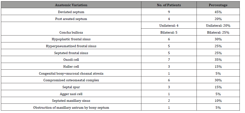
Table 2: Shows Incidence of anatomic variations of nose and Para nasal sinuses by Endoscopic examination (intra operative).
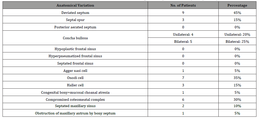
The study was conducted on 20 patients, 14 females and 6 males, their age ranged from 6 years to 45 years with a mean of age (23.45) (Table 1).
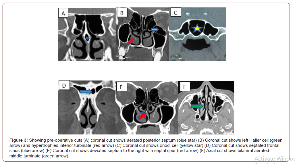
Regarding the incidence of anatomic variations by CT examination, it was found that the most common anatomic variation is the deviated septum which appears in 9 patients with percentage 45%. The second anatomic variation is Onodi cell which appears in 7 patients with percentage 35%, other variations as concha bullosa appear unilateral in 4 patients (20%) and bilateral in 5 patients (25%), posterior aerated septum in 4 patients (20%), hypo plastic frontal sinus in 6 patients (30%), hyper-Pneumatized frontal sinus in 5 patients (25%), septated frontal sinus in 5 patients (25%), Haller cell in 3 patients (15%), compromised osteomeatal complex in 6 patients (30%), septal spur in 3 patients (15%), septated maxillary sinus in 2 patients (10%), the least incidence of anatomical variations were congenital bony and mucosal atresia, agger nasi cell and obstruction of maxillary ostium by bony septum which appear only in one patient with percentage (5%) (Table 2).
Regarding the incidence of anatomic variations by endoscopic examination (intra operative), it was found that the most common anatomic variation is the deviated septum which appeared in 9 patients with percentage 45%. The second anatomic variation is the Onodi cell which appears in 7 patients with percentage 35%, followed by concha bullosa which was detected (intraoperative) unilateral in 4 patients (20%) and bilateral in 5 patients (25%), Haller’s cell in 3 patients (15%), compromised osteomeatal complex in 6 patients (30%), septal spur in 3 patients (15%) and septated maxillary sinus in 2 patients (10%). The least incidence of anatomical variations was congenital bony and mucosal atresia, agger nasi cell and obstruction of maxillary ostium by bony septum which appear only in one patient with percentage (5%). Other variations as posterior aerated septum, hypo-plastic frontal sinus, hyper-pneumatized frontal sinus and septated frontal sinus, couldn’t be detected intraoperative by the endoscope.
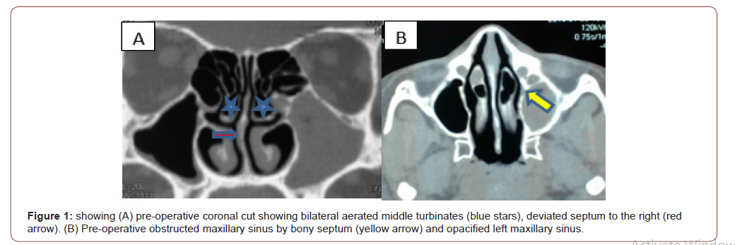
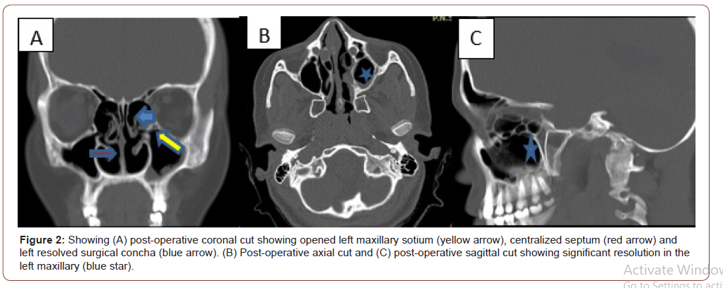
Case 1: Figure (1,2).
Case 2: Figure (3).
Conclusion
MDCT of the paranasal sinuses has improved the visualization of paranasal sinus anatomy and has allowed greater accuracy in evaluating paranasal sinus disease.
Acknowledgement
None.
Conflict of Interest
No conflict of interest.
References
- Kennedy DW, Zinreich J, Rosenbaum AE, Johns ME (1985) Functional endoscopic sinus surgery: Theory and diagnostic evaluation. Arch Otolaryngol 111: 576-582.
- Becker SP (1989) Anatomy for endoscopic sugery. Otolarygol Clin North Am 22: 677‑6
- Kasper KA (1936) Nasofrontal connection. A study based on one hundred consecutive dissections. Arch Otolaryngol 23: 322‑3
- Stammberger HR, Kennedy DW (1995) Paranasal sinus: Anatomic terminology and nomenclature. The anatomic terminology group. Ann Otol Rhinol Laryngol 167: 7‑
- Bolger WE, Butzin CA, Parsons DS (1991) Paranasal sinuses bony anatomic variantsand mucosal abnormalities: CT analysis for endoscopic surgery. Laryngoscope 101: 56‑
- Zinreich SJ, Kennedy DW, Rosenbaum AE, Gayler BW, Kumar AJ, et al. (1987) Paranasal sinuses. CT imaging requirements for endoscopic surgery. Radiology 163: 769‑7
- Laine FJ, Smoker WR (1992) The osteomeatal unit and endoscopic surgery: Anatomy, variation and imaging findings in inflammatory disease. AJR Am J Roentgenol 159: 849‑8
-
Balegh Abdel Hak, Moustafa Talaat. Anatomical Variations of Paranasal Sinuses on Multidetector Computed Tomography. On J Otolaryngol & Rhinol. 3(2): 2020. OJOR.MS.ID.000556.
-
Sinus surgery, Endo nasal, Rhinoscopy, Mucosal atresia, Paranasal sinus, Endoscopic surgery, Sphenoidectomy, Maxillary sinus, Frontal sinus, Rhinosinusitis, Concha bullosa.
-

This work is licensed under a Creative Commons Attribution-NonCommercial 4.0 International License.


