




 Case Report
An Interesting Case Report on Sinonasal Hemangiopericytoma: A Rare Entity
Senior specialist, Ministry of health, Sur hospital, Oman
Received Date: June 02, 2026; Published Date: June 09, 2026
Abstract
Hemangiopericytoma previously called glomangiopericytomas is a rare slowly progressive vascular tumor with borderline malignant potential, often seen in 6th to 7th decade of life. In this article we report the case of an elderly male who presented with heavy epistaxis the cause of which could not be determined clinically or with imaging. An intraoperative endoscopy revealed a friable mass at the roof of ethmoids. This later turned out to be a hemangiopericytoma. Epistaxis in elderly is often and commonly considered as due to hypertension or due to comorbidities like ischemic heart disease or diabetes mellitus. In this article we highlight such rare indolent tumors can also be the cause and should not be missed out during the detailed evaluation of the patients.
Keywords: Epistaxis, Hemangiopericytoma; Low grade malignant potential; Rare tumor
Introduction
In 1942, this tumor was first described by Stout and Murray. It is derived from mesenchymal cells with Zimmerman’s pericyte differentiation. The incidence of such tumors in the head and neck is about 15%. This is categorized as low grade malignant vascular tumor by WHO in 2005. It accounts for less than 0.5% of sinonasal primary neoplasm. It is most commonly seen in 6th to 7th decade of life. Literature reports CTNNB1 mutations. A rare association with oncogenic osteomalacia is also reported in the literature. In this article we describe a patient who presented to us with severe epistaxis and was later on diagnosed to have sinonasal hemangiopericytoma.
Case Report
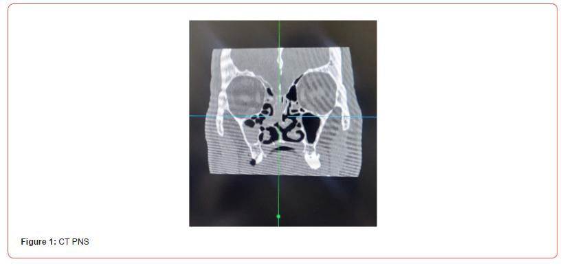
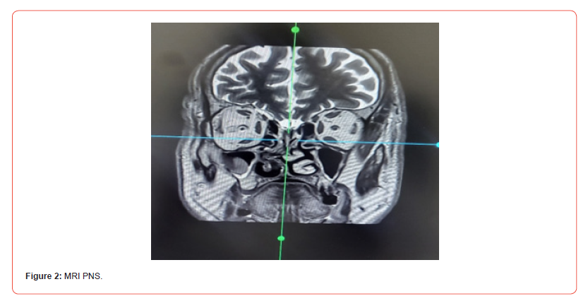
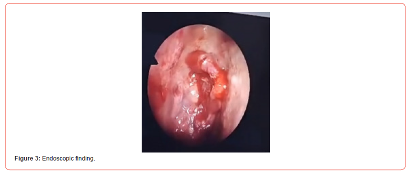
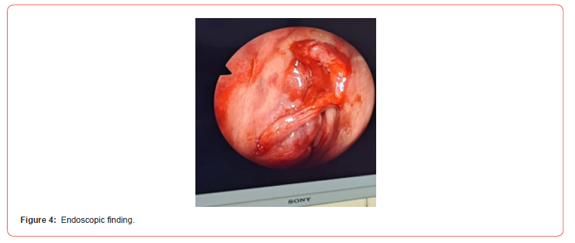
A 69year old elderly male presented to our emergency department with torrential nasal bleeding. Hemostasis was achieved with nasal packing for 2 days. He was not a known hypertensive although blood pressure recordings were noted to be high at the time of admission. He didn’t have any other comorbidities. When pack removal was done after 2 days, nasal bleeding on the right side was noted but no definite bleeding points were seen. Repacking was done again for 2 more days. Nasal endoscopy further on revealed a gross deviated nasal septum to right side. Clots were seen in middle meatus was removed partially. Computed tomography (figure 1) reported an opacification of ethmoidal sinus and clots in the nasal cavity. As the patient denied any nasal complaints before, ethmoidal sinus opacification was unjustified hence MRI (figure 2) with contrast was done and reported as normal. He was posted for septoplasty at the earliest assuming this gross deviated nasal septum touching lateral wall is the reason for this epistaxis. Interestingly, intraoperative endoscopy findings showed small fleshy friable mass at the ethmoidal area which bled on touch (figure 3 and 4). A biopsy was taken and almost complete removal of the lesion done with cauterization of the involved area. He was discharged on day 3 after nasal pack removal. Histopathology reported sinonasal hemangiopericytoma. This patient was referred to tertiary centre for further management as ours is a secondary care centre. MRI repeated there showed no lesion. The patient was further taken up for nasal endoscopy under general anaesthesia and doubtful area was excised and sent for histopathology analysis again. Intraop cerebrospinal fluid leak was noted and was repaired at the time. The second histopathology didn’t show any lesion. The patient was kept on follow up as these tumors have low grade malignant potential. The patient was followed up for 6 months and no recurrence was noted.
Discussion
Sinonasal hemangiopericytoma is a very rare entity. This is the first case of sinonasal hemangiopericytoma reported from this hospital. Grossly these lesions appear as fleshy reddish mass which bleeds on touch. These can present as severe nasal bleeding. Such small lesions can often be missed. Imaging studies done were unable to highlight these small lesions. The differential diagnosis for hemorrhagic lesion of the nasal cavity is extensive and includes glomus tumors, angioleimyoma, lobular capillary hemangioma, solitary fibrous tumor, angiofibroma, myopericytoma and so on.
These tumors are radioresistant but have an excellent prognosis when treated surgically as seen in our case also. From literature surgery remains the mainstay of treatment. However, large or highly vascularized tumor may need preoperative embolization prior to excision.
The prognosis of these tumors are good, though there is a less than 40% chance of recurrence. Recurrence most commonly occur in the 1st year due to incomplete excision [1-6].
Conclusion
Sinonasal hemangiopericytoma are slowly progressive tumors that can be highly vascular and can involve the sinonasal tract. Surgical excision is the mainstay of this type of radioresistant tumors. Long term follow up is required as there is chance of recurrence and metastasis.
Acknowledgement
None.
Conflicts of Interest
No conflicts of interest.
References
- Glomangiopericytoma: a case series with review of literature. Brazilian journal of otorhinolaryngology. 2022, 88(5): 817-820.
- Sinonasal hemangiopericytoma: A rare sinonasal tumor, SAGE Open Medical Case Reports Volume 11: 1-4 © The Author(s) 2023.
- Sinonasal hemangiopericytoma: Report of two cases and review of literature. International journal of surgery case reports/2022 May 25; 95: 107241.
- Nasal cavity, paranasal sinuses, nasopharynx. Other malignant tumors Sinonasal glomangiopericytoma
- Last author update: 20 July 2022 Last staff update: 20 July 2022 Copyright: 2004-2026, Pathology Outlines.com, Inc.
- Sinonasal hemangiopericytoma: a case report. Pages 216-223 | Acta Oto-Laryngologica Case Reports 2025, VOL. 10, no. 1, 216-223.
-

This work is licensed under a Creative Commons Attribution-NonCommercial 4.0 International License.




