



 Case Report
Infantile Osteopetrosis: A Rare Case Report
1PGT-OMFS, KRL General Hospital Islamabad, Pakistan
2Assistant Professor, KRL General Hospital Islamabad, Pakistan
3Professor, CDA Hospital Islamabad, Pakistan
4Assistant Professor, Bacha Khan Medical Complex Mardan, Pakistan
Received Date: April 13, 2026; Published Date: April 28, 2026
Introduction
The term osteopetrosis is derived from Greek words including ‘osteo’ that means bone and ‘petros’ that means stone. It was first described by Heinrich Ernst Albers-Schonberg in 1904. The other suggested names for osteopetrosis involving Albers-Schonberg disease or marble bone disease [1]. The pathophysiology of this rare hereditary disorder involves defective osteoclastic activity causing decreased osteoclastic carbonic anhydrase and chloride channel dysfunction resulting in sclerotic, thick and abnormal structure of bone that causes reduced strength and distorted architecture [2, 3, 4].
This case report is of infantile osteopetrosis concerned with multiple facial draining sinuses, its management in association with the diagnostic and therapeutic workup along with definitive treatment option.
Case Report
A 14 year old female patient presented to the department of Oral and Maxillofacial Surgery department of KRL General Hospital with complaint of pus discharge from face and mouth since one week. Patient had multiple facial draining sinuses and was a diagnosed case of infantile osteopetrosis. In addressing the patient’s history, parents revealed that the birth of the patient took place at home in District Jhang without any medical supervision. After 4-5 days of birth, parents noticed symptoms of difficulty seeing in their child. In order to address their concerns, parents went to PNS Hafeez Hospital Islamabad for an ophthalmology review. During routine examination, with multiple x-rays, the ophthalmologist at that point diagnosed the child with infantile osteopetrosis. The ophthalmologist reassessed the patient and repeated examinations and re-confirmed the diagnosis. Patient was admitted for one week in PNS Hafeez Islamabad and then discharged. Further medical treatment for the patient was done at Children Hospital Lahore and Faisalabad and DHQ Hospital Jhang where regular transfusions with whole blood was recommended and is still being followed till date. The CT Scan done on 08/04/2024 was suggestive of orbits showing mild left exophthalmus with left preseptal edema with an increased bone thickness posteriorly at the area of orbital apex. Currently patient is taking folic acid and vitamin D supplements and eye drops veganet and evotob eye ointment. (the history of presenting complaint is purely based on the narrative of attendants as they lacked the complete written documentation).
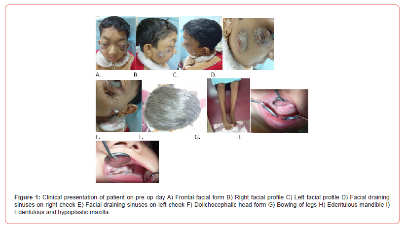
When patient was examined, on extraoral examination she had multiple draining sinuses involving midface, dolichocephalic head form, hypertelorism, frontal bossing, exophthalmus eyes bilaterally, bilateral blindness since birth, hearing intact, nasal discharge positive. On intraoral examination, patient had purulent discharge from left upper buccal vestibule, hypoplastic maxilla, no palatal erosion of bone, halitosis and failure of eruption of teeth (Figure 1).
Patient was admitted and routine examinations were done with antibiotic coverage including iv augmentin and iv flagyl started off initially. The complete blood count showed hemoglobin of patient as 8.7 g/dl for which blood transfusion was done. In baseline investigations, anti-HCV came out to be positive for which PCR for hepatitis was advised for confirmation. Pus c/s was done which was suggestive of growth of Proteus mirabilis.
Ultrasound abdomen was advised. In ultrasound abdomen, liver was enlarged measuring 17 cm in size. It showed normal homogeneous echotexture and parenchymal echogenicity. No focal lesion was seen. No intrahepatic cholestasis was noted. Spleen was normal in size and texture. No focal lesion was seen. Pancreas was normal in morphological and sonographic attributes recognized. No focal lesion was identified. Gall bladder was contracted. Status post meal, CBD was of normal caliber in extra-hepatic course. Portal Vein was of normal caliber at porta hepatic. Kidneys were normal position, shape and dimensions / cortical thickness with smooth contours observed. Parenchymal echopattern was normal with clear cortico-medullary differentiation. No cyst or mass was spotted. No stone or dilatation of the collecting system identified. Right kidney had bipolar length of 6.65 cm. Left kidney had bipolar length of 5.8 cm. No evidence of ascites or lymphadenopathy was recognized. Impression was of hepatomegaly.
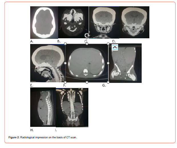
In radiological examination [Figure 2], CT scan head and neck, pelvis and abdomen was advised. The CT scan of head and neck region reported as bones of the skull, spine, the axial skeleton in visualized part of appendicular skeleton appeared diffusely thickened and hyperdense, suggestive of osteopetrosis. The intracerebral spaces were dilated, more so in the bilateral frontal horn region with the right frontal horn measuring 20.8 mm in left frontal horn measuring 22.5 mm. Prominence of the sulcal spaces were noted, predominantly around the frontal lobe region suggestive of brain atrophy along with effacement of rest of the sulci and gyri were noted. Suspicion of a rounded track in the midline, just below the hard palate could be diffusely thickened soft tissue or a discharging sinus. Significant narrowing of the optic canal was noted along with diffuse thinning of bilateral optic nerves. Bilateral mastoid air cells were sclerosed. Bilteral middle ear cavities were fairly well preserved. Flattening of clivus (platybasia)was noted with base of skull. Basilar invagination along with narrowing of the foramen magnum was noted.
In CT scan abdomen and pelvis, diffusely thickened and hyperdense sternum, spine, pelvic girdle and bilateral femur, were suggestive of osteopetrosis. There was no evidence of spinal canal stenosis. Liver was enlarged measuring 16.4 cm in CC dimension. Spleen was grossly enlarged measuring 16.8 × 10.3 × 10.8 cm (CC × AP × TR) with a splenic index of 900 and was reaching up to the left iliac fossa and causing mass-effect upon the left kidney and pancreas tail. The lungs were normal in volume. The heart and the great vessels were normal with no evidence of any soft and calcified plaque. No abnormal lymphadenopathy was seen in the root of the neck, mediastinum and axilla. No evidence pleural effusion was seen. The diaphragm was normal in contour with no evidence of hiatus hernia. The oesophagus, stomach, small and large gut loops were normal. Aorta and IVC were normal. No evidence of any lymphadenopathy was seen. Diffusely thickened and hyperdense bones, were suggestive of osteopetrosis. Significant hepatosplenomegaly; sequelae of osteopetrosis.
During the patient’s inpatient course, she underwent seizure like attack and severe headache explained by parents for which worthy Paediatrics Department of KRL Hospital was consulted, they advised ECG and urine R/E to rule out any cardiac event or meningitis as WBC count was raised to 24 denoting increased infection. Investigations came out to be normal. Patient’s antibiotic was switched from IV Augmentin to IV Meronem.
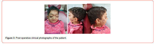
Patient was discharged with improved condition with regard to the presenting complaint of multiple draining facial sinuses (Figure 3) and referred to AFIP for consultation regarding bone marrow transplant, and recalled by paeds department KRL Hospital Islamabad for further workup for hepatitis C treatment.
Discussion
Infantile malignant osteopetrosis involves genetic disorders concerned with defective osteoclastic activity. Clinical presentation in patients with osteopetrosis involves transverse fractures of bone, myelophthistic anemia, extramedullary hematopoiesis with hepatosplenomegaly which in later stages may lead to acute leukemia, cranial nerve compression, diplopia, ends of long bones become bulbous with metaphyseal flare (Erlenmeyer flask deformity) and weakening of facial muscles [5- 7].
The radiographic appearance for osteopetrosis generally includes ‘bone in bone’ or ‘endobone’ appearance in bones, ‘sandwich’ for phalanges and ‘Rugger jersey spine’appearance mainly for vertebrae [8]. The histopathology of osteopetrosis involves classic lack of clear zone and ruffled borders in defective osteoclasts due to empty lacunae and calcification of cartilage that is spread within trabeculae. Apart from the clinical presentation, radiographic and histological features with genetic testing, evaluation of osteopetrosis can be done with laboratory tests showing increased levels of creatinine kinase BB and tartrateresistant acid phosphatase [9, 10].
On the basis of age, severity and patterns of genetic inheritance types of osteopetrosis involve autosomal recessive osteopetrosis also termed as malignant infantile osteopetrosis due to mutations in TCIRG1, CLCN7, OSTM1 and RANKL/RANK presenting in infancy causing osteoclast disruption cranial nerve compression and high mortality. The second type is intermediate autosomal recessive osteopetrosis due to mutations in CAII and PLEKHM1 presenting in childhood. The third variant is autosomal dominant osteopetrosis (Albers-Schonberg disease) being mildest form and the most common as well, is due to mutation in CLCN7 gene, having higher incidence in adolescents or adults involves increased fractures, cranial nerve involvement with mild anemia. The fourth variant is osteoclast poor osteopetrosis that is highly rare variant concerned with complete absence of osteoclasts due to RANKL/RANK mutations. Fifth category is of X-linked or SLC4A2deficiency [11, 12]. Autosomal dominant trait is more common than the autosomal recessive form I.e 1;20,000 and 1:250,000 births respectively [13, 14].
Medical management involves bone marrow transplant, interferon gamma therapy, neurological decompression for cranial nerve impingement, in cases of long bone fractures prolonged casting can be performed using plates and screws avoiding intramedullary devices, for degenerative joint disease total hip replacement can be done for end stage osteoarthritis [15, 16].
Acknowledgement
None.
Conflict of Interest
No Conflict of Interest.
References
- Kalenahalli Jagadish Kumar et al., (2013) Malignant infantile osteopetrosis. Indian J Hum Genet 19(1): 90-92.
- Zornitza Stark et al., (2009) Osteopetrosis. Orphanet J Rare Dis 20: 4: 5.
- Reddy Mh R (2011) Osteopetrosis (Marble Bone Disease): A Rare Disease in Children. Int J Clin Pediatr Dent 4(3): 232-234.
- Loría Cortés R, Quesada Calvo E, Cordero Chaverri C (1977) Osteopetrosis in children: a report of 26 cases. J Pediatr 91(1): 43-47.
- Tamer Ahmed EL-Sobky et al., (2016) A case of infantile osteopetrosis: The radioclinical features with literature update. Bone Rep 4: 11-16.
- G. Steward et al., (2003) Neurological aspects of osteopetrosis. Neuropathol Appl Neurobiol 29 (2): 87-97.
- K. Roopashri, R Gopakumar, B G (2008) Subhas Osteomyelitis in infantile osteopetrosis: a case report with review of literature. J Ind. Soc Pedodontics Prev Dent 26 (Supplement).
- Aya Laridi MD, et al. (2025) Radiological diagnosis of infantile osteopetrosis in a 1-year-old with macrocephaly and jaundice. Radiology Case Reports 20(9): 4226-4230.
- F Avijgan, F Abbasian, M Dehnavi, Z Sarchahi (2024) Malignant osteopetrosis of infancy: a case report. Int J Surg Case Rep 123: 110206.
- Sharma, S.N. Ingole, M.D. Deshpande, N. Kazi, D. Meshram, et al. (2020) A rare case of osteoclast-poor osteopetrosis (RANKL mutation) with recurrent osteomyelitis of mandible: a case report. Int J Clin Pediatr Dent 13 (6): 717
- B.G. Khorashadizadeh, Z. Soltani (2011) A case report of malignant infantile osteopetrosis. J Babol Univ Med Sci 13(5): 78-82.
- K. Bubshait, Z.E. Himdy, O. Fadaaq, H.I. Alshmas, D. Bubshait (2020) Malignant infantile osteopetrosis: a case report Cureus 12 (1): e6725.
- Thabet, M. Almajeedi, M. Mohammed, F. Ahmed (2022) Malignant infantile osteopetrosis in a 3-year-old Yemeni child: a case report.
- Lertwilaiwittaya, B. Suktitipat, P. Khongthon, W. Pongsapich, C. Limwongse, et al. (2021) Identification of novel mutation in RANKL by whole-exome sequencing in a Thai family with osteopetrosis; a case report and review of RANKL osteopetrosis. Mol Genet Genomic Med 9 (7): e1727.
- Gao, Q. Cheng, X. Zhang, G. Zhao (2020) Successful total hip arthroplasty for autosomal dominant osteopetrosis complicated by hip osteoarthritis: a case report and review of the literature. Exp Ther Med 19 (4): 2702-2706.
- Saima Gillani et al., (2017) Malignant Infantile Osteopetrosis. J Ayub Med Coll Abbottabad 29(2).
-

This work is licensed under a Creative Commons Attribution-NonCommercial 4.0 International License.




