 Case Report
Single Tooth Dento-Osseous Osteotomy for Malocclusion with The Ankylosed Tooth: A Case Report
Natsuko Hichijo, DDS, PhD1*, Yoshiyuki Mori, DDS, PhD2, Yuri Ofusa DDS3 and Tadahide Noguchi, DDS, PhD4
1Assistant Professor, Department of Dentistry, Oral and Maxillofacial Surgery, Jichi Medical University, Tochigi, Japan
2Professor, Department of Dentistry, Oral and Maxillofacial Surgery, Jichi Medical University Saitama Medical Center, Saitama, Japan
3Senior Resident, Department of Dentistry, Oral and Maxillofacial Surgery, Jichi Medical University, Tochigi, Japan
4Professor, Department of Dentistry, Oral and Maxillofacial Surgery, Jichi Medical University, Tochigi, Japan
Natsuko Hichijo, Department of Dentistry, Oral and Maxillofacial Surgery, Jichi Medical University, 3311-1 Yakushiji, Shimotsuke-shi, Tochigi-ken 329-0498, Japan.
Received Date: November 16, 2023; Published Date: November 27, 2023
Introduction
In orthodontic treatment, the ankylosed tooth will not move at all. Some reports have showed that orthodontists should pay attention to the permanent tooth that was traumatized during childhood [1-3]. However, it is very difficult for us to judge accurately whether the tooth is ankylosed [4, 5]. Shafer, et al. suggested that the diagnosis of ankylosis is based on the lack of tooth movement and a dull sound by percussion [6]. In addition, if the ankylosis infects the proximal root areas, we would be able to confirm it by periapical radiographs [6]. Though, these symptoms are not clear if the ankylosed area is small or located on the root surface [7]. Basically, we don’t have any other choice but to apply orthodontic forces to the affected tooth for the definitive diagnosis [4]. Therefore, we have to explain to the patient or parents about the possibilities that the tooth wouldn’t move and the alternatives before treatment
We encountered a patient who had been receiving orthodontic treatment at another hospital for over 5 years, but only her maxillary right central incisor did not move. Therefore, the severe overjet by the tooth remained and she hadn’t been able to finish her orthodontic treatment yet.
Here, we report the effective surgical intervention of a patient with malocclusion involved in the ankylosed tooth by single tooth dento-osseous osteotomy (STDO).
Case Report
A female patient, 23 years and 7 months of age, presented with a chief complaint that her maxillary right central incisor did not move. She hoped to correct the severe overjet (Figure 1). She had been treated for her malocclusion at another hospital for over five years when visiting our hospital, and her orthodontic treatment was almost finished, except for her maxillary right central incisor. Her referral letter stated that the tooth couldn’t be repositioned even with temporary anchorage devices, and it was likely to be caused by dental ankylosis. The tooth was damaged by trauma when in junior high school. Her orthodontist had no means of treatment and referred her to our hospital. He concluded that her maxillary right central incisor should require surgical intervention for alignment.
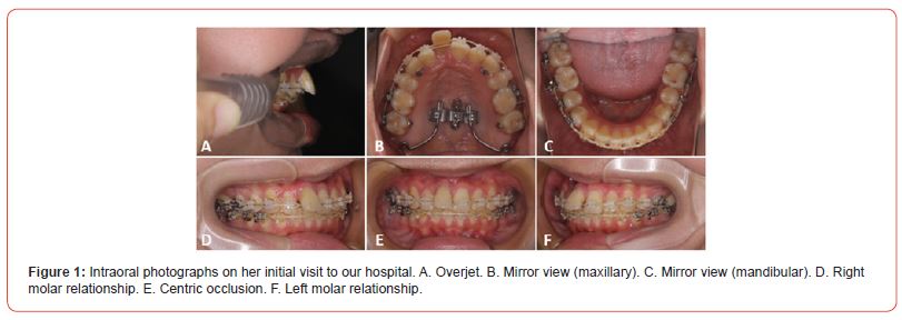
The tooth was highly labially inclined and appeared cloudy. It was a non-vital tooth without physiological perturbation and made a similar percussion sound close enough to another teeth. A dental radiographic image and computed tomography showed root resorption and undertreatment of dental pulp for her maxillary right central incisor (Figure 2). We also determined that dental ankylosis was the reason why alignment of her maxillary right central incisor couldn’t be achieved. The treatment objective was to correct the highly labially inclined tooth and establish the good upper arch form.
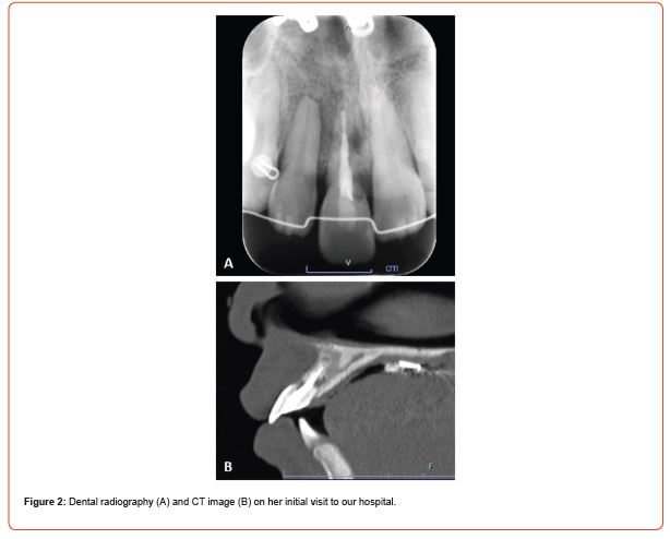
We conceived two alternatives for her treatment. The first alternative was surgical luxation to reposition the ankylosed tooth to the ideal position or angle. The second alternative was STDO that osteotomized bone block including her ankylosis could be rearranged properly. However, we hadn’t to despair of the first alternative in this case. Surgical luxation was to attempt to break the fusion between the cementum and bone [8]
The root of her ankylosis had already been resorbed partially and in a bad condition. If this alternative was chosen, there would be a strong possibility of the root fracture or that her tooth might have reankylosed during repair [8-10]. Therefore, we suggested STDO to her.
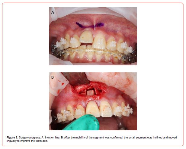
Initially, the upper arch wire was pulled out before surgery. STDO was performed with the same procedure as before 2, according to the protocol by Epker and Paulus [11]. A mucoperiosteal flap was reflected to expose the anterior maxilla from the left central incisor to the right lateral incisor (Figure 3A). Two separate subperiosteal tunnels were created inferiorly on both sides of the ankylosed tooth. Vertical osteotomies were performed from the alveolar crest 5mm above the root apex in a tapered form, and then horizontal osteotomy was performed by un ultrasonic bone-cutting device (Piezosurgery; Mectron Medical Technology, Carasco, Italy). After the mobility of the segment was confirmed, the small segment was inclined and moved lingually to improve the tooth axis (Figure 3B). Both oral surgeons and an orthodontist finally checked the new position of the segment and the corrected upper arch form, and the soft tissue incision was closed with surgical sutures. A new continuous 0.016×0.016 stainless steel archwire was placed in the brackets for stabilization in position.
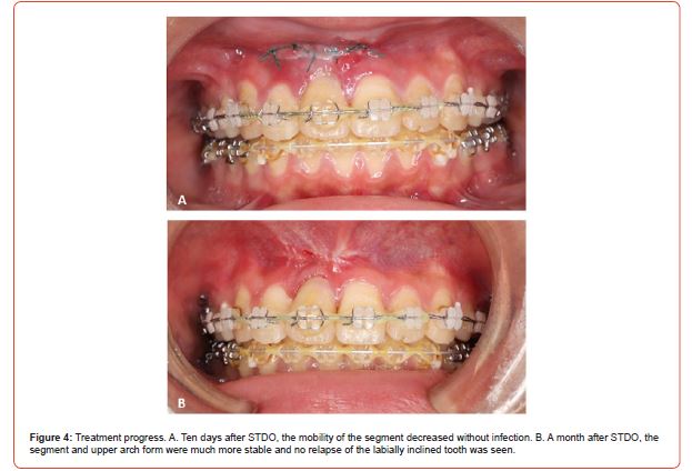
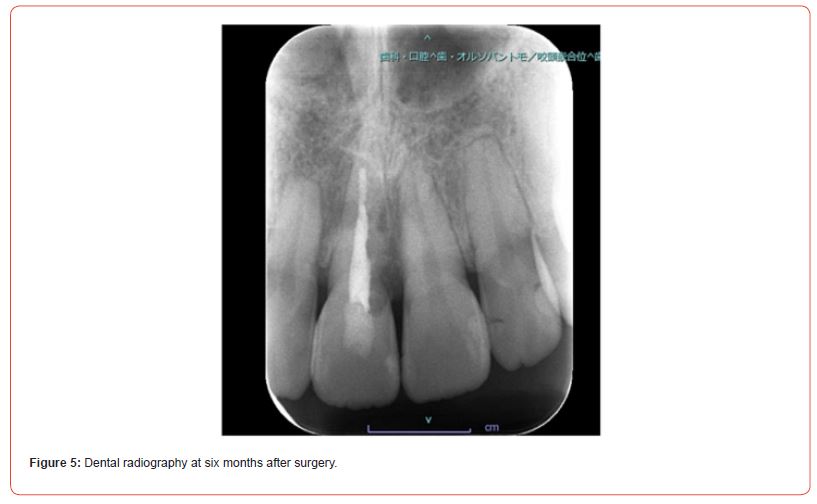
After 10 days of STDO, the mobility of the segment decreased without infection (Figure 4A). A month after STDO, the segment and upper arch form were much more stable and no relapse of the labially inclined tooth was seen (Figure 4B). Bone healing surrounding fracture lines was confirmed in the radiograph after 6 months of surgery, and a functional occlusion was well maintained (Figures 5, 6). Therefore, her edgewise appliances were removed in her original dental clinic.
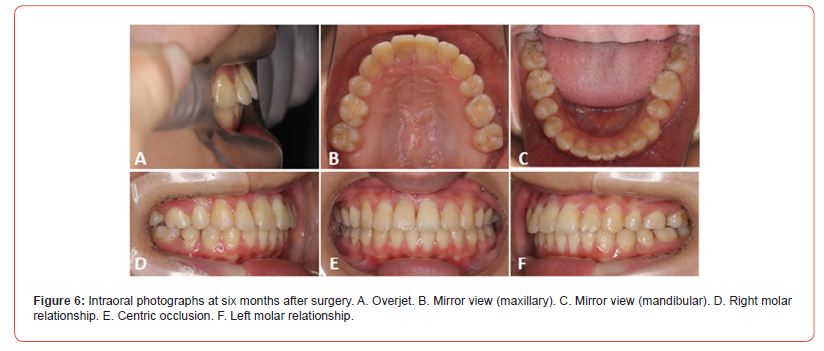
Posttreatment intraoral photos showed an excellent change, as compared with pretreatment ones. The severe overjet had disappeared, and her upper arch form was corrected (Figure 6). She was very satisfied with this treatment outcome.
Discussion
In this study, we reported the effective surgical intervention of a patient with the ankylosed tooth. Moreover, STDO contributed to provided sufficient improvement of the severe overjet by the ankylosed central incisor and shortening of the treatment duration.
STDO is surgical repositioning of the ankylosed tooth with surrounding alveolar bone under local anesthesia, without the need for hospitalization [11]. It is considered to be an effective approach to malposition of the ankylosed tooth because of the advantages of shortening of the treatment duration, the ability to move segments in any direction and stability [2, 11-13]. However, the single-tooth block segment has a limitation in the amount of movement due to the resistance of the attached gingiva and impairment of blood supply [2, 11-15]. Significant gingival recession is also reported [1, 2, 7]. Therefore, this approach needs technical proficiency.
Before her treatment, we explained sufficiently these risk to her and her parents. She had to be prepared to face the serious situation that the affected tooth would be extracted and she would need dental prosthesis treatment if the postoperative course was not good. However, they strongly hoped and started STDO. The oral surgeon who specialized in osteotomy for jaw deformity and has performed STDO previously was selected and performed for her [2, 16-18]. Because the occlusion was stable and the good upper arch form was retained for 6 months, her orthodontic appliances were removed.
This patient was not explained about the possibility of dental ankylosis of her tooth before initiation of orthodontic treatment. Thus, she never assumed that the longer-term treatment and the surgical procedure were required. As a result, she looked exhausted on her initial visit to our hospital. We hope that this case report will warn that treatment must be provided based on proper exams, diagnosis and informed consent so that patients can receive satisfactory treatment without anxiety. Moreover, we would like to highlight that this approach is very effective for improvement of malposition of the ankylosed tooth.
Acknowledgement
None.
Conflicts of Interest
No conflict of interest.
References
- Senışık NE, Koçer G, Kaya BÜ (2014) Ankylosed maxillary incisor with severe root resorption treated with a single-tooth dento-osseous osteotomy, vertical alveolar distraction osteogenesis, and mini-implant anchorage. Am J Orthod Dentofacial Orthop 146(3): 371-384.
- Ohkubo K, Susami T, Mori Y, Nagahama K, Takahashi N, et al. (2011) Treatment of ankylosed maxillary central incisors by single-tooth dento-osseous osteotomy and alveolar bone distraction. Oral Surg Oral Med Oral Pathol Oral Radiol Endod 111(5): 561-567.
- McNamara TG, O’Shea D, McNamara CM, Foley TF (2000) The management of traumatic ankylosis during orthodontics: a case report. J Clin Pediatr Dent 24(4): 265-267.
- Alessandri Bonetti G, Incerti Parenti S, Daprile G, Montevecchi M (2011) Failure after closed traction of an unerupted maxillary permanent canine: Diagnosis and treatment planning. Am J Orthod Dentofacial Orthop 140(1): 121-125.
- Andersson L, Blomlof L, Lindskog S, Feiglin B, Hammarstrom L (1984) Tooth ankylosis: clinical, radiographic and histological assessments. Int J Oral Surg 13(5): 423-431.
- Shafer WG, Hiue MK, Levy BM (1983) A textbook of oral pathology. (4th), Philadelphia: WB Saunders Pp. 540-541.
- Isaacson RJ, Strauss RA, Bridges Poquis A, Peluso AR, Lindauer SJ (2001) Moving an ankylosed central incisor using orthodontics, surgery and distraction osteogenesis. Angle Orthod 71(5): 411-418.
- Takahashi T, Takagi T, Moriyama K (2005) Orthodontic treatment of a traumatically intruded tooth with ankylosis by traction after surgical luxation. Am J Orthod Dentofacial Orthop 127(2): 233-241.
- Pithon MM (2016) Surgical Luxation and Orthodontic Traction of Ankylosed Upper First Molar. J Clin Orthod 50(5): 299-306.
- Paleczny G (1991) Treatment of the ankylosed mandibular permanent first molar: a case study. J Can Dent Assoc 57(9): 717-719.
- Epker BN, Paulus PJ (1978) Surgical--orthodontic correction of adult malocclusions: single-tooth dento-osseous osteotomies. Am J Orthod 74(5): 551-563.
- Bell WH (1973) Immediate surgical repositioning of one- and two-tooth dento-osseous segments. Int J Oral Surg 2(6): 265-272.
- Kawamura S, Kawamura H, Nagasaka H, Sato S, Motegi K, Ohmori Y, et al. (1992) Ankylosed Tooth Treated with Single-tooth Dento-osseous Osteotomy. Jpn J Jaw Deform 2(2): 132-138.
- Medeiros PJ, Bezerra AR (1997) Treatment of an ankylosed central incisor by single-tooth dento-osseous osteotomy. Am J Orthod Dentofacial Orthop 112(5): 496-501.
- Bell WH and McBride K (1976) Immediate surgical repositioning of anterior and posterior maxillary dento-osseous segments. J Oral Surg 34(10): 943-947.
- Mori Y, Susami T, Chikazu D, Saijo H, Sakiyama M, et al. (2009) Unilateral expansion of a narrow mandibular dental arch combined with bimaxillary osteotomies in a patient with hypoglossia. Int J Oral Maxillofac Surg 38(6): 689-693.
- Kawase Koga Y, Mori Y, Fujii Y, Kanno Y, Chikazu D, et al. (2016) Complications after intraoral vertical ramus osteotomy: relationship to the shape of the osteotomy line. Int J Oral Maxillofac Surg 45(2): 200-204.
- Susami T, Mori Y, Ohkubo K, Takahashi M, Hirano Y, et al. (2018) Changes in maxillofacial morphology and velopharyngeal function with two-stage maxillary distraction-mandibular setback surgery in patients with cleft lip and palate. Int J Oral Maxillofac Surg 47(3): 357-365.
-
Natsuko Hichijo, DDS, PhD*, Yoshiyuki Mori, DDS, PhD, Yuri Ofusa DDS and Tadahide Noguchi, DDS, PhD. Single Tooth Dento-Osseous Osteotomy for Malocclusion with The Ankylosed Tooth: A Case Reporta. On J Dent & Oral Health. 7(3): 2023. OJDOH. MS.ID.000664.
-
Orthodontic treatment, Tooth, Single tooth dento osseous osteotomy, Orthodontist, Oral surgeons, Dental ankylosis, Ankylosed tooth, Orthodontic appliances, Osteotomy for jaw, Dental clinic
-

This work is licensed under a Creative Commons Attribution-NonCommercial 4.0 International License.


