 Case Report
Prosthodontic Rehabilitation of Missing Molar using Friction Fit Zirconia Crown Over Dual One Piece Implants-Clinical Report and I Year Follow Up
Hariharan Ramakrishnan*
Professor, Department of Prosthodontics & Implantology, Ragas Dental College &Hospital, Chennai, India
ORCID: 0000-0003-4466-5744
Hariharan Ramakrishnan, Professor, Department of Prosthodontics & Implantology, Ragas Dental College &Hospital, Uthandi, Chennai, India. Email: abcv2005@yahoo.com
Received Date: January 14, 2021; Published Date: February 02, 2021
Abstract
The present patient report describes a missing mandibular molar rehabilitated by two one piece narrow diameter dental implants. Although the gold standard treatment is to place a regular diameter implant, unfortunately due to horizontal bone resorption, this option is not possible without lateral bone augmentation. In this situation narrow diameter implants are a viable alternative. immediate provisional crown is provided after implant placement and three months latter, replaced with partially transparent monolithic friction frit cement free zirconia crown. Follow up til the end of one year revealed healthy hard and soft tissues around implants.
Keywords: Alveolar bone loss; Dental implants; Immediate dental implant loading; Single tooth dental implants; Friction; Alveolar ridge augmentation
Introduction
Implantation is a preferred choice to replace a missing single tooth, avoiding adjacent vital teeth preparation and fixed prosthesis fabrication [1]. The most frequent mandibular molar to be replaced is the first mandibular molar. Implantation in the posterior area is a predictable procedure over time [2]. The low rate of complications in addition to the high long term success rate makes implant restoration a reliable solution to treat posterior partial edentulism [3]. The use of two implants to replace a single molar seems a logical treatment solution to avoid prosthetic complications associated with two part implants. yet, one significant disadvantage to the use of this concept is the limitation of the size of implants and their associated prosthetic components [4].
There are different definitions for the narrow diameter implant (NDI), starting from small body implant, implant with a reduced endosseous diameter and narrow body implant to reduced diameter implant. The diameter is always less than or equal to 3.5mm. Originally its use was reserved for the replacement of teeth with narrow clinical crowns and for limited interdental and interimplant spaces such as in the upper lateral or lower incisor areas [5]. However NDI finds another indication for its use, namely, with thin ridges. Indeed, following tooth loss bone collapses in a three dimensional pattern. The horizontal deficiency or width loss develops in a larger extent [6].
Here, the clinician has two options, either to perform horizontal ridge reconstruction procedures or to place an NDI in case of moderate horizontal bone loss [7]. In order to avoid invasive ridge management techniques, in cases of limited ridge width is the use of NDIs, therefore broadening their indications [8]. Most of the studies that evaluate the survival/success rate of NDIs in the posterior jaws, demonstrated an equivalent success rate to standard diameter implants [9]. The systematic review of Assaf, et al. [9] demonstrated that implant therapy using NDIs in the posterior jaw is a reliable modality provided that the clinician follows certain guidelines. These were as follows:
1. Bone thickness between 5 and 7mm.
2. Vertical bone length of 12mm above the inferior alveolar canal or 10 mm below the sinus allowing a placement of at least 10mm height NDI.
3. NDI position in bounded in molar region or free end saddle.
4. Bone quality type 1,2 or 3 according to the classification of Lekhhom and Zarb.
5. NDI with appropriate macro and microgeometry that is an external design allowing an acceptable initial stability and optimal surface preparation to enhance bone implant contact.
This paper describes the situation in which NDIs from Dentium implant system are used to replace missing 36 (Left mandibular first molar), as an alternative viable option in the treatment of moderately resorbed posterior ridges where lateral bone augmentation is not accepted by patient.
Clinical Report
The given table shows different parameters of treatment design (Table 1).
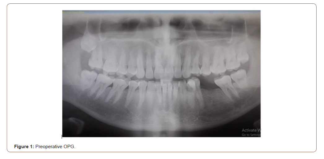
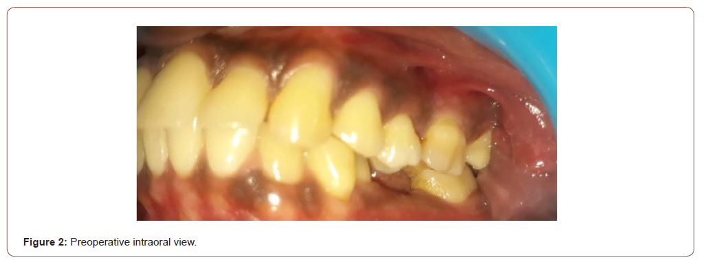
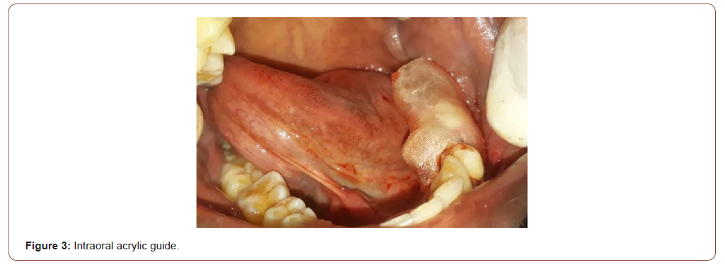
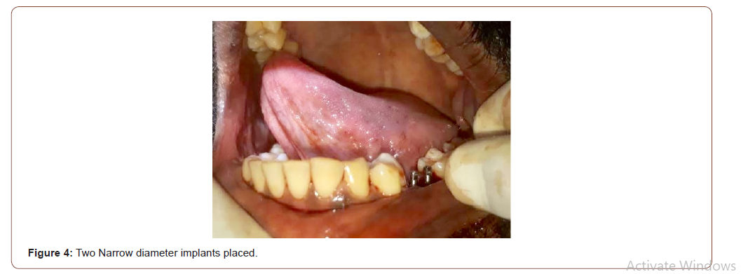
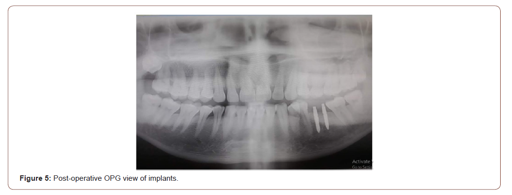
Thirty-seven-year-old male patient visited our department of prosthodontics and implantology for replacement of missing posterior tooth. During clinical and radiographic examination (Figure1), it is evident that the available bone height is 12mm and available width is 4.5mm only. There is no significant medical history. Patient is clearly explained of existing bone conditions and treatment options given including bone augmentation. Patient opted for implant therapy without bone augmentation. Patient consent for treatment is obtained. Patient is treated under strict sterile conditions. Pre surgical oral rinse with Chlorhexidine mouthwash undiluted (Rexidine plus, Septodont, India), followed by Povidone pre surgical clean up (Povidone iodine, India) of surgical area done. 2ml of local Anaesthesia (lignocaine with epinephrine, 1:100,000, Lignox, India) is used. Full thickness flap design and usual surgical protocols are followed. Two NDI are placed (Dentium, Slimline, South Korea,) according to manufacturer’s drilling recommendations and protocol. Hemostasis achieved immediately after surgery, and postoperative instructions are given to the patient. Primary stability is good for both implants (Figures 2-5).
Table 1:

Following surgery, antibiotic Moxclav 625mg tds for 3 days, Dolo 65Omg od (only to be taken if pain is of persisting nature) is prescribed. Immediate provisional implant crown using cold curing resin (DPI, cold cure, India) is provided. No provisional cement is used. Fit is good and clinically acceptable (Figure 6). Final prosthodontic rehabilitation is carried out 3 months after implant placement. Osseointegration is confirmed with X ray and clinical inspection carried out. Final impressions are made using one stage extra heavy and light body addition silicone impression material (Provil, Putty, light body, USA). Partially translucent monolithic zirconia crown is placed on the dual implants without use of any definitive cements. residual cements if any, left in the crevicular gingiva can lead to periimplantitis in future. in this patient since one piece implants are inserted, it is better to avoid definitive cements for insertion of permanent crown. friction fit of the permanent crown is said to be another reason for not chosing any kind of permanent cement. This fit is obtained in the laboratory by avoiding usage of any kind of spacers over implants on the master casts (Figures 7,8).
Follow up visits
Patient recalled for clinical examination after one month and then at end of one year. A panoramic x-ray taken (Figure 9). Implant success assessed according to the criteria defined by Busser, et al. [10] Implants are considered successful if the following criteria were met:
1. The absence of any peri-implant infections with suppuration.
2. The absence of persistent subjective complaints of pain.
3. The absence of continuous radiolucency around the implant.
4. The absence of any detectable implant mobility.
Discussion
The use of dental implants for single posterior tooth replacement has become a predictable treatment modality [11]. Single regular diameter implants might be incapable of predictably withstanding molar masticatory function and occlusal loading forces [12]. Wide diameter implants are not always a treatment option for replacing a single molar, especially when the buccolingual dimension is deficient. The use of two implants might also provide better prosthetic stability and prevent rotational forces on the prosthetic components [13]. The patient had no signs and symptoms for parafunction. The NDIs are restored with single zirconia crown. Strict occlusal considerations are applied such that slight contact in centric occlusion and no contact in lateral movement [9]. one significant barrier to the widespread use of this concept is the limitation of the size of implants and their associated prosthetic components. Nevetheless, when using narrow diameter implants 2 implants could be used even when the distance between adjacent teeth are limited [14].


The replacement of a single molar with 1 implant has been shown to be an effective treatment modality in short term studies [15,16]. However, this presents a biomechanical challenge. Occlusal forces are greatest in the molar region, leading to possible increased stress on the implant components as well as the surrounding bone [17]. logical solution to implant overload is the use of 2 implants to replace the roots of a missing molar [11]. The use of two implants provides more surface area for osseointegration and spreads the occlusal loading forces over a wider area while reducing a potential bending force that would exist in a single molar restoration [18- 20]. This clinical report provides an evidence for the usefulness of two narrow diameter dental implants serving as a viable treatment option providing good and predictable results. This patient is still under recall protocol and will be examined again at end of one year and subsequent visits.
Conclusion
In case of moderate horizontal bone resorption, NDI is a reliable option to replace a molar. Replacing a single missing molar with two narrow diameter implants will serve as a viable treatment option providing good and predictable long term results. Further observational and randomized controlled studies could provide deeper evidence based conclusions concerning the use of NDI in the posterior jaw.
Acknowledgement
None.
Conflict of Interest
The authors declare no conflict of interest.
References
- Schwartz-Arad D, Samet N, N Samet (1999) Single tooth replacement of missing molars: A retrospective study of 78 implants. J Periodontol 70: 449-454.
- McCaul LK, Jenkis WM, Kay EJ (2001) The reasons for the extraction of various tooth types in Scotland: A 15-year follow up. J Dent 29: 401-407.
- Muftu A Chapman RJ (1998) Replacing posterior with freestanding implants: Four-year prosthodontic results of a prospective study. J Am Dent Assoc 129: 1097-1102.
- Balshi TJ, Hernandez RE, Pryzlak MC (1996) A comparative study of one implant versus two replacing a single molar. Int J Oral Maxillofac Implants 11: 372-378.
- P Malo, M de Araujo Nobre (2011) Implants (3.3 mm diameter) for the rehabilitation of edentulous posterior regions: A retrospective clinical study with up to 11 years of follow-up. Clin Implant Dent Relat Res 13(2): 95-103.
- S Barter, P Stone, U Bragger (2012) A pilot study to evaluate the success and survival rate of titanium-zirconium implants in partially edentulous patients: results after 24 months of follow-up. Clin Oral Imp Res 23(7): 873-221.
- MC Cehreli, K Akca (2004) Narrow-diameter implants as terminal support for occlusal three unit FPDs: a biomechanical analysis. Int J Perio and Rest Dent 24(6): 513-519.
- V Arisan, N Bolukbas, S Ersanl, T Ozdemir (2010) Evaluation of 316 narrow diameter implants followed for 5-10 years: A clinical and radiographic retrospective study. Clin Oral Imp Res 21(3): 296-307.
- A Assaf, M Saad, M Daas, J Abdullah, R Abdallah (2015) Use of narrow-diameter implants in the posterior jaw: a systematic review. Imp Dent 24(3): 294-306.
- D Busser, J Wittneben, MM Bornstein, L Grutter, V Chapuis, UC Belser (2011) Stability of contour augmentation and esthetic outcomes of implant supported single crowns in the esthetic zone: 3-year result of a prospective study with early implant placement post extraction. J Periodontol 82(3): 342-349.
- Levin L, Laviv A, Schwatz-Arad D (2006) Long-term success of implants replacing a single molar. J Periodontol 77: 1528-1532.
- Balshi TJ, Wolfinger GJ (1997) Two implant supported single molar replacement: Interdental space requirements and comparison to alternative options. Int J Periodontics Restorative Dent 17: 426-435.
- Bakeen LG, Winkler S, Neff PA (2001) The effect of implant diameter, restoration design, andocclusal table variations on screw loosening of posterior single-tooth implant restorations. J Oral Implantol 27: 63-72.
- Ziv Mazor, Adi Lorean, Eitan Mijiritsky, Liran Levin (2012) Replacement of a molar with 2 narrow diameter dental implants. Implant Dent 21: 36-38.
- Schmitt A, Zarb GA (1993) The longitudinal clinical effectiveness of osseointegrated dental implants for single tooth replacement. Int J Prosthodont 6(7): 197-202.
- Laney WR, Jemt T, Harris D, Henry PJ, Krogh PH, et al. (1994) Osseointegrated implants for single-tooth replacement: progress report from a multicenter prospective study after 3 years. Int J Oral Maxillofac Implants 9(1): 49-54.
- Haraldson T, Carlsson GE, Ingervall B (1979) Functional state, Bite force and postural activity in patients with osseointegtrated oral implant bridges. Acta Odontol Scand 37(4): 195-206.
- Becker W, Becker BE (1995) Replacement of maxillary and mandibular molars with single endosseous implant restorations: a retrospective study. J Prosthet Dent 74(1): 51-55.
- Rangert B, Krogh PH, Langer B, Van Roeke lN (1995) Bending overload and implant fracture: a retrospective clinical analysis. Int J Oral Maxillofac Implants 10(3): 326-334.
- Bahat O, Handelsman M (1996) Use of wide implants and double implants in the posterior jaw: na clinical report. Int J Oral Maxillofac Implants 11(3): 379-386.
-
Hariharan Ramakrishnan. Prosthodontic Rehabilitation of Missing Molar using Friction Fit Zirconia Crown Over Dual One
Piece Implants-Clinical Report and I Year Follow Up. On J Dent & Oral Health. 4(1): 2021.
DOI: 10.33552/OJDOH.2021.04.000580.
-
Dental implants, Zirconia crown, Posterior area, Local Anaesthesia, Pain, Prosthetic components, Molar restoration, Crevicular gingiva, Teeth.
-

This work is licensed under a Creative Commons Attribution-NonCommercial 4.0 International License.


