 Research Article
Mineral Element Analysis of Rat Molar Enamel after Consumption of Sugary Drinks: An Energy Dispersive X-Ray Study
Asma Al-Jobair1* and Rita Khounganian2
1*Pediatric Dentistry and Orthodontics, College of Dentistry, King Saud University, Saudi Arabia
2Oral Medicine and Diagnostic Sciences, College of Dentistry, King Saud University, Saudi Arabia
Asma Al-Jobair, Pediatric Dentistry and Orthodontics, College of Dentistry, King Saud University, Saudi Arabia.
Received Date: May 05, 2025; Published Date: May 20, 2025
Abstract
Objectives: To investigate the effect of consumption of different sugary drinks on the surface configuration and mineral element content of rat molar enamel using scanning electron microscopy and energy dispersive X-ray (EDX) microanalysis system.
Methods: First mandibular molar teeth from 100 adult Sprague-Dawley rats were analyzed using scanning electron microscopy and energy dispersive X-ray spectroscopy (SEM-EDX). One hundred Sprague-Dawley rats were offered one of the following drinks ad libitum: chocolate milk, strawberry milk, banana milk, cola carbonated drink, citrus carbonated drink, orange carbonated drink, apple fruit drink, orange fruit drink, mango drink and distilled water. By the end of the experiment, rats were scarified, and the right side of the mandibles were removed and kept in 10% neutral buffered formalin solution for further analysis. The surface conformation of the teeth was studied and the mineral element contents (Na, Mg, P, Ca, and Ca/P) of the enamel for all the mandibular right first molars were analyzed using energy-dispersive X-ray microanalysis (EDX). The collected data were statistically analyzed using ANOVA and Tukey test.
Results: Among the experimental groups, enamel of the rats that consumed flavored milk contained more Ca and less P than the rats consumed carbonated drinks and fruit drinks with statistically significant differences. Within the experimental subgroups, enamel of the rats that consumed strawberry milk contained more Ca than the rats consumed all different carbonated drinks and fruit drinks with significant differences. However, P was found to be lower in the enamel of the rats consuming strawberry milk with significant difference to rats consuming orange carbonated and mango fruit drinks subgroups.
Conclusion: The enamel surface topography was affected following the consumption of different sugary drinks and altered the mineral element content of the teeth enamel.
Keywords: SEM, EDX, Carbonated drinks, Fruit drinks, Flavored milk, Experimental rats
Introduction
Nutrients are chemical substances required by the body to sustain basic functions and are optimally obtained by eating a balanced diet. Minerals are one of the six major classes of nutrients essential for human health. Minerals are inorganic micronutrients and could be classified as macrominerals or microminerals. Macrominerals including calcium, phosphorous, magnesium, sodium, potassium, and chloride are required in amounts greater than 100mg per day and play an essential role in human health [1]. Lack or abundance of these elements due to natural or man-made reasons can lead to critical clinical consequences [2].
Tooth enamel is the hardest and most resistant tissue in the body. It consists of 95% inorganic material (calcium hydroxyapatite crystals- Ca10(PO4)6(OH)2), 2% organic material (proteins such as amelogenin, enamelin, amelin, kallikrein-4 and ameloblastin), and 3% water [3]. As a result of the analyses conducted using different methods in tooth enameling, several chemical components are observed. These components include phosphorus (P), calcium (Ca), magnesium (Mg), zinc (Zn), lead (Pb), cobalt (Co), fluorine (F), iron (Fe), aluminum (Al), and selenium (Se). Inorganic structure of enamel consists of 36.1 Ca, 17.3 P, 3.0 carbon oxide, 0.5 Mg, 0.2 Na, 0.3 potassium (K), 0.016 F, 0.1 sulfur (S), 0.01 copper (Cu), 0.016 Zn, 0.003 silicon (Si). Some of these elements are taken in the tooth enamel from the oral environment during and after the mineralization and maturation period of the tooth [4].
The consumption of sugary drinks among children and adolescents is high and has increased during the past several decades. Consumption of carbonated drinks, fruit drinks and flavored milk is of professional health concern, because adverse nutritional and health effects have been linked with their consumption. Many studies suggested an association between soft drink consumption and dental caries and erosion, due to their effects on the surface configuration and mineral composition of the teeth enamel.
Dental erosion is the loss of tooth structure such as enamel and dentin by a chemical process that does not involve bacteria [5]. The loss of tooth structure may result in loss of tooth morphology, dentin hypersensitivity, and poor esthetics [6]. A recent meta-analysis showed that the overall estimated prevalence of erosive tooth wear in children below 7 years old is 39.64% [7]. By age five, dental erosion risk is influenced by the child’s gender and personal dietary choices [8]. Consumption of acidic food and drinks can increase the risk of developing dental erosion although there is no clear association between special diets and erosion. In people who consume acidic foods and drinks, a higher risk can be found for some specific comestibles. However, there is a lack of controlled epidemiological studies, making it difficult to generalize. Henceforth, there is a clear need for well-designed studies regarding this issue [9].
As some minerals play an important role in human health and have significant effects on dental health, changes in the density of some elements affect the teeth rendering them more susceptible to caries and erosion while others are protective against caries formation. Important elements such as calcium (Ca), phosphorus (P), Sodium (Na) and magnesium (Mg) have important effects on dental health. The Energy Dispersive X-ray (EDX) microanalysis is a technique of elemental analysis associated with scanning electron microscopy based on the generation of characteristic X-rays that detect the presence of elements in the specimens. The EDX can be considered as a useful tool in different biomedical fields and in all research that requires determination, endogenous or exogenous, in the tissue, cell or any other sample [10].
As consumption of drinks may alter the composition of enamel elements, the aim of this study was to investigate the effect of consumption of different sugary drinks on the surface configuration and mineral element content of rat molar enamel using scanning electron microscopy and energy-dispersive X-ray (EDX) microanalysis system.
Materials and Methods
One hundred Sprague -Dawley rats were offered one of the following drinks ad libitum (10 rats/group): chocolate milk, strawberry milk, banana milk, cola carbonated drink, citrus carbonated drink, orange carbonated drink, apple fruit drink, orange fruit drink, and mango fruit drink. In addition, 10 rats were offered distilled water to serve as a control group as previously described [11, 12]. The one hundred mandibular right first molar teeth that were removed from the jaws were coded and stored in 10% neutral buffered formalin (NBF) at room temperature until further use. All procedures were performed in accordance with the Guideline for the Care and Use of Laboratory Animals, after the approval of the Ethical Committee at the College of Dentistry Research Centre (CDRC), King Saud University (Project #1161).
For the SEM imaging, the teeth were washed with a normal saline solution, dried and carbon coated. The enamel surface was subjected to SEM analysis using a JEOL JSM-636OLA Analytic Scanning Electron Microscope (JEOL, Tokyo, Japan), at 20 kV using the backscattered electron detector. The scanning site was standardized for all of the teeth samples by focusing on the mid-buccal aspect of the crown. The enamel surface was visualized at different magnifications and accordingly captured, and the enamel surface composition analysis was performed in the same areas of the crown using EDX Spectrometry. The same SEM unit (JEOL JSM-636OLA Analytic Scanning Electron Microscope) was used to record the element percentage rates of Na, Mg, P, and Ca of the enamel for all the lower right first molars with a count rate of between 10000 and 15000 counts. Measuring time was 50s (live seconds) at a resolution of 119 eV and specimen tilt of 0.0. On each tooth one spot measurement (spot size 2nm) was made in a clinically sound enamel surface in the center of the buccal enamel of each tooth. The element content of Na, Mg, P, Ca, and Ca/P was calculated as the relative amount of the total element content (100%) in weight percent. The element mass percentage rates of Na, Mg, P, and Ca of all the recorded minerals in the test and control groups were statistically analyzed using ANOVA and Tukey test. The statistical significance was set at P ≤ 0.05.
Results
Scanning Electron Microscopic Analysis
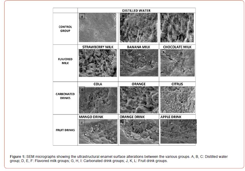
The rat mandibular right molar teeth demonstrated topographical variations and irregularities after the consumption of sugary drinks in comparison to those consuming distilled water as shown in Figure 1 (A -L).

The enamel surface, in the distilled water group appeared containing dense and orderly network of enamel rods that varied in appearance between the shallow and well defined rod types. The characteristic rod and interrod enamel structure was apparent, they were uniformly oriented on the enamel surface (Figure1 A- C).
There was an apparent observation of the loss of the surface enamel in the flavored milk drinks group with increased porosity (Figure 1 D-F) that exhibited multiple wavy irregularities in the form of crests, folds, depressions and valleys, with the appearance of enamel cracks (Figure 1 E, F). Some areas were stacked with microorganisms (Figure 1 E, F).
In the carbonated drinks group, surface discontinuity with increased porosity and irregularities of the enamel surface was readily observed with loss of enamel rods. The core of the enamel rod was mainly lost, with the appearance of the gaps between the rod and interrod boundaries giving the empty honeycomb appearance due to the demineralization of the surface enamel and the appearance of the openings of the dentinal tubules (Figure 1 G, H, I).
In the fruit drinks group, the enamel surface revealed surface roughness due to peaks, folds and depressions with evident microporosities that were packed with microorganisms. Demineralization of the rod and interrod appeared in some zones whereas other areas, the rods appeared shallow and/or well defined. (Figure1 J, K, L).
Mineral Element Analysis
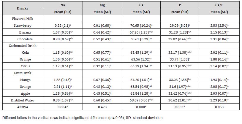
Within the groups, the mean value of sodium concentration in molar enamel of rats that consumed carbonated and fruit drinks was higher than in rats that consumed flavored milk and distilled water as shown in table 1.
Table 1:Statistical Differences of the Mineral Content between Different Groups.

On the other hand, the mean value of magnesium concentration in molar enamel of rats that consumed distilled water was higher than in rats consuming other drinks but was not statistically significant.
The mean value of calcium concentration in molar enamel of rats that consumed flavored milk was higher than in rats that consumed carbonated and fruit drinks with significant difference, however, no significant difference was detected in calcium value of enamel between rats that consumed flavored milk and the control group. Furthermore, the mean value of phosphorus concentration in molar enamel of rats that consumed flavored milk was higher than the rats that consumed distilled water but was not statistically significant. In addition, no statistically significant difference was detected in Ca/P ratio in rat enamel consuming different drinks (table 1).
Regarding the mineral concentration within the sub-groups, table 2 demonstrates that the enamel concentration of sodium was lower in rats that consumed strawberry flavored milk with significant difference to mango and apple drinks sub-groups. However, there was no significant difference in the enamel concentration of magnesium between the sub-groups.
Calcium concentration was the highest in rat enamel consuming strawberry flavored milk with significant difference to all other sub-groups except distilled water groups as shown in table 2. Additionally, phosphorus level was the lowest in rat enamel consuming strawberry flavored milk with significant difference to rats that consumed orange carbonated and mango drink sub-groups. In addition, no statistically significant differences were detected in Ca/P ratio in rat enamel between different sub-groups (table 2).
Table 2:Statistical Differences of the Mineral Content within the Different Sub-groups.

Discussion
The present study investigated the mineral element content of rat molar enamel after consumption of commonly used sugary drinks and beverages.
SEM is a powerful magnification tool with several advantages for analyzing the ultrastructure and elemental composition of dental hard tissues. It provides quick and detailed morphological and compositional data with minimal sample preparation. It allows the examination of surface morphology at the nanometer level leading to high-resolution digital images. The EDX spectroscopy attachment along with the backscattered SEM serves as a useful combination in examining elemental composition at the ultrastructural level [13].
The topographical ultrastructural architecture of the enamel surfaces in most groups appeared irregular and porous with various configurations of the enamel rods, that appeared shallow and well defined compared to the distilled water group that exhibited a smoother surface with regular orientation of the enamel rods. However, SEM provides only a subjective estimation of the surface roughness [14].
The surface enamel is composed of enamel prisms or rods, rod sheaths, and cementing interprismatic substance. Enamel prisms or rods are the basic structural component of enamel. They originate at the dentino‑enamel junction and extend through the thickness of the enamel surface [15]. Ultra structurally, the surface of the rods is visible as a result of the abrupt variations in the crystal orientation from one rod to the other [15, 16].
According to a number of literature resources, there are various forms of prisms and a variety of options regarding their orientation to each other. A prism head can appear above the enamel surface, lie on the same level with slight contouring, or be a cavity, thus forming different patterns or forms [15, 17]. Majority of the enamel rods in the distilled water group were smooth with some grooves, and few showed abundant microporosities within the flavored and fruit drink groups. Another secondary pattern seen in some of the carbonated drink group and fruit drink groups was the exposed enamel rods.
According to Beniash, et al. they stated that enamel surfaces can be classified into prismatic and aprismatic varieties. The crystals in the prismatic enamel are not aligned in a specific direction, whereas aprismatic enamel features crystals that are parallel to each other [18]. These differences should be taken into consideration when using native enamel for studies. Aprismatic enamel is predominantly found in the cervical, proximal, and occlusal areas, forming a layer with higher mineralization [18, 19]. Consequently, these regions were avoided in the present research as they lack a distinctive structural pattern, and the study was carried out on the buccal third of the molars.
The exposure of the tooth to the various acidic sugary drinks rendered the surface to increased irregularities and porosities which in turn may elevate the susceptibility of enamel to bacterial adhesion and biofilm formation, which could then potentially yield to demineralization and the accumulation of plaque leading to caries initiation [18, 20].
Previous researchers have revealed that bacterial colonization initiates and spreads through the enamel surface irregularities including porosities, depressions, perikymata, cracks. In vivo data have shown that rough surfaces significantly facilitate plaque retention and can harbor bacteria by up to 25 times more than their smooth counterparts [21]. High enamel surface roughness attracts the adherence of bacterial plaque, leading to a radical decrease in pH, chemical dissolution of the enamel, and the occurrence of dental caries. The lesser liability of the enamel to irregularities and roughness will remarkably decrease the plaque accumulation [22].
Furthermore, the surface roughness of enamel is strongly influenced by the mechanical and chemical processes that continuously occur in the oral cavity [23]. It is very challenging to accurately replicate natural biological conditions, which underscores the need for more detailed subsequent studies that closely approximate the in situ conditions of the oral cavity.
Minerals contained in the inorganic portion of a tooth should occur in strictly defined concentrations, as they determine its hardness, resistance to environmental influence and appropriate direction of biochemical transformations, among others. The absence of some minerals in the tooth tissue may affect the content of other minerals and result in greater tooth vulnerability to dental caries and other pathological agents [24].
In this experiment, sodium was found to be higher in the enamel of rats consuming carbonated and fruit drinks. In these animals, caries was found to be higher compared to animals consuming flavored milk [11, 12]. Similar to previous studies where they showed that Na mean concentrations in saliva were significantly higher in caries group [25, 26]. This might be attributed to the fact that acids attack the mineral phase of teeth resulting in demineralization and Na is diffused from the crystal apatite thereby increasing the levels of Na in the enamel of the teeth.
On the other hand, the mean value of calcium concentration in molar enamel of rats that consumed carbonated and fruit drinks was significantly lower than in rats that consumed flavored milk. Milk contains anticariogenic substances, such as Ca, P, casein, proteins, and fats. The Ca concentration of milk and the level of Ca and P ions in the saliva could affect the balance between demineralization and remineralization of enamel [27].
Although Ca value was higher in rat teeth consuming flavored milk, no significant difference was detected in calcium value of enamel between rats that consumed flavored milk and control group. This means that prolonged exposure to carbonated, and fruit drinks could lead to significant calcium loss. Carbonated and fruit drinks used in this study had a pH ranging between 2-3, thus could be the cause for direct enamel demineralization, which was revealed by the low level of calcium in the teeth of rats consuming carbonated and fruit drinks. Teeth exposed to carbonated and fruit drinks diffuse the hydroxyapatite out of the enamel and break it down into Ca2+ and PO43- ions. In addition, these sugary drinks also contain very high levels of phosphate with no calcium. High phosphate consumption is likely to extract calcium from the teeth to facilitate enamel erosion and thereby propagating cariogenesis [28].
Moreover, no significant differences were detected in the calcium concentration between teeth rat consuming carbonated drinks and teeth rat consuming fruit drinks. The acid contained in the carbonated drinks used in this study contains three kinds of acids, namely citric acid, phosphoric acid, and carbonic acid. The acid that mostly reduces the concentration of calcium is citric acid, which is the acid that is mainly contained in fruit drinks [29].
The presence of these minerals in different concentrations in the enamel of rat teeth indicated that these mineral elements present in drinks, foods, and water are incorporated throughout the tooth layers during the pre-eruptive period of tooth development and are further absorbed by the outer enamel surface during the demineralization and remineralization processes that occurs during the post-eruptive periods within the oral cavity leading to the vulnerability of the teeth to erosion and later on the increased porosity and opening of the interrod spaces allows the invasion of bacteria through the eroded enamel surface promoting dental caries over a period of time.
Conclusion
Based on the results of this study, it could be concluded that the enamel surface topography was affected following the consumption of different sugary drinks and altered the mineral element content of the teeth enamel.
The consumption of sugary drinks with high sugar content and acidity can contribute to detrimental oral health and may also affect general health. Therefore, it is necessary to educate parents about the harmful effects of different types of sugary drinks and advise them to consume more milk, water and sugar free fresh juices instead.
Acknowledgement
This research is supported by the College of Dentistry Research Center, King Saud University as funded research #1161.
Conflict of Interest
No Conflict of Interest.
References
-
<
- Morris AL, Mohiuddin SS. Biochemistry, Nutrients. In: StatPearls [Internet]. Treasure Island (FL): StatPearls Publishing; 2025 Jan. 2023 May 1.
- Pathak MU, Shetty V, Kalra D (2016) Trace elements and oral health: A systematic review. Journal of Advanced Oral Research. 7(2): 12-20.
- Nanci A (2013) Ten Cate’s Oral Histology: Development, Structure, and Function. (8th), St. Louis: Elsevier Mosby 7: 122-164.
- Shashikiran ND, Subba Reddy VV, Hiremath MC (2007) Estimation of trace elements in sound and carious enamel of primary and permanent teeth by atomic absorption spectrophotometry: An in vitro study. Indian J Dent Res 18: 157-162.
- Imfeld T (1996) Dental erosion. Definition, classification and links. Eur J Oral Sci 104(2 Pt 2): 151-155.
- Taji S, Seow WK (2010) A literature review of dental erosion in children. Aust Dent J 55(4): 358-475.
- Yip K, Lam PPY, Yiu CKY (2022) Prevalence and Associated Factors of Erosive Tooth Wear among Preschool Children—A Systematic Review and Meta-Analysis. Healthcare 10(3): 491.
- Gatt G, Attard N (2022) Risk prediction models for erosive wear in preschool-aged children: a prospective study. BMC Oral Health 22: 312.
- Schlueter N, Luka B (2018) Erosive tooth wear - a review on global prevalence and on its prevalence in risk groups. Br Dent J 224(5): 364-370.
- Scimeca M, Bischetti S, Lamsira HK, Bonfiglio R, Bonanno E (2018) Energy Dispersive X-ray (EDX) microanalysis: A powerful tool in biomedical research and diagnosis. Eur J Histochem 62(1): 2841.
- Al-Jobair A, Khounganian R (2015) Evaluating the cariogenic potential of flavored milk: an experimental study using rat model. J Contemp Dent Pract 16(1): 42-47.
- Al-Jobair, Asma M, Rita M Khounganian (2016) Comparison of the Carcinogenicity of Carbonated Soft Drinks and Fruit flavored Drinks Using Experimental Rats. Research & Reviews: Journal of Dental Sciences 4(3): 4-9.
- Syed S, Yassin SM, Almalki AY, Ali SAA, Alqarni AMM, et al. (2022) Structural Changes in Primary Teeth of Diabetic Children: Composition and Ultrastructure Analysis. Children (Basel) 9(3): 317.
- Teutle Coyotecatl B, Contreras Bulnes R, Rodríguez Vilchis LE, Scougall Vilchis RJ, Velazquez Enriquez U, et al. (2022) Effect of Surface Roughness of Deciduous and Permanent Tooth Enamel on Bacterial Adhesion. Microorganisms 10: 1701.
- Stavrianos C, Papadopoulos C, Vasiliadis L, Dagkalis P, Stavrianou I, et al. (2010) Enamel structure and forensic use. Res J Biol Sci 5: 650‑
- Zamudio Ortega CM, Contreras Bulnes R, Scougall Vilchis RJ, MoralesLuckie RA, Olea Mejía OF, et al. (2024) Morphological, chemical and structural characterization of deciduous enamel: SEM, EDS, XRD, FTIR and XPS analysis. Eur J Paediatr Dent 15: 275‑
- Beniash E, Stifler CA, Sun CY, Jung GS, Qin Z, et al. (2019) The hidden structure of human enamel. Nat Commun 10: 4383.
- Lippert F, Parker DM, Jandt KD (2004) In vitro demineralization/remineralization cycles at human tooth enamel surfaces investigated by AFM and nanoindentation. J Colloid Interface Sci 280: 442-448.
- Akasapu A, Hegde U, Murthy PS (25018) Enamel surface morphology: An ultrastructural comparative study of anterior and posterior permanent teeth. J Microsc Ultrastruct 6: 160-164.
- Quirynen M, Bollen CML (1995) The influence of surface roughness and surface-free energy on supra-and subgingival plaque formation in man: A review of the literature. J Clin Periodontol 22: 1-14.
- Karan S, Kircelli BH, Tasdelen B (201) Enamel surface roughness after debonding: Comparison of two different burs. Angle Orthod 80: 1081-1088.
- Serbanoiu DC, Vartolomei AC, Ghiga DV, Moldovan M, Sarosi C, et al. (2024) Comparative Analysis of Enamel Surface Roughness Following Various Interproximal Reduction Techniques: An Examination Using Scanning Electron Microscopy and Atomic Force Microscopy. Biomedicines 12: 1629.
- Bialek M, Zyska A (2014) The biomedical role of zinc in the functioning of the human organism. Pol J Public Health.124: 160-163.
- Benghasheer, Hala Fathallah (2013) Salivary Sodium and Potassium in Relation to Dental Caries in a Group of Multiracial School Children 3(1): 307-313.
- Han Shan Lin, Jia Rong Lin, Suh Woan Hu, Hsiao Ching Kuo, Yi Hsin Yang (2014) Association of dietary calcium, phosphorus, and magnesium intake with caries status among schoolchildren. Kaohsiung J Med Sci 30(4): 206-212.
- Moynihan P, Petersen PE (2004) Diet, nutrition and the prevention of dental diseases. Public Health Nutrition 7(1a): 201-226.
- Johansson AK, Johansson A, Birkhed D, Omar R, Baghdadi S, Carlsson GE (1996) Dental erosion, soft-drink intake, and oral health in young Saudi men, and the development of a system for assessing erosive anterior tooth wear. Acta Odontol Scand 54(6): 369-378.
- Tahmassebi JF, Duggal MS, Malik Kotru G, Curzon ME (2006) Soft drinks and dental health: A review of the current literature. J Dent 34(1): 2-11.
- Rirattanapong P, Vongsavan K, Surarit R (2013) Effect of soft drinks on the release of calcium from enamel surfaces. Southeast Asian J Trop Med Public Health 44(5): 927-930.
-
Asma Al-Jobair* and Rita Khounganian. Mineral Element Analysis of Rat Molar Enamel after Consumption of Sugary Drinks: An Energy Dispersive X-Ray Study. On J Dent & Oral Health. 8(4): 2025. OJDOH.MS.ID.000695.
-
Teeth, Oral environment, Carbonated drinks, Tooth morphology, Cavity, Tooth, Dental caries, Tooth tissue, Flavored milk
-

This work is licensed under a Creative Commons Attribution-NonCommercial 4.0 International License.


