 Research Article
Effect of Height Differences on the Retention and Wear Behaviour of Ball Attachment System Over Mandibular Dual Implants
Manimala M2, Vidhya J1*, Azhagarasan NS3, Jayakrishnakumar S3 , Hariharan Ramakrishnan3 and Vallabh Mahadevan3
1Reader, Department of Prosthodontics & Implantology, Ragas Dental College &Hospital, Chennai, India
2Department of Prosthodontics & Implantology, Ragas Dental College &Hospital, Uthandi, Chennai, India.
3Professors, Department of Prosthodontics & Implantology, Ragas Dental College &Hospital, Chennai, India ORCID Number: 0000-0003-4466-5744
Vidhya J, Reader, Department of Prosthodontics & Implantology, Ragas Dental College &Hospital, Uthandi, Chennai, India.
Received Date: December 25, 2020; Published Date: February 03, 2021
Abstract
Purpose of the study: To comparatively evaluate the effect of height differences on the retention and wear behaviour of ball attachment system over mandibular dual implants.
Materials and methods: Two groups of twenty samples each containing polymerised master block of dual implant analogues at same height in Group 1 and dual implant analogues of different heights in Group 2 were fabricated along with ball abutments, nylon cap and metal housing. Qualitative analysis of samples for wear factor were carried out using stereomicroscope and quantitative analysis of retention force at baseline, 360, 720, 1080, 1440 insertion-removal cycles were determined using Universal testing machine. statistical analysis were carried out using independent ‘t’ test and paired sample ‘t’ test.
Results: Group I test samples, the sample mean obtained at baseline, 360, 720,1080, and 1440 cycles were 24.73N, 23.49N, 22.46N, 21.07N, & 19.28N respectively. Group II test samples, the sample mean obtained at baseline, 360, 720,1080 & 1440 cycles were 27.13N 25.80N, 24.16N, 22.73N and 21.85N respectively. comparison of the mean retention force of the test samples at baseline, 360 (3 months), 720 (6 months), 1080 (9 months) and after 1440 (12months) insertion-removal cycles within Group 1 and Group II using Paired sample ‘t’-test was statistically significant at all levels.
Conclusion: Retention force was found to be higher on dual implants placed at different heights in mandibular anterior region and is statistically significant and corroborated by the Lesser surface deformations of attachment system.
Keywords: Dental implants; Denture retention; Mandibular prosthesis; Prosthesis retention; Titanium
Introduction
Edentulism is considered as a poor health outcome and it often compromises the quality of life of edentulous people [1]. Complete edentulism is a condition that is most prevalent among the elderly population [1,2]. It has a negative impact on the masticatory function, speech, esthetics and on the quality of life. The prosthetic management of the completely edentulous patient has been a major challenge in dentistry [3,4]. Conventional complete denture have been a standard and classical treatment option for the completely edentulous patients for more than a century [1,3,5]. Conventional complete denture is commonly provided to completely edentulous patients to restore masticatory function and esthetics [1,5]. The complete denture wearers often encounter problems like difficulty in mastication, discomfort during speech along with poor stability and retention [1,5,6]. They are usually satisfied with the upper denture but the majority of them often struggle with the lower denture due to the lack of retention and stability [1,7].
Alveolar ridge resorption is one of the important factors which is associated with loss of stability and retention in lower denture. It also reduces the amount of underlying tissue available for denture support [1,7]. The resorption of the residual alveolar ridges is a chronic, continuous, life-long catabolic process of bone remodelling. The rate of reduction in size of the residual ridge is maximum in the first three months and then gradually tapers off [8]. According to Boucher, during the first year after tooth extraction, the reduction in the residual ridge height in the mid sagittal plane is 2-3mm for maxilla and 4-5mm for mandible and the annual rate of reduction in ridge height is 0.1-0.2mm for mandible and this reduction in height of the ridge is four times less in the maxilla [1,8,9]. Many studies have shown that the poor retention and stability could be managed using a fixed prosthesis supported by five or six implants or by the fabrication of an overdenture to implants [9]. However, placing six implants in an atrophic mandible is not always possible. Therefore, the concept of using two or four implants to support an overdenture was introduced.
Overdenture is a therapeutic approach which is directed in improving the oral function in elderly edentulous patients [10]. The concept of overdenture initially involved fixing mechanical attachments to teeth roots to enhance retention and stability of conventional complete dentures [6,9]. With the evolution of implant overdentures, there is reduction in displacement of the prosthesis due to lateral forces leading to better retention and stability improving the masticatory function and overall quality of life [5,11,12]. Crum, Rooney [8] reported an average of 5.2mm loss in the alveolar ridge height in denture patients compared to overdenture patients where there is 0.6mm loss which is relatively low. According to McGill consensus, two-implant overdentures has been accepted as the gold standard treatment care for the edentulous mandible [5,13]. Various factors contribute to the success of an implant supported overdentures, including the fit and precision of dentures and the retentive capacity of its attachment system to provide long duration of function. Therefore, Retention is considered as one of the most significant factors in determining patient satisfaction in removable prosthodontics [9,14] and is defined as the force that resists withdrawal along the path of insertion and stabilizes the overdenture during its function [14,15].
Also, the choice of attachment is primarily dependent on the retention required, jaw morphology and anatomy, condition of the mucosal ridge, oral function and patient compliance for recall appointments. There are different attachments to retain implant supported overdentures. They were classified on the basis of their flexibility, geometrical shape and cross section, casting precision and manufacturing procedures. In one of the major classifications, these attachments are divided into splinted and unsplinted systems. Splinted systems includes the bar attachments whereas unsplinted systems includes magnets, ball types and locators [5,7,16,17].
Within these systems, self-aligning attachments (locators) and ball attachments have been frequently used due to their simplicity. Specifically, ball attachments are considered as one of the simplest type of attachments for clinical usage. It provides varying degrees of resiliency in both vertical and horizontal directions. Also, the specific design of the ball attachments may influence the amount of its free movement thereby limiting its resiliency [1,5,11,18]. The retention and the longevity of the attachments have been studied as a common problem in many clinical and in vitro studies. There are lot of studies evaluating the retentive force of different attachment systems in mandibular two-implant overdentures simulating different periods of clinical usage [6,7,11,19]. Some studies have comparatively evaluated the retention capacity of ball, bar and locator attachments in implant overdentures [19]. Studies have also compared retention between locator, ball and magnetic attachments [20].
Many studies have compared the retentive capacity of different color codings of a single attachment system [14,21]. Few authors have studied the retentive forces of the attachments by placing implants at different angulations [14,20]. Previous studies have evaluated the retentive force using micro material testing machine (MTM) [9,15], a dental mastication simulator [14,21], CS-Dental testing machine [16], load cell (cyclic fatigue machine) [22], Imada device (IM) [23,24] and Universal testing machine (UTM) [6,11,24] and among these methods Universal testing machine (UTM) has been accepted as reliable and valuable instrument to test the retention forces in vitro [6,11]. One of the main factors of retention loss is the change induced on the components of the attachment systems as a result of wear [6,11]. The factors which are associated with the clinical wear of attachments includes masticatory forces, parafunctions, temperature and composition of saliva, products used for the maintenance of denture and the presence of food residues [22]. The wear of overdenture retentive mechanisms has been identified as the most common prosthodontic complications which is about 33% [2].
In addition to the retentive forces, few studies have also evaluated the amount of wear and its effects in the attachment systems. Wear have been assessed using Metallographic microscope [25], Micro material testing machine (MTM) [15,26-28], Coordinate measuring machine (CMM) [18], Scanning electron microscopy (SEM) [14,29], and stereo microscopy [6,17]. The positioning of the implant overdenture attachments is very important for two-implant overdentures. This is because during pathological overloading, the bone around the implants becomes deformed and resorbed due to the increased stress and strain gradients [5]. This situation may cause incompatibility of the components of the implant system and microfracture of the implant [5]. Clinicians predicted that the two independent implants must be positioned at the same occlusal height, parallel to the occlusal plane [24]. However, placement of implants in the same occlusal height in completely edentulous patient is not always possible since the alveolar bone resorbs with different types of resorption patterns [5]. The rate of residual ridge resorption differs from person to person and even at different times and sites in the same person. Unlike in maxilla, the speed of bone loss in mandible is different in different parts of the jaw, distal parts of the residual ridge disappear faster than the anterior parts, which may affect the symmetry of the remaining bone height resulting in difficulty in placing the implants in position [8].
Few studies have evaluated the retention force of attachment systems retained by single or two implants and also at different abutment heights [30]. Ozan O and Ramoglu S have compared two different attachment systems in two-implant overdentures by evaluating the stress distribution in peri-implant site and on attachments by positioning implants in different height levels using the 3D Finite Element Analysis method [5]. Currently, studies comparatively evaluating the retention and wear behaviour of two-implant overdenture attachments with implants positioned at different height levels are lacking. In light of the above, the aim of the present in vitro study was to evaluate the effect of implant height differences on the retention and wear behaviour of ball attachment system in mandibular two-implant overdentures. The null hypothesis for the present study was that implant height difference does not affect the retention and wear behavior of the dual implant overdenture attachments.
Materials and Methods
A rectangular block of dimensions 60mm x 20mm x 10mm, made of plaster of paris (Ramaraju Mills ltd., India) was custom made to serve as an index for the fabrication of wax blocks of similar dimensions and then to be converted into heat cure acrylic resin blocks of uniform dimensions to be used in this study. Addition silicone impression material of putty and light body consistencies (Aquasil, Denstply, USA) were used for obtaining the index in a single step procedure. The putty was hand mixed with equal quantities of base and catalyst to obtain homogenous dough. Light body material in a cartridge was attached to the auto mixing gun (Heraeus Kulzer, Dormagen, Germany). A spiral mixing tip (yellow 70mm , Adenta, USA) was attached to the cartridge tip and material was injected gently over the custom- made plaster block. The mixed putty was also placed over the plaster block and left undisturbed until set. After setting, the plaster block was removed from the index and the mold space area was inspected for defects and acceptability. The putty index thus obtained was used to fabricate the test samples of standardized dimensions for this study.
a) Master wax blocks(n=2),
b) Prosthetic wax blocks (n=20).
Modeling wax (Hindustan manufacturer, Hyderabad, India) (was melted and poured into the mold space created by the putty and was allowed to cool. After the wax had completely hardened, the wax blocks were retrieved carefully and placed at room temperature. Twenty-two such wax blocks of 60mm x 20mm x 10mm dimensions were fabricated in which two blocks were used as master blocks and twenty blocks were used as prosthetic blocks to conduct the study. Out of the two master blocks, in one block a height of 2mm was increased with wax on one half of the block exactly measured from the centre of the block. Similarly, out of the twenty prosthetic blocks, ten blocks were increased to a height of 2mm as it was done for the master blocks. Two implant analogs with the diameter of 4mm and height of 12.7mm (Norris dental implants, Israel) were positioned parallel to the insertion-removal path and to one another, on each of the master blocks using a dental surveyor (seshin precison Ind., co, korea). The master blocks were placed on the surveying platform of the surveyor and stabilized. The surveying platform was made parallel to the floor. The implant analogs were positioned into the wax blocks at a distance of 22mm from each other with equal distance from the centre of the block. The implant analogs were submerged into the wax block up to the crest module of the implant analogs (2 Master blocks with implant analogs and 20 Prosthetic blocks).
The two master blocks with the implant analogs and the twenty prosthetic blocks were fabricated using heat polymerized acrylic resin. The ball abutments of diameter 2.5mm and height 2mm (Noris Dental implants, Israel) were screwed into all the implant analogs embedded in the master blocks. Modelling wax was used to block the undercuts around the ball abutments in the master blocks. The metal housing of 5mm diameter and 3.2mm height along with nylon cap insert of standard retention (Noris Dental Implants) was placed over the ball abutments for picking it up onto the prosthetic block. The prosthetic blocks were now drilled with acrylic burs to create space for the metal housing with nylon cap inserts (Figure 1). In order to prevent the acrylic adhesion of two blocks, a thin layer of separating medium (Tejpal pharma and surgical, India), is applied on the master blocks. Clear autopolymerising acrylic resin (RR cold cure, DPI, India), was poured into the space created and the metal housings with the nylon cap insert placed over the ball abutments in the master blocks were picked up in the prosthetic blocks. Upon setting, the master block with ball abutment was retrieved from the prosthetic blocks with the metal housing and nylon cap insert. Twenty prosthetic blocks thus obtained were divided into two groups of ten blocks each according to the height difference.
1. Group I (n =10) Test samples for the implant analogs placed at same height.
2. Group II (n = 10) Test samples for the implant analogs placed at different height.
Each test sample was placed over the imaging platform of the stereo microscope prior to retention testing. Images were captured in a computer-controlled software system in 40x magnification at an object lens distance of about 13mm and the same magnification had been used for all the test samples (Figure 2). A total of twenty test samples were tested individually in the Universal testing machine (INSTRON 8874) (Figure 3) to measure the force required to separate the prosthetic block from the master block. Both the master block with ball abutment-implant analog assembly and the prosthetic test samples were positioned on the machine table and secured tightly into the upper and lower clamps of the universal testing machine. Engagement and disengagement of the attachments were carried out at right angles to the horizontal level of the blocks. The testing machine was programmed to apply 1440 cycles of insertion-removals. Assuming that a patient removes and inserts his prosthesis four times a day, the retention force values were noted at baseline, after 360 cycles (simulating 3 months of clinical use), 720 cycles (simulating 6 months of clinical use), 1080 cycles (simulating 9 months of clinical use) and after 1440 cycles (simulating one year of clinical use) The test samples were kept moist with artificial saliva throughout the testing as it acts as lubricant to simulate potential in-vivo conditions. The tests were conducted in an open room at room temperature.


50mm/min at a frequency of 0.8hz and the retention force values were obtained in a computer-controlled software which is being attached to the testing machine. Each test sample was placed in the imaging platform of the stereo microscope after the completion of 1440(12 months) insertion-removal cycles at 40x magnification to assess the wear on the surface of the test samples. The image was captured in the computer controlled software attached to the system. The maximum vertical dislodging force required to separate the two blocks were recorded (in Newtons) at a crosshead speed of 50mm/min at a frequency of 0.8hz and the retention force values were obtained in a computer controlled software which is being attached to the testing machine. Each test sample was placed in the imaging platform of the stereo microscope after the completion of 1440(12 months) insertion-removal cycles at 40x magnification to assess the wear on the surface of the test samples. The image was captured in the computer controlled software attached to the system (Figure 4, 5).


test and a P value of <0.05 was considered statistically significant. Surface wear was assessed using descriptive analysis.
Results
For Group I test samples, the sample mean of the retention force obtained at baseline, 360, 720, 1080, and 1440 cycles were 24.73N, 23.49 N, 22.46N, 21.07N, & 19.28N respectively (Table 1). For Group II test samples, the sample mean of the retention force obtained at baseline, 360, 720, 1080 & 1440 cycles were 27.13N, 25.80N, 24.16 N, 22.73N& 21.85 N respectively (Table 2). Overall comparison of the mean retention force, statistically significant difference was observed between the two test groups. Group II exhibited significantly higher retention force values compared to Group 1 (Table 3). comparison of the mean retention force of the test samples at baseline, 360 (3 months), 720 (6 months), 1080 (9 months) and after 1440 (12months) insertion-removal cycles within Group 1 and Group II using Paired sample ‘t- test was statistically significant at all levels. (Table 4,5). Percentage loss of the mean retention force of the test samples at 360 (3 months), 720 (6 months), 1080 (9 months) and after 1440 (12 months) insertionremoval cycles of Group I and 2 were tabulated (Table 6,7).
Table 1:Retention force of overdenture specimen with ball attachment system retained by two implants placed in the same height at baseline, 360 (3 months), 720 (6 months), 1080 (9 months) and after 1440 (12 months) insertion removal cycles in Group I.
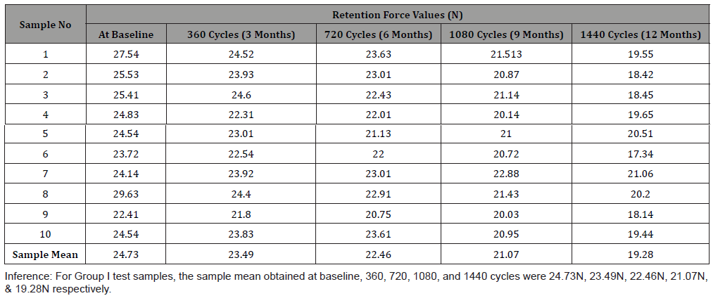
Table 2:Retention force of overdenture specimen with ball attachment system retained by two implants placed in different height at baseline, 360 (3 months), 720 (6 months), 1080 (9 months) and after 1440 (12 months) insertion removal cycles in Group II.
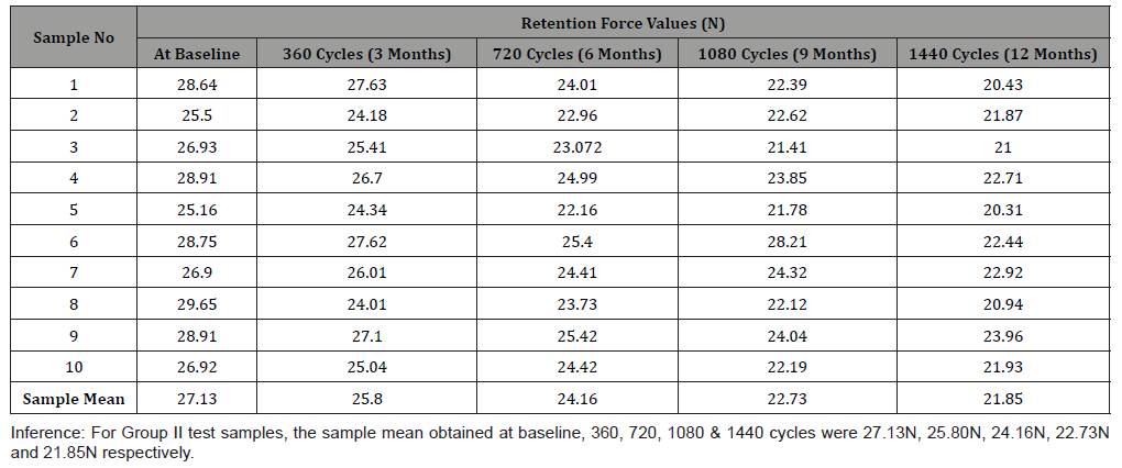
Table 3:Comparison of the mean retention force of the test samples at baseline, 360 (3 months), 720 (6 months), 1080 (9 months) and after 1440 (12 months) insertion-removal cycles between Groups I & II using Independent sample ‘t’-test.
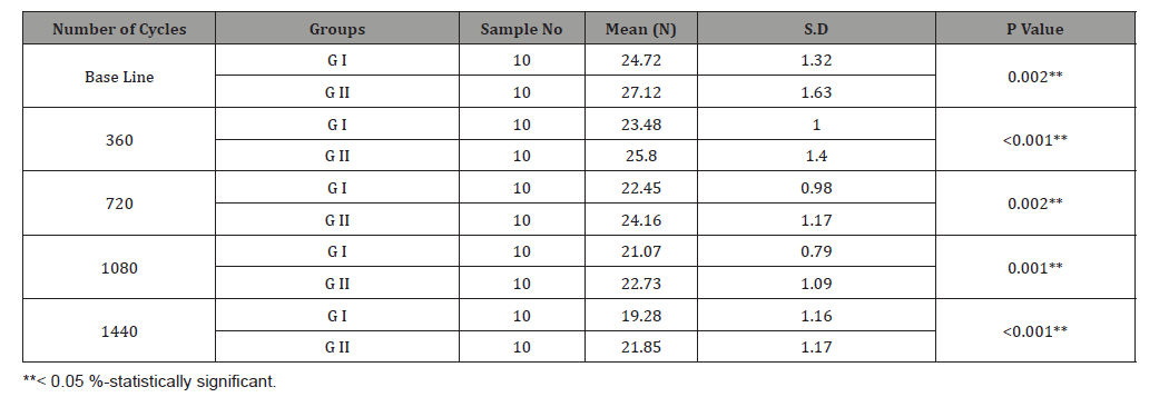
Table 4:Comparison of the mean retention force of the test samples at baseline, 360 (3 months), 720 (6 months), 1080 (9 months) and after 1440 (12 months) insertion-removal cycles within Group I using Paired sample ‘t’-test.
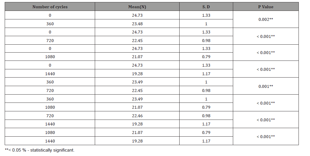
Table 5:Comparison of the mean retention force of the test samples at baseline, 360 (3 months), 720 (6 months), 1080 (9 months) and after 1440 (12months) insertion-removal cycles within Group II using Paired sample ‘t’-test.
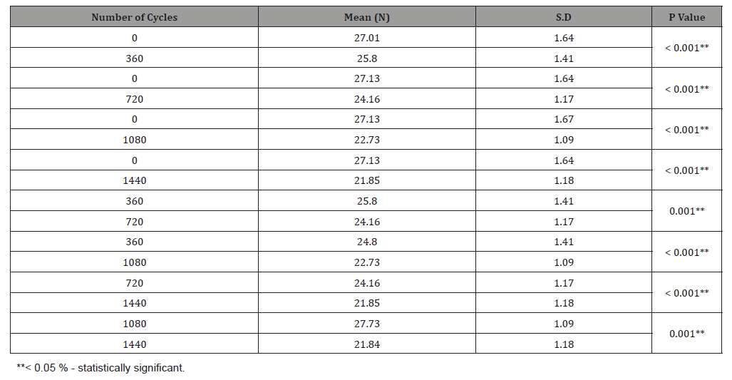
Table 6:Percentage loss of the mean retention force of the test samples at 360 (3 months), 720 (6 months), 1080 (9 months) and after 1440 (12 months) insertion- removal cycles in Group I.

Table 7:Percentage loss of the mean retention force of the test samples at 360 (3 months), 720 (6 months), 1080 (9 months) and after 1440 (12 months) insertion removal cycles in Group II.

Discussion
In the completely edentulous patient, alveolar bone can be resorbed with different types of resorption patterns which may affect the symmetry of the bone height in the anterior mandible [5,8]. Resorption rate of the residual alveolar ridge differs from one person to other and at different sites and times in the same patient [8]. All these factors may affect the symmetry of the bone height in the mandible. In those cases, implants could be placed at different heights according to the literatures [5]. Different number of cycles had been used in different studies, like 10,000 cycles equivalent to a corresponding time of use of 9 years [7], 5500 cycles (3 years) [6,7], 14,600 cycles (which represents 10 years of clinical use)[7] and 1080 cycles (1 year)[11]. In the present study , the test samples were subjected to 1440 insertion-removal cycles which corresponds to one year of clinical usage of dentures,11 considering an average of four removals per day in accordance with previous studies [7,12] and the retention force was measured at baseline, 360, 720, 1080 and 1440 insertion-removal cycles.
Previous studies have employed demineralised water, saline solution and 0.9% isotonic sodium chloride solution maintained at 22-degree celcius to simulate in vivo conditions during their testing procedure [7]. In the present study, artificial saliva was maintained throughout the testing procedure at room temperature to simulate a wet oral environment, also to act as a protective and lubricant layer between the components of the attachments used as mentioned in some previous studies [6,27]. Many studies evaluated the retentive force of overdenture attachments in different dislodgment speeds (900 cm/min to 0.5 mm/min domain). (0.5mm/min to 150 mm/ sec), 120mm/min [24]. Sarnat proposed a crosshead speed of 50mm/min [24] and said that it is as close to the speed of the movement of real overdenture removal from its retention elements when vertical force is applied [6,11] and therefore it was used in the present study. The retention force test was carried out with the above mentioned parameters.
Ozan O and Ramoglu S have evaluated the stress distribution on the peri-implant bone and on different attachments by placing implants at different height levels in mandibular two-implant overdentures and concluded that decreased stress values in the peri-implant bone were obtained with the models with increased height difference [5]. Ying, et al. studied about the influence of the height of the stud attachments and concluded that the greater height of the stud attachments exerted the highest lateral force on the implant and greatest denture displacement [33]. A study on evaluation of retentive force of different locator abutment heights done by Sia, et al. [21] showed that groups with 0mm (32.3N), 2mm (37.1N), 4mm (41.9N) height difference have lower retention than the group with 6mm (53.6 N) height difference. This was due to increased friction or rotational path of dislodgement. In the attachments with 6mm height difference between each other, it was also reported that during testing, separation of the 2 attachments from the 2 abutments did not occur at the same time, but often one followed the other. Thus, he concluded that the different height levels of the abutments might have provided a rotational path of dislodgement, thereby requiring higher retentive force [21].
In the present study, implants placed at different height showed a significantly higher retention values than the implants at same height. This might be due to the different height levels of the implant which have provided rotational path of dislodgement thereby requiring higher retention force comparatively which correlates with the study done by Sia, et al. [21] with different abutment heights which was mentioned above. The reason for this situation need to be explored further in future studies. Studies similar in design with the present study are lacking in literature to enable further comparisons. According to previous studies, most of the attachment systems showed a common trend toward a reduction in the retention force [11,31]. Memarian, et al. [32] evaluated the retentive properties of two commercially available ball attachments (straumann and Rhein83) and showed there was gradual and significant decrease in the retention force after 5500 cycles. Arora, mittal [11] evaluated the retention force values of different attachments like locator, ball/O-Ring and ball/nylon-cap simulating 1 year of clinical usage and shown that all attachments lose retention over time. Therefore, repeated insertion-removal cycles led to a gradual and continuous loss of retention as mentioned in the above studies.
In the present study, regardless of the high retention values at the beginning of the study in both the groups, there is gradual decrease of retention values which is in agreement with the abovementioned previous studies.
Some studies showed 18.7% loss of retention of O-rings after simulated 30 months of clinical use and in other study, it was 16.6% retention loss after simulated 6 months of clinical use and 57.1% loss after 24 months [2]. In a study by Arora, Mittal [11] the retention force values of different attachments like locator, ball/ORing and ball/nylon-cap simulating 1 year of clinical usages were evaluated and retention loss was maximum for ball/o-ring (76.6%) and minimum for ball/nylon cap (18.4%) and locator (20.2%). In the present study, the overall percentage loss of retention force after 1440 cycles in Group I and Group II was 21.9 % and 19.33 % respectively. This study has also revealed that there was percentage loss which was slightly higher than those observed in previous studies. This might be due to variations in the company of the nylon cap employed or the height difference of the implant analogs. However, this needs to be evaluated further in the future studies.
Many previous studies have reported that, the retention force of about 5 to 7 N was enough to stabilize the overdentures during function [6,11,12]. All the test samples of both groups have retention force values more than the above mentioned value and therefore it was considered satisfactory to stabilize the overdenture. However, reduced retention characteristic may be desirable in some patients with poor manual dexterity [11], who may have difficulty during insertion and removal of overdenture. Therefore, under such specific situations, matching the retentive level of the attachments to the physical condition and required needs of the patient should be considered while planning treatment. In the present study, stereo microscopic images showed wear on the surface of the attachments of all the test samples of both groups at the end of the study after completion of 1440 insertion removal cycles which is in line with the above mentioned studies.
Also, the implants placed at different height have shown less wear deformations than the implants placed at same height. Therefore, it was concluded that, the implants placed at different height had less wear which corroborates with the greater retention obtained with it and the implants placed at same height had more wear which corroborates with the lesser retention obtained. From the results obtained and within the limitations of the present study, it can be concluded that, the implant height difference has its effect on both the retention and wear of the overdenture attachments used. Thus, the null hypothesis of this study was rejected. The present study had some limitations. Factors like parafunctions, temperature of saliva and its composition, products used for cleansing dentures as well as the presence of food residues may influence the parameters tested above which are difficult to simulate in vitro. Under clinical conditions, the forces exerted on the attachments are more complex, with tridimensional forces often occurring. Further studies are needed to evaluate the retention force by placing implants at various height differences, simulating longer periods of clinical usage.
Conclusion
Retention force was found to be higher on dual implants placed at different heights in mandibular anterior region and is statistically significant and corroborated by the Lesser surface deformations of attachment system over these implants.
Clinical Significance
Placement of implants at different heights remains a viable clinical option and should be considered for enhanced retention of ball attachment system.
Acknowledgement
None.
Conflict of Interest
The authors declare no conflict of interest.
References
- Sharma S, Makkar M, Teja SS, Singh P (2017) Implant-supported overdenture: a review. J Pharm Biomed Sci 07(7): 270-277.
- Valente MLC, Shimano MVW, Agnelli JA, Reis AC (2018) Retention force and deformation of an innovative attachment model for mini-implant-retained overdenture. J Prosthet Dent 121(1): 129-134.
- Alqutaibi AY, Kaddah AF (2016) Attachments used with implant supported overdenture. IDMJAR 2: 1-5.
- Rutkunas V, Mizutani H (2004) Retentive and stabilizing properties of stud and magnetic attachments retaining mandibular overdenture-An in vitro study. Stomatol 6(3): 85-90.
- Ozan O, Ramoglu S (2015) Effect of implant height differences on different attachment types and peri implant bone in mandibular two-implant overdentures:3D Finite element study. J Oral Implantol 41(3): e50-e59.
- Reda KM, El-Torky IR, EL-Gendy MN (2016) In vitro retention force measurement for three different attachment systems for implant-retained overdenture. J Indian Prosthodont Soc 16: 380-385.
- Kobayashi M, Srinivasan M, Ammann P, Perriard J, Ohkubo C, et al. (2013) Effects of in vitro cyclic dislodging on retentive force and removal torque of three overdenture attachment systems. Clin Oral Implants Res 25(4): 426-434.
- Samyukta, Abirami G (2016) Residual ridge resorption in complete denture wearers. J Pharm Sci Res 8(6): 565-569.
- Rutkunas V, Mizutani H, Takahashi H (2005) Evaluation of stable retentive properties of overdenture attachments. Stomatologija 7(4): 115-120.
- Chung KH, Chung CY, Cagna DR, Cronin RJ (2004) Retention characteristics of attachment system for implant overdentures. J Prosthodont 13: 221-226
- Arora S, Mittal S (2017) Comparative evaluation of alteration in retention force values of different attachment systems for implant overdenture over various time intervals:an in vitro study. IJIRAS 4(4): 1-6.
- Guttal SS, Nadiger RK, Abhichandani S (2012) Effect of insertion and removal of tooth supported overdentures on retention strength and fatigue resistance of two commercially available attachment systems. Int J Prosthodont Restor Dent 2(2): 47-51.
- Daou EE (2013) Stud attachments for the mandibular implant-retained overdentures: Prosthetic complications-A literature review. Saudi Dent J 25: 53-60.
- Choi JW, Bae JH, Jeong CM, Huh JB (2017) Retention and wear behaviors of two implant overdenture stud-type attachments at different implant angulations. J Prosthet Dent 117: 628-635.
- Rutkunas V, Mizutani H, Takahashi H, Iwasaki N (2011) Wear simulation effects on overdenture stud attachments. Dent Mater J 30(6): 845-853.
- Aroso C, Silva AS, Ustrell R, Mendes JM, Braga AC, et al. (2016) Effect of abutment angulation in the retention and durability of three overdenture attachment systems: An in vitro study. J Adv Prosthodont 8: 21-29.
- Shayegh SS, Hakimaneh SMR, Baghani MT, Shidfar S, Kashi FK, et al. (2017) Effect of Interimplant Distance and Cyclic Loading on the Retention of Overdenture Attachments. J Contemp Dent Pract 18(11): 1078-1084.
- Vafaee F, Fotovat F, Firuz F, Soufiabadi S, Roshanaei G, et al. (2016) The amount of wear in attachment of implant-supported overdentures in mandible. J Dent Mater Tech 5(4): 181-88.
- Shastry T, AnupamaNM, Shetty S, Nalinakshamma M (2016) An in vitro comparative study to evaluate the retention of different attachment systems used in implant-retained overdentures. J Indian Prosthodont Soc 16: 159-166.
- Yang TC, Maeda Y, Gonda T, Kotecha S (2011) Attachment systems for implant overdenture: Influence of implant inclination on retentive and lateral forces. Clin Oral Implants Res 22: 1315-1319.
- Sia PKS, Masri R, Driscoll CF, Romberg E (2016) Effects of locator abutment height on the retentive values of pink locator attachment: An in vitro study. J Prosthet Dent 117(2):283-288.
- Carvalho ER, Figueiral MH, Fonseca P, Vaz MA, Branco FM (2014) In vitro study of the insertion and disinsertion effect on retention of two attachment systems of an overdenture of two implants. Revista Odonto Ciencia 29(1): 1-5.
- Fromentin O, Lassauzay C, Nader S A, Feine J, Albuquerque (2010) Testing the retention of attachments for implant overdentures-validation of an original force measurement system. J Oral Rehabil 37: 54-62.
- Jalalian S, Ansari lari H, Atashrazm P, Fatemi S (2015) In vitro evaluation of the effect of disto-labially inclined implant and Locator attachment on retention and longevity of implant-supported overdentures. J Res Dent Sci 11(4): 196-205.
- Rodrigues RCS, Faria ACL, Macedo AP, Sartori IAM, deMattos MG C, et al. (2009) An in vitro study of non-axial forces upon the retention of an O-ring attachment. Clin Oral Implants Res 20: 1314-1319.
- Amarsanaa D, Mizutani H, Takahashi H, Igarashi Y (2009) Stability of mandibular overdentures before and after attachment wear-An in vitro study. Ann Jpn prosthodont Soc 1: 77-84.
- Pigozzo MN, Mesquita MF, Henriques GEP, Vaz LG (2009) The service life of implant-retained overdenture attachment systems. J Prosthet Dent 102: 74-80.
- Rutkunas V, Mizutani H, Takahashi H (2006) Influence of attachment wear on retention of mandibular overdenture. J Oral Rehabil 34: 41-51.
- Mahross HZ, Baroudi K (2015) Evaluation of retention and wear behavior for different designs of precision attachments. Oral Health Dent Manag 14(4): 244-249.
- Mattia PR, BotegaDM, Zani SR, Rivaldo EG, Frasca LCF (2016) Evaluation of retentive force of attachment systems for overdentures retained by one or two implants. stomatos 22(42): 15-22.
- Scherer MD, McGlumphy EA, Seghi RR, Campagni WV (2013) Comparison of retention and stability of implant-retained overdentures based upon implant number and distribution. Int J Oral Maxillofac Implants 28(6): 1619-1628.
- Memarian M, Zarrati S, Karimi S, Bahrami M (2018) Comparative evaluation of retentive properties of two compatible ball attachments in mandibular implant-retained overdentures: An in vitro study. J Dent Tehran 15(2): 106-115.
- Ying Z, Gonda T, Maeda Y (2017) Influence of attachment height and shape on lateral force transmission in implant overdenture treatment. Int J Prosthodont 30: 586-590.
-
Manimala M, Vidhya J, Azhagarasan NS, Jayakrishnakumar S, Hariharan R, Vallabh M. Effect of Height Differences on the
Retention and Wear Behaviour of Ball Attachment System Over Mandibular Dual Implants. On J Dent & Oral Health. 4(2): 2021.
DOI: 10.33552/OJDOH.2021.04.000581 .
-
Lower denture, Oral function, Prosthetic blocks, Dental implants, Denture retention, Edentulous patients, Teeth roots, Prosthodontic complications, Dual implants.
-

This work is licensed under a Creative Commons Attribution-NonCommercial 4.0 International License.


