
 Research Article
Artificial Intelligence in CBCT for Dental Implantology: Current Applications, Limitations, and Future Perspectives - A Literature Review
Former Head Section of Oral and Maxillofacial Radiology, Lebanese University, Beirut, Lebanon
Received Date: September 29, 2026; Published Date:October 07, 2026
Abstract
Artificial intelligence (AI) is increasingly being integrated into cone-beam computed tomography (CBCT)-based workflows in dental implantology. This structured narrative literature review evaluated current AI applications in implant dentistry, critically assessed their technical and clinical limitations, and discussed future perspectives for integration into implant planning and treatment workflows. A literature search was conducted using PubMed/MEDLINE, Scopus, Web of Science, and Embase. Evidence was synthesized according to clinically relevant domains, including anatomical segmentation, identification of edentulous regions and implant sites, bone assessment, virtual implant planning, comparison with expert planning, workflow optimization, and outcome prediction. AI demonstrated encouraging performance in CBCT segmentation, anatomical and bone assessment, and selected components of virtual implant planning. Automated systems showed substantial agreement with expert-generated plans in selected clinical settings and consistently reduced planning time while providing high repeatability. However, performance varied according to anatomical complexity, imaging conditions, software environment, and the specific task evaluated. Limited external validation, heterogeneous datasets and evaluation methods, and the gap between technical performance and demonstrated clinical benefit remain important barriers to generalizability. Current evidence supports AI primarily as a decision-support tool that complements professional judgment. Future research should prioritize multicenter prospective validation, standardized evaluation frameworks, multimodal data integration, explainable and uncertainty-aware models, and clinically meaningful outcomes.
Introduction & Background
Dental implant therapy is a well-established treatment modality for the rehabilitation of partially and completely edentulous patients. Successful implant treatment requires accurate preoperative assessment and three-dimensional planning, including evaluation of available bone volume and quality, identification of relevant anatomical structures, and determination of implant position according to surgical and prosthetic requirements. Conebeam computed tomography (CBCT) has consequently become an essential imaging modality in contemporary implant dentistry because it provides three-dimensional information on maxillofacial anatomy that cannot be adequately obtained from conventional two-dimensional radiographs [1, 2].
Despite these advantages, interpretation of CBCT datasets can be complex and time-consuming, and manual image interpretation and segmentation may be affected by observerdependent variability [3, 4]. Artificial intelligence (AI), particularly machine learning and deep learning, has emerged as a promising approach for automated analysis of medical and dental imaging data. Deep-learning architectures can extract complex image features and spatial relationships from volumetric datasets and have been investigated for automated detection, classification, and segmentation of teeth, bone, periapical lesions, the mandibular canal, and the maxillary sinus [4-12].
The combination of AI and CBCT is particularly relevant to implant dentistry. Reported applications include automated anatomical segmentation, assessment of alveolar bone dimensions and density, localization of critical anatomical structures, identification of potential implant sites, virtual implant positioning, and integration of CBCT data with intraoral surface scans [10-13]. AI-assisted digital workflows may also contribute to surgical guide design, navigation, treatment-outcome prediction, and assessment of postoperative complications [13-19].
Nevertheless, translation from promising technical performance to reliable routine clinical use remains incomplete. AI performance may be influenced by training-dataset characteristics, CBCT acquisition parameters, imaging devices, algorithms, software platforms, and patient populations. Limited external validation and heterogeneity among available systems may restrict generalizability, while algorithmic transparency, data privacy, cybersecurity, regulatory requirements, and medicolegal responsibility remain important considerations [20-22]. A critical synthesis is therefore needed to distinguish established applications from emerging technologies. This review examines the current applications of AI in CBCT-based dental implantology, critically evaluates their technical and clinical limitations, and discusses future perspectives for integration into implant planning and treatment workflows.
Review Materials and Methods
Review Design
The present study was conducted as a structured narrative literature review addressing the current applications, limitations, and future perspectives of artificial intelligence (AI) applied to cone-beam computed tomography (CBCT) in dental implantology. A narrative approach was selected because the available literature encompasses considerable methodological diversity, including differences in study design, imaging datasets, AI architectures, training and validation procedures, clinical objectives, and performance measures. Such heterogeneity makes a qualitative and clinically oriented synthesis particularly suitable for examining both the translational potential and the limitations of emerging AI technologies [23].
Literature Search Strategy
A structured literature search was initiated on August 9, 2026. The electronic search focused on PubMed/MEDLINE, Scopus, Web of Science, and Embase. The search strategy was constructed around three principal conceptual domains: artificial intelligence, CBCT imaging, and dental implantology.
Medical Subject Headings (MeSH), when applicable, and free-text terms were combined using Boolean operators. Search terminology included combinations of the following concepts: “artificial intelligence,” “machine learning,” “deep learning,” “neural networks,” “convolutional neural networks,” “cone-beam computed tomography,” “CBCT,” “dental implants,” “implant dentistry,” “implant planning,” “image segmentation,” “anatomical structure identification,” “treatment planning,” and “clinical decision support” [23-25].
The search syntax was adapted to the indexing characteristics of each database. To broaden literature identification, the reference lists of relevant original studies and review articles were also examined manually for additional publications related to AIassisted CBCT applications in implant dentistry.
Because AI-based imaging technologies are developing rapidly, particular attention was given to recent publications. Earlier studies were nevertheless retained when they provided relevant methodological foundations, described the development of an important AI application, or contributed substantially to understanding the evolution of AI-assisted CBCT analysis.
Eligibility Criteria
Publications were considered eligible when they investigated an AI-based or machine-learning approach involving CBCT imaging and demonstrated direct or potential relevance to dental implantology.
The review considered clinical, retrospective, prospective, validation, and experimental studies whose objectives were related to clinically meaningful implant applications. Relevant areas included automated or semi-automated segmentation of maxillofacial structures, identification of anatomical landmarks and structures at risk, assessment of alveolar bone characteristics, implant site evaluation, virtual implant positioning, selection of implant dimensions, assistance with drilling protocols, image registration, surgical planning, and AI-supported clinical decisionmaking [24-26].
Studies were considered relevant when AI constituted a substantial component of the imaging or decision-support process rather than an incidental computational tool.
Publications were excluded when they did not involve AIrelated methods, did not incorporate CBCT imaging, or had no clear relationship to implant diagnosis, planning, placement, or implant-related decision-making. Studies dealing exclusively with other dental disciplines were excluded unless their findings had a direct and identifiable application to CBCT-based implantology. Editorials, letters to the editor, conference abstracts without sufficient methodological information, and publications for which adequate information could not be obtained were not considered primary evidence.
Study Selection
Retrieved publications were evaluated progressively for relevance to the objectives of the review. Titles and abstracts were initially examined to identify potentially eligible studies. Articles considered relevant at this stage underwent full-text assessment, with particular attention to the relationship among AI methodology, CBCT imaging, and implant-related clinical applications.
Duplicate records and publications outside the predefined scope were excluded. Because the objective was to provide a structured narrative synthesis rather than a quantitative systematic review, study selection emphasized methodological relevance, clinical applicability, and contribution to understanding the present capabilities and limitations of AI-assisted CBCT in implant dentistry [23, 25].
Data Extraction and Thematic Classification
Relevant information was extracted from the selected publications using a predefined descriptive framework. The information of interest included study characteristics, type and size of imaging datasets, CBCT-related parameters when reported, AI methodology and model architecture, training and validation procedures, target anatomical or clinical task, reference or comparison standard, performance metrics, major findings, and limitations reported by the investigators [24].
Particular attention was paid to the distinction between model development and clinically validated applications. Information regarding independent or external validation, when available, was also considered because of its importance in evaluating model generalizability.
For narrative synthesis, the evidence was organized into clinically relevant thematic domains reflecting the principal applications identified in the literature. These included AI-assisted anatomical segmentation and structure recognition, bone and implant-site assessment, preoperative implant planning, automated virtual implant positioning, surgical and drilling decision support, multimodal image integration, and emerging predictive or decisionsupport applications [23, 25, 26].
Assessment of Methodological Quality
Given the heterogeneity of the included literature and the narrative nature of the present review, methodological quality was considered through a critical appraisal of the principal characteristics influencing the reliability and clinical transferability of the reported findings. Particular attention was given to dataset size and composition, definition of reference standards, training and validation procedures, independence of test datasets, availability of external validation, reported performance measures, and potential sources of bias.
The interpretation of AI performance also considered whether the investigated systems had been evaluated exclusively under controlled experimental conditions or had undergone validation in clinically representative datasets.
Evidence Synthesis
Because of substantial heterogeneity among the available studies in terms of AI architectures, CBCT datasets, clinical tasks, validation procedures, reference standards, and reported outcome measures, quantitative pooling was not considered appropriate for the objectives of this review. The findings were therefore synthesized narratively [23].
The synthesis focused first on established and emerging clinical applications of AI in CBCT-based implantology. Reported performance was interpreted alongside methodological limitations rather than considered in isolation. Particular consideration was given to dataset heterogeneity, risk of overfitting, external validity, generalizability across CBCT devices and acquisition protocols, model interpretability, reproducibility, and the degree of human supervision required.
Finally, the available evidence was examined from a translational perspective to identify unresolved methodological and clinical challenges and to outline future directions for the integration of AI into CBCT-based implant diagnosis, treatment planning, and clinical decision-support workflows.
Results
Included studies varied substantially in design, imaging datasets, AI architectures, and performance measures. Findings were organized into seven clinical domains: CBCT segmentation and anatomical detection; edentulous-region and implant-site detection; bone assessment; virtual implant planning and implant dimension selection; AI versus human/expert planning; efficiency, repeatability, and workflow optimization; and outcome prediction and clinical decision support.
The main characteristics and findings of the studies contributing to these thematic domains are summarized in Table 1.
Table 1:Main characteristics and findings of studies evaluating artificial intelligence applications in CBCT-based dental implantology
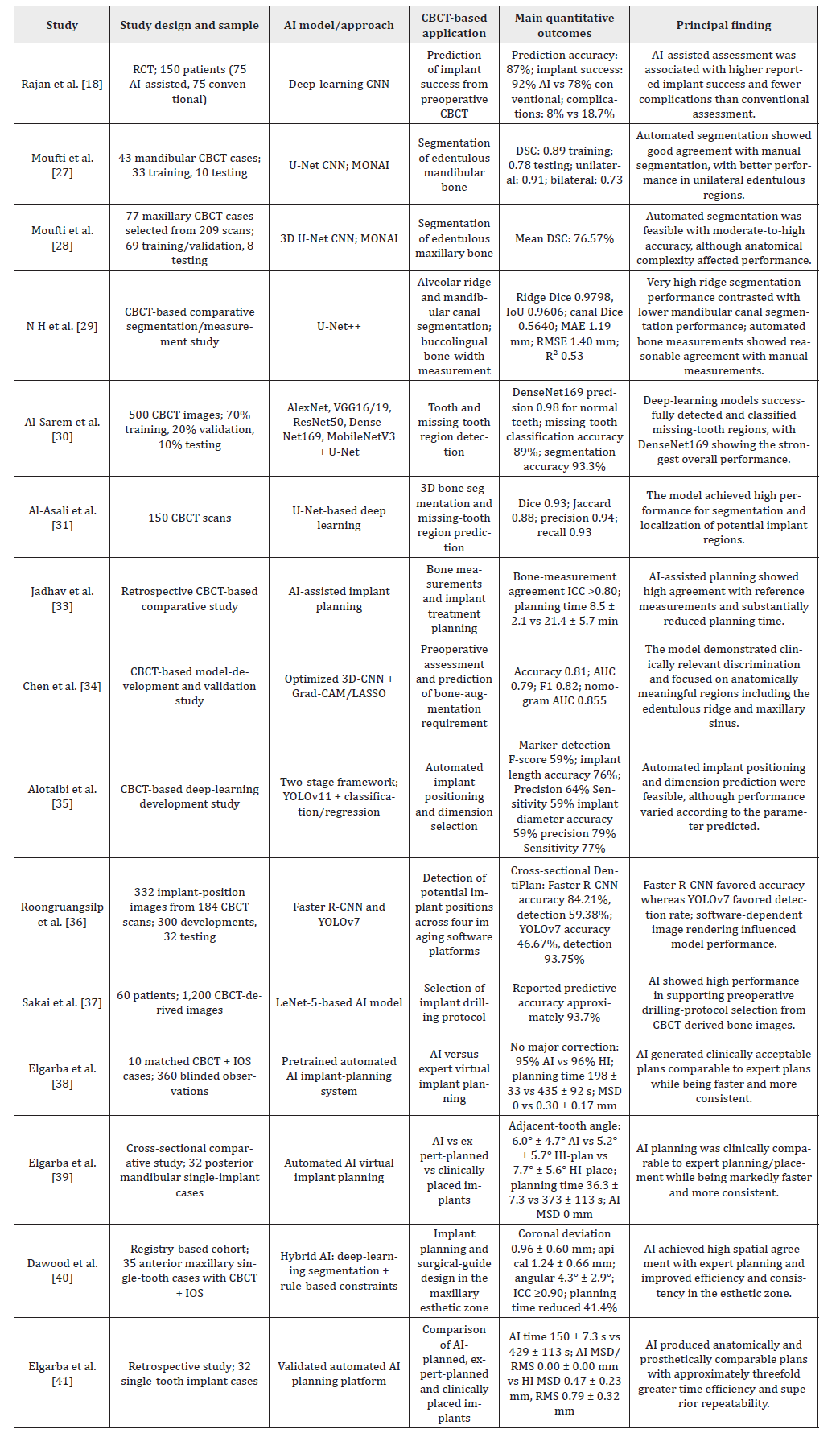
Abbreviations: AI, artificial intelligence; AUC, area under the receiver operating characteristic curve; CBCT, cone-beam computed tomography; CNN, convolutional neural network; DSC, Dice similarity coefficient; Grad-CAM, gradient-weighted class activation mapping; HI, human intelligence; ICC, intraclass correlation coefficient; IoU, intersection over union; IOS, intraoral scan; LASSO, least absolute shrinkage and selection operator; MAE, mean absolute error; MONAI, Medical Open Network for AI; MSD, mean surface distance; R-CNN, region-based convolutional neural network; RCT, randomized controlled trial; RMS, root mean square; RMSE, root mean square error; YOLO, You Only Look Once; 3D-CNN, three-dimensional convolutional neural network.
AI-based CBCT segmentation and anatomical structure detection
AI-based segmentation of CBCT images has shown high performance in automatically delineating anatomical structures relevant to implant planning. Moufti et al. [27] developed a U-Netbased model to automatically segment edentulous mandibular bone. The mean Dice similarity coefficient (DSC) was 0.89 for the training dataset and 0.78 for the testing dataset, with higher segmentation performance for unilateral than bilateral edentulous regions (0.91 vs. 0.73).
More recently, the same research group extended this approach to maxillary edentulous regions. In a dataset of 77 CBCT examinations, the automated model achieved a mean DSC of 76.57% [28].
Discrepancies between automated and manual segmentation were mainly observed in narrow bone regions and areas with irregular sinus-floor anatomy; in some cases, the automated model provided more precise anatomical delineation than manual segmentation.
High segmentation performance has also been reported for specific mandibular structures. Using a U-Net++ architecture, N H et al. [29] obtained a Dice score of 0.9798, an intersection-over-union of 0.9606, a precision of 0.9820, and a recall of 0.9778 for alveolar ridge segmentation. In contrast, mandibular canal segmentation yielded a lower Dice score of 0.5640.
At a broader level, a recent systematic review including 31 studies and 11,432 CBCT examinations reported consistently high performance for AI-based automated segmentation of maxillofacial structures, with Dice coefficients frequently exceeding 0.98 and substantial reductions in processing time compared with manual segmentation [24].
AI-based detection of edentulous regions and implant sites
AI-based approaches have also been developed to identify edentulous regions in CBCT images automatically and to facilitate the localization of potential implant sites. Al-Sarem et al. [30] evaluated a deep-learning approach using 500 CBCT examinations for three-dimensional detection of missing-tooth regions. The proposed system was designed to automatically identify edentulous areas and provide spatial information relevant to subsequent implant-site planning.
Subsequent investigations extended automated analysis to segment and localize missing-tooth regions. Al-Asali et al. [31] evaluated 150 CBCT scans using an AI-based segmentation approach designed to identify edentulous regions and support implant-site localization. The model demonstrated the feasibility of automatically delineating missing-tooth regions on threedimensional CBCT datasets, thereby providing a structured anatomical basis for subsequent implant-planning procedures.
Across the available evidence, automated detection performance appeared to vary according to anatomical location. A previously published quantitative synthesis reported an overall accuracy of approximately 96% for mandibular edentulous regions and 83% for maxillary regions [32].
AI-based bone assessment for implant planning
AI-based analysis of CBCT images has been investigated for the automated assessment of alveolar bone dimensions relevant to implant planning. N H et al. [29] evaluated automated bucco-lingual bone-width measurements following AI-based segmentation of the mandibular alveolar ridge. Compared with manual measurements, the automated approach yielded a mean absolute error (MAE) of 1.19 mm, a root mean square error (RMSE) of 1.40 mm, and an R² value of 0.53, indicating measurable correspondence between AIgenerated and manually obtained bone-width measurements.
Jadhav et al. [33] directly compared AI-assisted and manual bone measurements on CBCT images. Mean bone height was 12.06 ± 1.62 mm with AI versus 12.36 ± 1.78 mm manually, whereas mean bucco-lingual width was 7.18 ± 0.82 mm versus 7.55 ± 0.98 mm, respectively. The corresponding mean differences were -0.30 mm for bone height and −0.37 mm for bucco-lingual width. Agreement between the two approaches showed an ICC >0.80.
Beyond linear bone measurements, Chen et al. [34] investigated AI-assisted preoperative assessment using a three-dimensional convolutional neural network (3D-CNN). The optimized model achieved an accuracy of 0.81, an area under the receiver operating characteristic curve (AUC) of 0.79, and an F1-score of 0.82. Grad- CAM visualization showed that model attention was concentrated at clinically relevant regions, including the maxillary sinus floor adjacent to missing-tooth sites.
AI-driven virtual implant planning and implant dimension selection
AI-based systems have increasingly been investigated for automated or semi-automated virtual implant planning, including the selection of implant dimensions and the determination of clinically relevant treatment parameters. Alotaibi et al. [35] evaluated an AI-based model for predicting implant length and diameter from preoperative imaging data. For implant length prediction, the model achieved an accuracy of 76%, precision of 64%, and sensitivity of 59%. For implant diameter prediction, the corresponding values were 59%, 79%, and 77%, respectively. These findings demonstrated variable performance according to the implant dimension being predicted.
Roongruangsilp et al. [36] compared two AI architectures, Faster R-CNN and YOLOv7, for dental implant planning across four implant-planning software environments. Faster R-CNN demonstrated higher overall accuracy across the evaluated imaging conditions, whereas YOLOv7 showed comparatively lower but more consistent performance across different image-quality settings.
AI has also been evaluated as a decision-support tool for selecting surgical parameters associated with implant placement. Sakai et al. [37] developed a model to support selection of the implant drilling protocol. The trained model achieved an overall accuracy of approximately 93.7-93.8%. Sensitivities for drilling protocols A, B, and C were 97.5%, 95.0%, and 85.0%, respectively, while the corresponding AUC values were 98.6%, 98.6%, and 99.4%.
Taken together, these studies evaluated AI at different stages of virtual implant planning, ranging from implant dimension prediction to software-assisted planning and drilling-protocol selection [35, 36, 37].
AI versus human/expert implant planning
Several studies directly compared AI-generated and humangenerated implant plans across spatial, dimensional, and clinical planning parameters. Elgarba et al. [38] compared fully automated AI-generated implant plans with those produced by human experts. Major corrections were not required in 95% of AI-generated plans and 96% of human-generated plans. However, 35% of AI plans versus 56% of human plans required no modification, while 48% of the evaluations rated AI-generated plans as equivalent or superior to human-generated plans.
In a subsequent investigation, Elgarba et al. [39] compared AI-generated plans, human-generated plans, and the implants actually placed clinically. No statistically significant differences were observed among the three approaches for implant location and angulation or for the selected implant dimensions, including diameter and length (p > 0.05).
Jadhav et al. [33] similarly compared AI-assisted and manual CBCT-based implant planning. AI-assisted planning showed a mean angular deviation of 1.90 ± 0.83°, compared with 3.99 ± 1.09° for manual planning, and a mean linear deviation of 0.43 ± 0.15 mm, compared with 1.01 ± 0.38 mm, respectively. Both differences were statistically significant (p < 0.0001).
Clinical applicability was further evaluated by Dawood et al. [40] in the maxillary esthetic zone. AI-assisted planning resulted in mean coronal, apical, and angular deviations of 0.96 ± 0.60 mm, 1.24 ± 0.66 mm, and 4.3 ± 2.9°, respectively. Established clinical thresholds were met in 94.3% of cases for coronal deviation, 85.7% for apical deviation, and 94.3% for angular deviation. Agreement between AI- and human-generated plans was high across spatial coordinates, with intraclass correlation coefficients (ICC) ≥0.90.
Overall, the comparative studies showed substantial agreement between AI-assisted and expert implant planning for several spatial and dimensional parameters, while the magnitude and direction of differences varied according to the evaluated outcome [33, 38, 39, 40].
Clinical efficiency, repeatability and workflow optimization
Beyond planning accuracy, AI-assisted implant planning has been evaluated in terms of time efficiency, reproducibility, and workflow performance. Elgarba et al. [41] reported a marked reduction in planning time with fully automated AI-based planning compared with human planning. Mean planning time was 150 ± 7.3 seconds for AI versus 429 ± 113 seconds for human-generated plans (p < 0.001). The automated approach also demonstrated repeatability in repeated planning procedures, with mean surface distance (MSD) and root mean square (RMS) values of 0, indicating identical repeated outputs under the evaluated conditions.
Dawood et al. [40] similarly reported that AI-assisted planning reduced median planning time by 41.4% compared with human planning (p < 0.0001). The automated system demonstrated complete consistency across repeated planning procedures, while maintaining high agreement with expert-generated plans.
Jadhav et al. [33] observed a comparable improvement in workflow efficiency. Mean planning time was 8.5 ± 2.1 minutes with AI assistance versus 21.4 ± 5.7 minutes with manual planning. Clinician confidence scores were also higher during AI-assisted planning (82.1 ± 9.2 versus 68.9 ± 11.0; p < 0.001).
Collectively, these comparative studies consistently reported shorter planning times with AI-assisted approaches, together with high repeatability or consistency of automated planning outputs [33, 40, 41].
AI-assisted outcome prediction and clinical decision support
Beyond image analysis and implant positioning, researchers have also investigated AI-based approaches as decision-support tools for predicting treatment-related outcomes. Rajan et al. [18] reported an implant success rate of 92% in the AI-assisted group compared with 78% in the non-AI group, while complication rates were 8% and 18.7%, respectively. The AI model achieved a reported predictive accuracy of 87% for treatment outcomes.
At the evidence-synthesis level, recent umbrella reviews have reported high performance of AI for several implant-related image-analysis and planning tasks, whereas prediction of longerterm treatment outcomes has generally shown more variable performance. Alfaraj et al. [42], for example, reported outcomeprediction performance ranging approximately from 62.4% to 80.5% across the evidence synthesized.
Performance Metrics
Performance was reported using metrics from the included studies. Dice similarity coefficient (DSC) and intersection over union (IoU) quantify overlap between automated and reference segmentations, with values nearer 1 indicating greater agreement [29]. Accuracy is the proportion of correct predictions; precision is the proportion of correct positive predictions; recall (sensitivity) is the proportion of true positives identified; and the F1-score combines precision and recall [30, 34]. Mean absolute error (MAE) and root mean square error (RMSE) quantify prediction-reference differences, with lower values indicating smaller errors [29]. R² describes explained variability, intraclass correlation coefficient (ICC) assesses measurement agreement or reproducibility, and area under the receiver operating characteristic curve (AUC) measures discrimination, with values nearer 1 indicating better performance [29, 33, 34].
For repeated three-dimensional planning, mean surface distance (MSD) and root mean square (RMS) quantified spatial deviation; lower values indicated greater similarity [38, 41].
Discussion
The findings of this review indicate that artificial intelligence is progressively expanding the role of CBCT from a primarily diagnostic imaging modality toward a more integrated component of digital implant planning. Current applications range from anatomical segmentation and detection of edentulous regions to bone assessment, implant dimension selection, automated virtual positioning, workflow optimization, and outcome prediction. However, these applications have not reached the same level of maturity. The most consistent evidence concerns image-based tasks and selected components of implant planning, whereas greater variability remains for complex anatomical structures, implant dimension prediction, and longer-term outcome prediction [23-26, 32, 42, 43]. AI in CBCT-based implantology should therefore be viewed as progressing from relatively structured image-analysis tasks toward increasingly complex clinical decision support rather than as a uniformly validated technology across the entire implant workflow.
Recent literature has also addressed clinical validation, trustworthy and explainable AI, informed consent, and the development of integrated AI-assisted dental workflows [44-48].
Automated segmentation represents one of the most technically advanced applications. High performance has been reported for clearly defined structures such as the alveolar ridge, whereas results remain less consistent for anatomically complex or poorly contrasted structures. This was particularly evident for mandibular canal segmentation, which showed lower performance than alveolar ridge segmentation [29]. Differences were also observed between maxillary and mandibular edentulous regions [27, 28, 32]. These findings highlight that segmentation accuracy cannot be interpreted independently of anatomical complexity. Small errors involving narrow residual ridges, the maxillary sinus floor, or the mandibular neurovascular canal may influence subsequent measurements or implant positioning. Consequently, high Dice or overlap coefficients should not automatically be interpreted as evidence of clinical safety; region-specific errors and their clinical consequences should also be considered.
AI-based measurements of available implant bone showed encouraging agreement with manual measurements, although performance varied among models and anatomical conditions [29, 33]. The clinical significance of measurement error depends strongly on the anatomical safety margin. Errors that may be acceptable in sites with abundant bone could become relevant near the mandibular canal, maxillary sinus, adjacent roots, or a thin buccal cortical plate. Statistical agreement and clinical acceptability should therefore remain distinct concepts, and AI-generated measurements should continue to undergo clinician verification in anatomically constrained sites.
The available evidence also demonstrates a transition from anatomical analysis toward automated virtual implant planning. Implant positioning requires integration of bone availability with anatomical limitations, prosthetic requirements, implant dimensions, restorative emergence, surgical access, and, when necessary, augmentation procedures. Current AI systems can assist with several of these components, but performance remains taskdependent [35, 37, 43]. For example, implant length and diameter prediction showed unequal accuracy [35], whereas AI-assisted drilling-protocol selection achieved substantially higher predictive performance [37]. Success in one planning task therefore cannot be extrapolated to the entire implant-planning process.
Direct comparisons between AI-generated and expertgenerated plans are particularly relevant clinically. Recent studies demonstrated substantial spatial and dimensional agreement between automated and clinician-generated plans [33, 38-41], including selected posterior mandibular and maxillary estheticzone cases. These findings suggest that AI can generate clinically plausible initial treatment proposals under defined conditions. Nevertheless, available studies generally involve selected indications and populations. Their results cannot yet establish autonomous competence in more complex situations such as severe atrophy, anatomical abnormalities, extensive imaging artifacts, multiple adjacent implants, or advanced augmentation procedures.
Reproducibility should also be distinguished from accuracy. Automated systems have demonstrated very high repeatability, including identical outputs when the same cases were processed repeatedly [38-41]. This may reduce inter- and intra-operator variability, but a consistently reproduced plan is not necessarily a correct plan. Systematic errors arising from image quality, segmentation, registration, or decision rules may also be reproduced consistently. Repeatability is therefore an important component of reliability but cannot replace independent anatomical and clinical validation.
Time efficiency represents another consistent advantage. Comparative studies reported substantial reductions in planning time while maintaining clinically acceptable agreement with expertgenerated plans [33, 38-41, 43]. Automated generation of an initial treatment proposal may allow clinicians to devote more attention to verification, risk assessment, prosthetic considerations, and individualized decision-making. The role of AI should therefore be understood as supporting and restructuring professional involvement rather than eliminating it.
Outcome prediction remains more challenging. Although favorable findings have been reported for AI-assisted prediction of implant success and complications [18], broader evidence suggests greater variability for prognostic applications than for segmentation or structured planning tasks [19, 42]. Implant outcomes depend on numerous interacting biological, surgical, prosthetic, behavioral, and patient-related factors that extend beyond CBCT information. More reliable predictive systems will therefore likely require multimodal data integration rather than reliance on imaging alone.
Limitations Of Current Ai Applications In CBCTBased Implantology
Several limitations currently restrict the generalizability of AI applications in CBCT-based implantology. Many studies rely on relatively small, retrospective, single-center, or selected datasets, while independent external validation remains limited [23-26, 32, 42]. Restricted datasets may not adequately capture anatomical variation, bone resorption patterns, pathological conditions, restorative materials, image artifacts, or uncommon clinical situations. Class imbalance may further produce high overall performance while concealing clinically important failures.
Technical heterogeneity represents another major challenge. CBCT datasets vary according to scanner, field of view, voxel size, exposure parameters, reconstruction algorithms, preprocessing procedures, and software environments. AI performance may therefore differ across devices and implant-planning platforms [36]. Validation across multiple scanners, acquisition protocols, reconstruction conditions, and software systems is required before broad clinical generalizability can be assumed [23, 25, 32, 36, 49].
Methodological heterogeneity also limits direct comparison between studies. Different reference standards, datasets, validation strategies, and performance metrics are used, and measures such as Dice coefficients, IoU, accuracy, AUC, MAE, RMSE, ICC, and spatial deviations assess different aspects of model performance. Furthermore, the definition of ground truth may itself vary because expert annotations and implant plans are influenced by clinician experience, implant systems, surgical philosophies, and prosthetic objectives. Standardized evaluation protocols, calibrated expert reference standards, independent testing datasets, and clinically verified outcomes should therefore be prioritized.
A further limitation is the gap between technical accuracy and demonstrated clinical effectiveness. High segmentation performance, agreement with expert planning, or reduced processing time does not necessarily imply improved surgical safety, fewer complications, greater implant survival, or better patient-reported outcomes. Much of the current evidence remains focused on algorithmic performance rather than prospective clinical benefit [23-26, 32, 42]. Prospective multicenter studies are therefore needed before technical performance can be translated into claims of improved patient care.
Ethical, Legal, and Regulatory Considerations
The increasing integration of AI into diagnosis and implant planning introduces ethical and medico-legal issues that must accompany assessments of technical performance. Explainability remains an important concern because deep-learning systems may generate accurate outputs without providing clinicians with a readily interpretable rationale. Explainable AI approaches may help clinicians identify potential errors and understand the factors influencing a recommendation [20, 45, 46].
Human oversight therefore remains fundamental. AI systems should currently be regarded as decision-support technologies rather than autonomous decision-makers. Clinicians must retain the ability to accept, modify, or reject AI-generated recommendations after considering the complete biological, surgical, prosthetic, and patient-specific context [45].
Informed consent and data governance also require attention. When AI materially contributes to diagnosis or treatment planning, patients should receive clear information about its role, benefits, limitations, and ongoing clinician supervision [22, 47]. At the same time, integration of CBCT data with intraoral scans, clinical records, photographs, or cloud-based systems increases data sensitivity. It reinforces the need for appropriate anonymization, cybersecurity, access control, storage, transfer, and secondary-use policies [21, 45, 50].
Liability may become increasingly complex when treatment decisions arise from interactions between clinician judgment and algorithmic recommendations. Documentation of the AI system used, its intended purpose, relevant version, clinician review, and modifications to proposed plans may therefore become important elements of responsible clinical implementation. Emerging regulatory frameworks similarly emphasize trustworthy, transparent, and human-centered AI [45].
Future Perspectives
Future progress in AI-assisted CBCT implantology should focus increasingly on real-world generalizability rather than isolated improvements in performance metrics. Larger, heterogeneous, multicenter datasets incorporating different patient populations, anatomical conditions, CBCT devices, acquisition protocols, software platforms, and implant indications are required. Independent external validation, standardized benchmark datasets, and harmonized reporting frameworks would allow more meaningful comparison between algorithms [23-26, 32, 42, 44, 49].
Future systems are also likely to evolve beyond CBCT-only analysis toward multimodal digital implant workflows. Integration of CBCT with intraoral scans, prosthetic information, facial and soft-tissue data, clinical records, and longitudinal information could support a more comprehensive virtual representation of the patient [43, 48]. Rather than independently automating segmentation, measurement, or implant positioning, integrated systems may combine diagnosis, prosthetically driven planning, surgical-guide design, navigation, and outcome assessment within a unified decision-support environment.
Multimodal integration may be particularly valuable for outcome prediction, for which anatomical imaging alone is insufficient. Combining radiographic information with systemic, periodontal, surgical, prosthetic, behavioral, and longitudinal data may improve individualized risk assessment. Increasing model complexity, however, will reinforce the importance of explainability, uncertainty estimation, and clinician oversight.
Future algorithms should therefore ideally communicate not only a proposed result but also the confidence associated with that result. Identification of low-confidence cases could alert clinicians to situations requiring additional scrutiny or conventional assessment [45, 46].
Prospective clinical trials should increasingly evaluate endpoints beyond technical accuracy, including changes in treatment planning, operative time, surgical complications, implant-positioning accuracy, clinician workload, patient-reported outcomes, prosthetic complications, and long-term implant survival. Post-deployment monitoring will also be important to detect changes in algorithm performance when systems are exposed to populations or imaging conditions different from those used during development [45, 50].
Ultimately, the most plausible future model is not fully autonomous implant dentistry but an increasingly sophisticated collaboration between clinician and AI. AI is well suited to standardized, repetitive, and computationally intensive tasks, whereas clinicians remain responsible for integrating biological, surgical, prosthetic, ethical, and patient-specific considerations. Integrated digital workflows should therefore be designed to enhance rather than replace clinical judgment [43, 45, 48].
The present review should itself be interpreted in light of its narrative design. The available literature remains highly heterogeneous in AI architectures, dataset characteristics, imaging protocols, clinical tasks, reference standards, and performance metrics, limiting direct comparison among applications. Moreover, rapid technological evolution means that new models and validation studies may modify the evidence base within a relatively short period. The present synthesis should therefore be regarded as a contemporary assessment of a continuously evolving field.
Overall, current evidence supports a cautiously progressive role for AI in CBCT-based dental implantology. Automated segmentation, anatomical analysis, preliminary implant planning, and workflow optimization have reached encouraging levels of performance, and AI-generated plans can approach expert planning in selected clinical situations while reducing planning time [24, 33, 38-44]. Nevertheless, performance remains dependent on anatomy, task complexity, imaging conditions, software environment, and dataset characteristics, while outcome prediction and fully autonomous decision-making remain less mature. The most appropriate current framework is therefore a human-centered collaborative model in which AI supports computationally intensive aspects of implant planning while the clinician retains responsibility for interpretation, verification, and the final treatment decision.
Conclusion
Artificial intelligence is emerging as a valuable adjunct to CBCT-based dental implantology, with particularly promising applications in anatomical segmentation, bone assessment, virtual implant planning, and workflow optimization. Current evidence indicates that AI-assisted systems can improve efficiency, reproducibility, and, in selected clinical situations, achieve planning outcomes comparable to those of experienced clinicians. However, performance remains dependent on anatomical complexity, imaging conditions, software environments, and the quality and representativeness of training datasets, while external validation and evidence of direct clinical benefit remain limited. Future progress should therefore prioritize multicenter and prospective validation, standardized evaluation frameworks, multimodal data integration, explainable and uncertainty-aware models, and assessment of clinically meaningful outcomes. At present, AI should be regarded as a decision-support technology rather than an autonomous substitute for professional expertise, with clinicians retaining responsibility for verification, interpretation, and final treatment decisions.
Acknowledgement
None.
Conflicts of Interest
No conflicts of interest.
References
- Ladeira DB, da Cruz AD, de Almeida SM (2015) Digital panoramic radiography for diagnosis of the temporomandibular joint: CBCT as the gold standard. Braz Oral Res 1806-83242015000100303.
- Guerrero ME, Noriega J, Jacobs R (2014) Preoperative implant planning considering alveolar bone grafting needs and complication prediction using panoramic versus CBCT images. Imaging Sci Dent 44: 213-220.
- Wanderley VA, Leite AF, de Faria Vasconcelos K (2022) Impact of metal artefacts on subjective perception of image quality of 13 CBCT devices. Clin Oral Investig 26: 4457-4466.
- Lim HK, Jung SK, Kim SH, Cho Y, Song IS (2021) Deep semi-supervised learning for automatic segmentation of inferior alveolar nerve using a convolutional neural network. BMC Oral Health 7: 630.
- Hung K, Montalvao C, Tanaka R, Kawai T, Bornstein MM (2020) The use and performance of artificial intelligence applications in dental and maxillofacial radiology: A systematic review. Dentomaxillofac Radiol 49: 20190107.
- Altalhi AM, Alharbi FS, Alhodaithy MA (2023) The Impact of Artificial Intelligence on Dental Implantology: A Narrative Review. Cureus 30: 47941.
- Sorkhabi MM, Khajeh MS (2019) Classification of alveolar bone density using 3-D deep convolutional neural network in the cone-beam CT images: A 6-month clinical study. Measurement 148: 106945.
- Setzer FC, Shi KJ, Zhang Z, Yan H, Yoon H, et al. (2020) Artificial Intelligence for the Computer-aided Detection of Periapical Lesions in Cone-beam Computed Tomographic Images. J Endod 46: 987-993.
- Issa J, Olszewski R, Dyszkiewicz Konwińska M (2022) The Effectiveness of Semi-Automated and Fully Automatic Segmentation for Inferior Alveolar Canal Localization on CBCT Scans: A Systematic Review. Int J Environ Res Public Health 19: 560.
- Kurt Bayrakdar S, Orhan K, Bayrakdar IS, Bilgir E, Ezhov M, et al. (2021) A deep learning approach for dental implant planning in cone-beam computed tomography images. BMC Med Imaging 21: 86.
- Ni FD, Xu ZN, Liu MQ (2024) Towards clinically applicable automated mandibular canal segmentation on CBCT. Journal of Dentistry 144: 104931.
- Macrì M, Dalbis V, Dalbis G (2024) The Role and Applications of Artificial Intelligence in Dental Implant Planning: A Systematic Review. Bioengineering (Basel) 11: 778.
- Pinter GT, Decker R, Szenasi G, Barabas P, Huszar T (2022) Dynamic Navigation for Dental Implant Placement. J Vis Exp 13.
- Stünkel R, Zeller AN, Bohne T, Böhrnsen F, Wedi E, et al. (2022) Accuracy of intraoral real-time navigation versus static, CAD/CAM-manufactured pilot drilling guides in dental implant surgery: an in vitro study. Int J Implant Dent 8: 41.
- Tao B, Shen Y, Sun Y, Huang W, Wang F, et al. (2020) Comparative accuracy of cone-beam CT and conventional multislice computed tomography for real-time navigation in zygomatic implant surgery. Clin Implant Dent Relat Res 22: 747-755.
- Feher B, Lettner S, Heinze G, Karg F, Ulm C, et al. (2020) An advanced prediction model for postoperative complications and early implant failure. Clin Oral Implants Res 31: 928-935.
- Yadalam PK, Trivedi SS, Krishnamurthi I (2022) Machine learning predicts patient tangible outcomes after dental implant surgery. IEEE Access 10:131481-131488.
- Rajan RSS, Kumar HSK, Sekhar A, Nadakkavukaran D, Feroz SMA, et al. (2024) Evaluating the Role of AI in Predicting the Success of Dental Implants Based on Preoperative CBCT Images: A Randomized Controlled Trial. J Pharm Bioallied Sci 16: 5886-5888.
- Zhu Y, Du M, Li P, Lu H, Li A, et al. (2025) Prediction models for the complication incidence and survival rate of dental implants-a systematic review and critical appraisal. Int J Implant Dent 11: 5.
- Blackman J, Veerapen R (2025) On the practical, ethical, and legal necessity of clinical Artificial Intelligence explainability: an examination of key arguments. BMC Med Inform Decis Mak 5: 111.
- Murdoch B (2021) Privacy and artificial intelligence: challenges for protecting health information in a new era. BMC Med Ethics 15: 122.
- Chau M, Rahman MG, Debnath T (2025) From black box to clarity: Strategies for effective AI-informed consent in healthcare. Artif Intell Med 167: 103169.
- Acerra A, Aliberti A, Amato A, Eccellente A, Santurro A, et al. (2026) Artificial Intelligence in Implant Dentistry: Clinical Validity, Diagnostic Performance, Surgical Planning, and Medico-Legal Implications—A Narrative Review. Dent J (Basel) 14: 389.
- Iacob AM, Verdecchia A, García Mesa Y, Spinas E, Cobo T (2026) Effectiveness of automated segmentation of maxillofacial structures in cone-beam computed tomography images using artificial intelligence: A systematic review. Int Orthod 24: 101081.
- Vázquez Sebrango G, Anitua E, Macía I, Arganda Carreras I (2025) The role of artificial intelligence in implant dentistry: a systematic review. Int J Oral Maxillofac Surg 54: 1098-1122.
- Revilla León M, Gómez Polo M, Vyas S, Barmak BA, Gallucci GO, et al. (2023) Artificial intelligence applications in implant dentistry: A systematic review. J Prosthet Dent 129: 293-300.
- Moufti MA, Trabulsi N, Ghousheh M, Fattal T, Ashira A, et al. (2023) Developing an Artificial Intelligence Solution to Autosegment the Edentulous Mandibular Bone for Implant Planning. Eur J Dent 17: 1330-1337.
- Moufti MA, Alhalabieh T, Mohammad K, Alfailakawi T, Alshimmari T, et al. (2026) Developing an Artificial Intelligence Solution to Auto segment the Edentulous Maxillary Bone for Implant Planning. Eur J Dent 20(04): 1353-1359.
- N H, Ahmed J, Sujir N, Verma T, Vakharia K, et al. (2026) Comparison of artificial intelligence methods with manual measurements for mandibular bucco-lingual bone width assessment using cone-beam computed tomography images. Oral Radiol 42: 774-783.
- Al-Sarem M, Al-Asali M, Alqutaibi AY, Saeed F (2022) Enhanced Tooth Region Detection Using Pretrained Deep Learning Models. Int J Environ Res Public Health 21: 15414.
- Al-Asali M, Alqutaibi AY, Al-Sarem M, Saeed F (2024) Deep learning-based approach for 3D bone segmentation and prediction of missing tooth region for dental implant planning. Sci Rep 16: 13888.
- Alqutaibi AY, Algabri R, Ibrahim WI, Alhajj MN, Elawady D (2025) Dental implant planning using artificial intelligence: A systematic review and meta-analysis. J Prosthet Dent 134:1619-1629.
- Jadhav P, P S, Jain S, Parveen H, Likhithaswamy HR, Ur Rahman SA (2026) Artificial Intelligence Versus Human Expertise in Implant Treatment Planning: A Retrospective Cone-Beam Computed Tomography-Based Comparative Study. Cureus 18: e109410.
- Chen F, Chen H, Hai D (2025) AI-assisted preoperative surgical planning for dental implant. J Transl Med 24: 104.
- Alotaibi S, Alsomali M, Alghamdi S, Alfadda S, Alturaiki I, et al. (2025) Automatic placement of simulated dental implants within CBCT images in optimum positions: a deep learning model. Med Biol Eng Comput 63: 2325-2339.
- Roongruangsilp P, Narkbuakaew W, Khongkhunthian P (2025) Performance of two different artificial intelligence models in dental implant planning among four different implant planning software: a comparative study. BMC Oral Health 25: 984.
- Sakai T, Li H, Shimada T (2023) Development of artificial intelligence model for supporting implant drilling protocol decision-making. J Prosthodont Res 67: 360-365.
- Elgarba BM, Fontenele RC, Mangano F, Jacobs R (2024) Novel AI-based automated virtual implant placement: Artificial versus human intelligence. J Dent 147: 105146.
- Elgarba BM, Fontenele RC, Dawood EA, Lahoud P, Meeus J, et al. (2025) Clinical Feasibility of AI-Driven Automated Virtual Dental Implant Placement: A Cross-Sectional Comparative Study. Clin Implant Dent Relat Res 27: 70111.
- Dawood EA, Elgarba BM, Fontenele RC, Jacobs R (2026) Clinical Applicability of Artificial Intelligence-Driven Implant Planning and Surgical Guide Design in the Maxillary Esthetic Zone: A Registry-Based Cohort Study. Clin Oral Implants Res 37: 994-1007.
- Elgarba BM, Dawood EA, Fontenele RC, Lahoud P, Meeus J, et al. (2026) Automating virtual dental implant planning: can artificial intelligence match clinical expertise? Dentomaxillofac Radiol 1: 421-430.
- Alfaraj A, Limones Á, Ahmad S (2026) Harnessing AI in prosthodontics and implant dentistry: An umbrella review of systematic evidence. J Prosthodont 35: 127-142.
- Elgarba BM, Fontenele RC, Tarce M, Jacobs R (2024) Artificial intelligence serving pre-surgical digital implant planning: A scoping review. J Dent 143:104862.
- Che SA, Yang BE, Park SY (2025) Clinical evaluation of AI-based three-dimensional dental implant planning: A multicenter study. J Dent 162: 106066.
- Ducret M, Wahal E, Gruson D, Amrani S, Richert R, et al. (2024) Trustworthy Artificial Intelligence in Dentistry: Learnings from the EU AI Act. J Dent Res 103: 1051-1056.
- Muhammad D, Bendechache M (2024) Unveiling the black box: A systematic review of Explainable Artificial Intelligence in medical image analysis. Comput Struct Biotechnol J 12: 542-560.
- Roganović J (2025) Developing a consent checklist for AI in dentistry: Thematic analysis and pilot survey validation. Digit Health 11: 20552076251393227.
- Kadempour A, Wathanapong P, Cantatore A, Mirafzali S, Haku Mizuhara K, et al. (2026) Artificial intelligence-driven workflow synchronizing interdisciplinary dentistry: Narrative review. J Prosthodont Res 10.2186/jpr.JPR_D_25_00287.
- Fontenele RC, Gaêta Araujo H, Jacobs R (2025) Cone beam computed tomography in dentistry: Clinical recommendations and indication-specific features. J Dent 159: 105781.
- Tuygunov N, Samaranayake L, Khurshid Z, Rewthamrongsris P, Schwendicke F, et al. (2025) The Transformative Role of Artificial Intelligence in Dentistry: A Comprehensive Overview Part 2: The Promise and Perils, and the International Dental Federation Communique. Int Dent J 75: 397-404.
-

This work is licensed under a Creative Commons Attribution-NonCommercial 4.0 International License.