 Case Report
Treatment of a Rare Case of a Congenitally Missing Permanent Maxillary Canine
Hardus Strydom, Cape Town, South Africa.
Received Date:July 03, 2025; Published Date: July 14, 2025
Abstract
Dental agenesis is a common phenomenon with the third molars being the most common congenitally missing tooth. The maxillary canines are commonly impacted or ectopic in position and in extremely rare cases the maxillary canine may be congenitally missing. This presents the treating clinician with various aesthetic, psychological, and functional challenges that require treatment. This paper presents such a rare case of a unilateral congenitally missing maxillary canine in a non-syndromic male patient and presents the reader with various treatment options to be considered.
Keywords:Agenesis, Permanent maxillary canine
Introduction
Canines play an important protective [1, 2], and functional [3] role if in the correct occlusal position. Its development or odontogenesis starts with the enamel organ and will go through the known bud, cap and bell stages of dental development [4, 5]. Various factors, genetic and environmental, play a role in the regulation of odontogenesis in general and interference may result in the developmental absence (agenesis) of one or multiple teeth [6, 7]. Hypodontia has been reported to have a prevalence of 6.4% (95% CI: 5.7, 7.2) with the most commonly congenitally missing tooth (excluding third molars) being the mandibular second premolar, followed by the maxillary lateral incisor and then the maxillary second premolar [8]. However, the prevalence vary greatly across different populations with rates reported from as low as 2.8% in a study conducted on Malaysian patients [9] to as high as 12.6% reported in a study conducted on Bavarian patients [10], indicating influences from geographic, racial and ethnic factors.
The maxillary canine, if present, goes through the developmental stages and will clinically erupt between ages 10-12 [11] with known standard deviations [12, 13]. Though congenitally missing teeth is a common dental anomaly [14] agenesis of a maxillary canine is extremely rare [15, 16]. A differential diagnosis for a missing permanent canine, following clinical investigation, should always include a possible ectopic position or impaction of the canine. Treatment is needed to address the functional, aesthetic, and psychological problems associated with canine agenesis. This treatment requires an interdisciplinary approach [17-19] and include options such as substitution, dental Autotransplantation or prosthetic replacement of the congenitally missing tooth [20- 23]. The individual malocclusion, dental morphology and skeletal maturation all need to be carefully considered before selecting the best treatment option for each individual case.
Cases presenting with good functional occlusions may indicate, and only require, prosthetic replacement of the missing tooth. A prosthetic replacement may be fixed or removable, and may be either a tooth supported, or an implant supported prosthesis. An implant supported fixed prosthetic replacement of a single missing tooth has a 5-year survival rate of 98.3% (CI: 96.8%–99.1%) [24] and has the major advantages of preservation of the adjacent teeth, and the dentoalveolar process, but an alternative must be considered in cases where the surgical procedure of implant placement is contraindicated. These may include medical health conditions, insufficient bone volume or age. Disadvantages to the implant supported prosthetic replacement of a missing tooth include the financial burden and ankylosis. An implant supported prosthetic replacement can be compared to an ankylosed tooth [25- 28] and early placement should be avoided. Facial growth slows down after puberty, but residual growth still occurs. Forsberg and colleagues showed a significant increase in facial height between the ages of 25 and 45 [29] and continued vertical dentoalveolar growth in adults was confirmed by Tallgren and Sollow [30]. The jury is still out with regards to the ideal time to place an implant supported crown in the aesthetic zone, but early implant placement will result in infra-occlusion of the implant supported crown and should be avoided.
Tooth supported fixed prosthetic replacement of the missing tooth is a possible alternative and has 5-year survival rate of 94.7% (CI: 94.1%-96.9%) [24] and 88.7% after 10 years [31]. Possible complications for this alternative include loss of vitality, abutment tooth fracture, and secondary caries on the abutment tooth or teeth [32].
An alternative to prosthetic replacement of the missing tooth is dental Autotransplantation [33], a technique sensitive procedure, described as early as the 1700s [34], that becomes an option in cases where the patient requires a dental extraction of another tooth that can then act as a donor tooth. This tooth must be assessed in terms of form and function when determining its viability as donor. Various biological principles, including extra-alveolar time, periodontal ligament cell survival, damage to the dental follicle and Hertwig epithelial root sheath, effect of tooth position relative to the socket and splinting method, are critical for positive outcomes [35]. The survival rate for this procedure has been reported to range from 75% to 98% with the stage of root formation playing an important role [36-39]. Transplanted teeth with open apices may allow revascularization and a greater chance of survival compared to transplants with completed root formation at the time of the transplant [37]. Revascularization in transplants with mature roots and closed apices is unlikely and additional endodontic treatment is indicated for these teeth. This very technique sensitive procedure has many advantages with the most significant being continued eruption with dentoalveolar growth in growing patients [40], but it does not go without complication. These include replacement resorption (ankylosis), root resorption, and pulpal necrosis for those transplanted teeth that did not undergo root canal treatments at the time of transplantation [41, 42].
Substitution as treatment option for a congenitally missing lateral incisors has been widely published [23, 43-45]. Robertsson and Mohlin reported superior periodontal health for substitution cases compared to prosthetic replacement cases. They also found that patients report higher aesthetic satisfaction with substitution compared to those receiving prosthetic replacements [46]. This was corroborated by De-Marchi, et al. who studied smile attractiveness through the eyes of dentists and laypersons [47].
In this article we present a case report of a unilateral congenitally missing maxillary canine treated using substitution to avoid the need for prosthetic replacement of the missing tooth.
Case Presentation
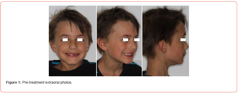
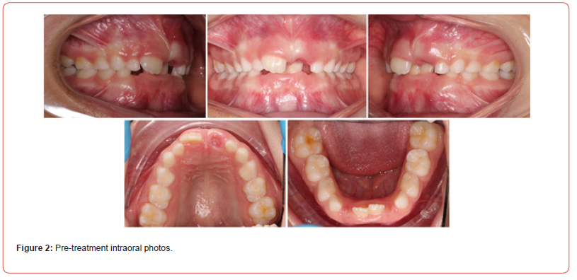
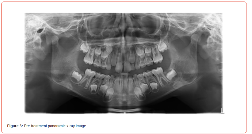
A healthy seven-year-old boy presented at the author’s orthodontic practice with a slightly convex profile, competent lips, and normal smile line (Figure 1). He had an Angle class II/1 (1/2 pw) malocclusion with a 6mm overbite and palatal contact in the early mixed dentition (Figure 2). A removable appliance was used to address the deep bite, and the first radiographic examination was done a year later and revealed the radiographic absence of the left maxillary canine (Figure 3). The decision was made to substitute the congenitally missing maxillary canine with the first maxillary premolar and use coronoplasty to avoid prosthetic replacement of the missing tooth.
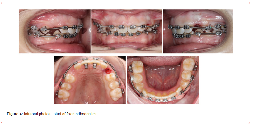
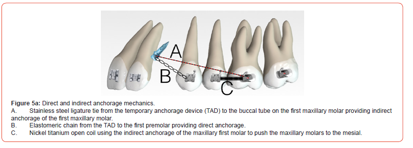
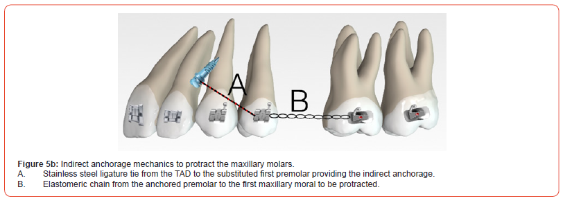
Treatment was started with a fixed orthodontic appliance (MiniMaster American orthodontics) to utilize the available leeway space and avoid the need for any additional dental extractions (Figure 4). The left maxillary deciduous canine was removed and both dental arches were leveled and aligned. A temporary anchorage device (Vector TAS mini screw 6mm ORMCO) was placed distal to the left lateral incisor root as soon as a stainless steel archwire could be placed. The protraction of the teeth in the second quadrant was then started with a combination of direct and indirect anchorage (Figure 5). The direct traction was done from the temporary anchorage device (TAD) to the first premolar. The indirect anchorage was achieved by ligature tie from the TAD to the first molar that then allowed push mechanics from the anchored first molar to the second premolar. The mechanics was adjusted as soon as the first premolar reached the canine position in class I occlusion. The second premolar was now ligature tied to the TAD and elastomeric chain used to protract the molars into class I occlusion (Figure 5b).
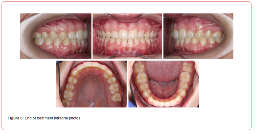
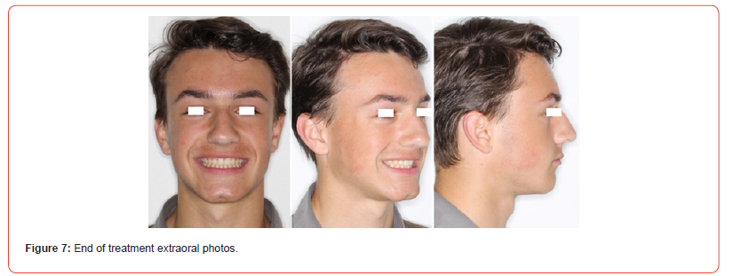
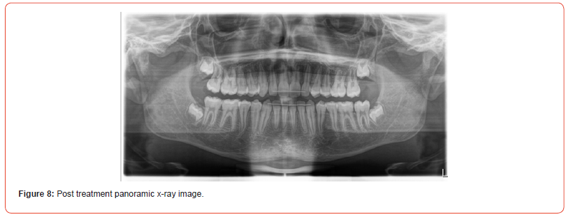
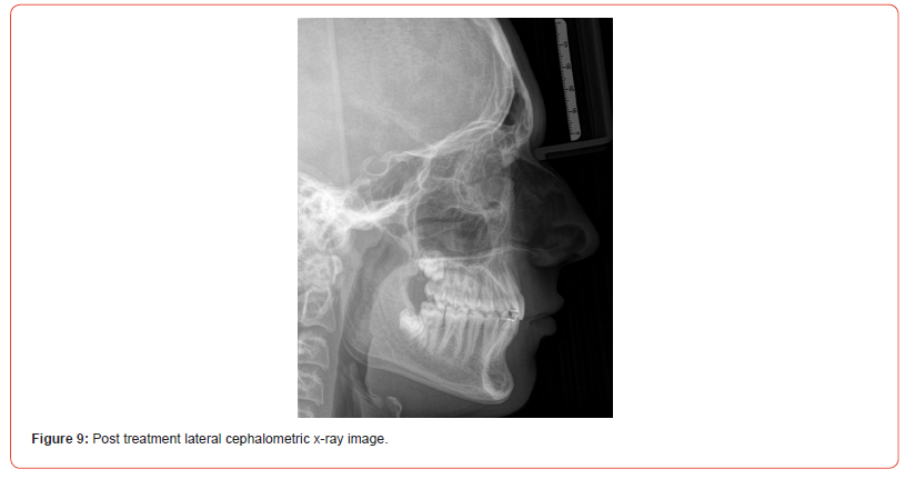
The case was finished with the canines and molars on the righthand side in class I relationship. The left maxillary first premolar was adjusted with composite to resemble a canine and finished in class I occlusion good gingival zenith, and the molar on the lefthand side in a full unit class II (Figure 6). The case was finished with a balanced, functional occlusion and favorable facial aesthetics (Figure 7).
Discussion
Congenitally missing canines are rare with reported incidences ranging from 0.18% (16) to 0.45% [48]. The indication to treat has a functional, aesthetic and emotional motivation [49] with options that include prosthetic replacement, Autotransplantation and dental substitution [20-23]. Factors like the condition of the predecessor, tooth size and shape, skeletal age, the patient’s malocclusion as well as the patient and/or parent’s preferences should be carefully considered during treatment planning [50]. Prosthetic replacement of the missing tooth may be an option but has its disadvantages when treating younger patients. It requires a temporary fixed or removable prosthesis, that may require preparation of abutment teeth, to allow maturation of the dentoalveolar process before a permanent prosthetic replacement can be placed. The dentoalveolar process rely on mechanical load through intermittent bite forces. Edentulous areas have a lack of functional forces transmitted via teeth and the periodontal ligament to bone. This lack of stimulus leads to bone loss or resorption [51]. Localized progressive resorption of the dentoalveolar process will thus continue in the edentulous area and may require augmentation procedures to ensure successful treatment and aesthetic results with the final prosthesis [52, 53]. Substitution is a viable treatment option and should be considered where possible.
The presented case shows a young patient with healthy teeth adjacent to the congenitally missing maxillary canine, and a third molar developing in a favorable position. There was no indication for dental extractions, which meant that Autotransplantation was excluded. Prosthetic replacement of the missing canine would require preparation of healthy teeth to act as abutment teeth, and potentially also require removal of his maxillary left third molar at a later stage.
We decided to proceed with a substitution treatment with coronoplasty of the maxillary first premolar to act as the substitute for the missing canine. The patient presented without an increased overjet and required additional anchorage to allow mesialization of the teeth in his second quadrant to protect the maxillary incisor position. Temporary anchorage devices (TADs) have become an integral part of the progressive orthodontist’s armamentarium [54, 55] The anchorage was reinforced with the addition of a temporary anchorage device (TAD) placed distal to the maxillary lateral incisor root. Baik et al. showed mesial drift of the mandibular third molars during mesialization of second molars to substitute lost first molars [56]. In the presented case the maxillary left third molar drifted mesial following mesial movement of the first and second maxillary molars and premolars. A limited treatment may be indicated to optimize the occlusal contact of the maxillary left third molar at a later stage, but there is no need for wisdom tooth removal.
Conclusion
Substitution presents a viable alternative to prosthodontic replacement of the missing permanent maxillary canine. It remains a treatment that requires interdisciplinary care and aims to address aesthetics and psychosocial needs at an early age.
Acknowledgement
None..
Conflict of Interest
The author declares no financial interest exist.
References
- Beyron H (1964) Occlusal Relations and Mastication in Australian Aborigines. Acta Odontol Scand 22: 597-678.
- Kahn AE (1977) The importance of canine and anterior tooth positions in occlusion. J Prosthet Dent 37(4): 397-410.
- Clark JR, Evans RD (2001) Functional occlusion: I. A review. J Orthod 28(1): 76-81.
- Hovorakova M, Lesot H, Peterka M, Peterkova R (2018) Early development of the human dentition revisited. J Anat 233(2): 135-145.
- Jheon AH, Seidel K, Biehs B, Klein OD (2013) From molecules to mastication: the development and evolution of teeth. Wiley Interdiscip Rev Dev Biol 2(2): 165-182.
- Meade MJ, Dreyer CW (2023) Tooth agenesis: An overview of diagnosis, aetiology and management. Jpn Dent Sci Rev 59: 209-218.
- Larmour CJ, Mossey PA, Thind BS, Forgie AH, Stirrups DR (2005) Hypodontia--a retrospective review of prevalence and etiology. Part I. Quintessence Int 36(4): 263-270.
- Khalaf K, Miskelly J, Voge E, Macfarlane TV (2014) Prevalence of hypodontia and associated factors: a systematic review and meta-analysis. J Orthod 41(4): 299-316.
- Nik-Hussein NN (1989) Hypodontia in the permanent dentition: a study of its prevalence in Malaysian children. Aust Orthod J 11(2): 93-95.
- Behr M, Proff P, Leitzmann M, Pretzel M, Handel G, et al. (2011) Survey of congenitally missing teeth in orthodontic patients in Eastern Bavaria. Eur J Orthod 33(1): 32-36.
- Haavikko K (1970) The formation and the alveolar and clinical eruption of the permanent teeth. An orthopantomographic study. Suom Hammaslaak Toim 66(3): 103-170.
- Hurme VO (1949) Ranges of normalcy in the eruption of permanent teeth. J Dent Child 16(2): 11-15.
- Hägg U, Taranger J (1986) Timing of tooth emergence. A prospective longitudinal study of Swedish urban children from birth to 18 years. Swed Dent J 10(5): 195-206.
- Rakhshan V (2013) Meta-analysis and systematic review of factors biasing the observed prevalence of congenitally missing teeth in permanent dentition excluding third molars. Prog Orthod 14: 33.
- Rózsa N, Nagy K, Vajó Z, Gábris K, Soós A, et al. (2009) Prevalence and distribution of permanent canine agenesis in dental paediatric and orthodontic patients in Hungary. Eur J Orthod 31(4): 374-379.
- Fukuta Y, Totsuka M, Takeda Y, Yamamoto H (2004) Congenital absence of the permanent canines: a clinico-statistical study. J Oral Sci 46(4): 247-252.
- Gill DS, Barker CS (2015) The multidisciplinary management of hypodontia: a team approach. Br Dent J 218(3): 143-149.
- Johal A, Huang Y, Toledano S (2022) Hypodontia and its impact on a young person's quality of life, esthetics, and self-esteem. Am J Orthod Dentofacial Orthop 161(2): 220-227.
- Laing E, Cunningham SJ, Jones S, Moles D, Gill D (2010) Psychosocial impact of hypodontia in children. Am J Orthod Dentofacial Orthop 137(1): 35-41.
- Nirola A, Bhardwaj SJ, Wangoo A, Chugh AS (2013) Treating congenitally missing teeth with an interdisciplinary approach. J Indian Soc Periodontol17(6): 793-795.
- Punj A, Yih J, Rogoff GS (2021) Interdisciplinary management of nonsyndromic tooth agenesis in the digital age. J Am Dent Assoc 152(4): 318-328.
- Krassnig M, Fickl S (2011) Congenitally missing lateral incisors--a comparison between restorative, implant, and orthodontic approaches. Dent Clin North Am 55(2): 283-299, viii.
- Kokich VO Jr, Kinzer GA (2005) Managing congenitally missing lateral incisors. Part I: Canine substitution. J Esthet Restor Dent 17(1): 5-10.
- Pjetursson BE, Valente NA, Strasding M, Zwahlen M, Liu S, et al. (2018) A systematic review of the survival and complication rates of zirconia-ceramic and metal-ceramic single crowns. Clin Oral Implants Res 29 Suppl 16: 199-214.
- Jemt T, Ahlberg G, Henriksson K, Bondevik O (2006) Changes of anterior clinical crown height in patients provided with single-implant restorations after more than 15 years of follow-up. Int J Prosthodont 19(5): 455-461.
- Odman J, Gröndahl K, Lekholm U, Thilander B (1991) The effect of osseointegrated implants on the dento-alveolar development. A clinical and radiographic study in growing pigs. Eur J Orthod 13(4): 279-286.
- Thilander B, Odman J, Gröndahl K, Lekholm U (1992) Aspects on osseointegrated implants inserted in growing jaws. A biometric and radiographic study in the young pig. Eur J Orthod 14(2): 99-109.
- Thilander B, Odman J, Gröndahl K, Friberg B (1994) Osseointegrated implants in adolescents. An alternative in replacing missing teeth? Eur J Orthod 16(2): 84-95.
- Forsberg CM, Eliasson S, Westergren H (1991) Face height and tooth eruption in adults--a 20-year follow-up investigation. Eur J Orthod 13(4): 249-254.
- Tallgren A, Solow B (1991) Age differences in adult dentoalveolar heights. Eur J Orthod 13(2): 149-156.
- Schmidlin K, Schnell N, Steiner S, Salvi GE, Pjetursson B, et al. (2010) Complication and failure rates in patients treated for chronic periodontitis and restored with single crowns on teeth and/or implants. Clin Oral Implants Res 21(5): 550-557.
- Sailer I, Makarov NA, Thoma DS, Zwahlen M, Pjetursson BE (2015) All-ceramic or metal-ceramic tooth-supported fixed dental prostheses (FDPs)? A systematic review of the survival and complication rates. Part I: Single crowns (SCs). Dent Mater 31(6): 603-623.
- Dokova AF, Lee JY, Mason M, Moretti A, Reside G, Christensen J (2024) Advancements in tooth Autotransplantation. J Am Dent Assoc 155(6): 475-483.
- Pape HD, Heiss R (1976) [History of tooth transplantation]. Fortschr Kiefer Gesichtschir. 20: 121-125.
- Fong CC, Agnew RG (1958) Transplantation of teeth: clinical and experimental studies. J Am Dent Assoc 56(1): 77-86.
- Rohof ECM, Kerdijk W, Jansma J, Livas C, Ren Y (2018) Autotransplantation of teeth with incomplete root formation: a systematic review and meta-analysis. Clin Oral Investig 22(4): 1613-1624.
- Chung WC, Tu YK, Lin YH, Lu HK (2014) Outcomes of auto transplanted teeth with complete root formation: a systematic review and meta-analysis. J Clin Periodontol 41(4): 412-423.
- Machado LA, do Nascimento RR, Ferreira DM, Mattos CT, Vilella OV (2016) Long-term prognosis of tooth Autotransplantation: a systematic review and meta-analysis. Int J Oral Maxillofac Surg 45(5): 610-617.
- Sicilia Pasos J, Kewalramani N, Pena Cardelles JF, Salgado Peralvo AO, Madrigal Martinez Pereda C, Lopez Carpintero A (2022) Autotransplantation of teeth with incomplete root formation: systematic review and meta-analysis. Clin Oral Investig 26(5): 3795-3805.
- Czochrowska EM, Stenvik A, Zachrisson BU (2002) The esthetic outcome of auto transplanted premolars replacing maxillary incisors. Dent Traumatol 18(5): 237-245.
- Louropoulou A, Andreasen JO, Leunisse M, Eggink E, Linssen M, et al. (2024) An evaluation of 910 premolars transplanted in the anterior region-A retrospective analysis of survival, success, and complications. Dent Traumatol 40(1): 22-34.
- Tsukiboshi M (2002) Autotransplantation of teeth: requirements for predictable success. Dent Traumatol 18(4): 157-180.
- Khalil A, Alrehaili R, Almatrodi R, Koshak A, Tawakkul B, et al. (2024) Congenitally Missing Lateral Incisors: Prioritizing Space Closure Whenever Feasible. Cureus 16(11): e74471.
- Zachrisson BU, Rosa M, Toreskog S (2011) Congenitally missing maxillary lateral incisors: canine substitution. Point. Am J Orthod Dentofacial Orthop 139(4): 434, 6, 8 passim.
- Kiliaridis S, Sidira M, Kirmanidou Y, Michalakis K (2016) Treatment options for congenitally missing lateral incisors. Eur J Oral Implantol 9 Suppl 1: S5-24.
- Robertsson S, Mohlin B (2000) The congenitally missing upper lateral incisor. A retrospective study of orthodontic space closure versus restorative treatment. Eur J Orthod 22(6): 697-710.
- De-Marchi LM, Pini NI, Ramos AL, Pascotto RC (2014) Smile attractiveness of patients treated for congenitally missing maxillary lateral incisors as rated by dentists, laypersons, and the patients themselves. J Prosthet Dent 112(3): 540-546.
- Davis PJ (1987) Hypodontia and hyperdontia of permanent teeth in Hong Kong schoolchildren. Community Dent Oral Epidemiol 15(4): 218-220.
- Silva Meza R (2003) Radiographic assessment of congenitally missing teeth in orthodontic patients. Int J Paediatr Dent 13(2): 112-116.
- Cho SY, Lee CK, Chan JC (2004) Congenitally missing maxillary permanent canines: report of 32 cases from an ethnic Chinese population. Int J Paediatr Dent 14(6): 446-450.
- Jonasson G, Skoglund I, Rythen M (2018) The rise and fall of the alveolar process: Dependency of teeth and metabolic aspects. Arch Oral Biol 96: 195-200.
- Schwarz F, Ramanauskaite A (2022) It is all about peri-implant tissue health. Periodontol 2000 88(1): 9-12.
- McKenna GJ, Gjengedal H, Harkin J, Holland N, Moore C, et al. (2022) Effect of Autogenous Bone Graft Site on Dental Implant Survival and Donor Site Complications: A Systematic Review and Meta-Analysis. J Evid Based Dent Pract 22(3): 101731.
- Park JH, Shin K (2020) An Overview of Clinical Applications for Temporary Anchorage Devices (TADs). Temporary Anchorage Devices in Clinical Orthodontics. pp. 1-15.
- Patel KS, Parmar I, Digumarthi UK, Parmar J, Quraishi A, et al. (2025) Temporary Anchorage Device: A Narrative Review. Cureus 17(4): e81617.
- Baik UB, Kang JH, Lee UL, Vaid NR, Kim YJ, et al. (2020) Factors associated with spontaneous mesialization of impacted mandibular third molars after second molar protraction. Angle Orthod 90(2): 181-186.
-
Hardus Strydom*. Treatment of a Rare Case of a Congenitally Missing Permanent Maxillary Canine. On J Dent & Oral Health. 9(1): 2025. OJDOH.MS.ID.000705.
-
: Maxillary canine, Missing tooth, Odontogenesis, Implant, Endodontic treatment, Root resorption, Periodontal health, Prosthetic replacement, Maxillary premolar, Orthodontist’s armamentarium
-

This work is licensed under a Creative Commons Attribution-NonCommercial 4.0 International License.


