 Case Report
Treatment of Dentigerous Cyst in Pediatric Patients: Two Case Reports
Emre Oğuzbal1, Elif Sepet1*, Sezer Şenkardeş2, Burak Yazıcı2 and Emine Tuna Akdoğan2
1Department of Pediatric Dentistry, Faculty of Dentistry, Istanbul Kent University, Istanbul, Turkey
2Department of Oral and Maxillofacial Surgery, Faculty of Dentistry, Istanbul Kent University, Istanbul, Turkey
Elif Sepet, Department of Pediatric Dentistry, Faculty of Dentistry, Istanbul Kent University, Istanbul, Turkey.
Received Date:February 22, 2024; Published Date:March 04, 2024
Abstract
Introduction: Dentigerous cysts are benign developmental lesions of the jaws that grow asymptomatically, usually associated with an impacted
tooth. These lesions can cause many complications such as pain, paresthesia, resorption, and tooth displacement over time. The treatment approach
in dentigerous cysts generally consists of surgical interventions including marsupialization and enucleation methods. In this study, the treatment
of dentigerous cysts in 2 pediatric patients was presented and it was aimed to discuss the outcomes of treatment methods in the light of evidence
based dentistry.
Case report: The patients were referred to Istanbul Kent University, Department of Pedodontics, for routine check-ups. In the first case,
the panaromic radiograph and computed tomography examinations showed large, circular, well-defined uniocular areas covering the crowns of
mandibular left and right third molars in an 11-year-old boy. The second case was a 13-year-old girl with swelling in the right mandible region. The
panaromic radiograph and computed tomography examinations showed a large, circular, well-defined cyst covering the crown of the mandibular
right second premolar. The teeth were extracted, and the enucleation technique was performed in both cases. The lesions were diagnosed as
dentigerous cysts according to the pathology results. Long-term clinical and radiographic follow-ups of the patients were planned.
Conclusion: Treatment of dentigerous cysts in pediatric patients requires a multidisciplinary approach, and many factors are effective in
treatment planning.
Keywords:Dentigerous cyst; Pediatric dentistry; Enucleation
Introduction
Dentigerous cysts, also known as follicular cysts, are benign developmental lesions of the jaws that grow asymptomatically [1]. It forms at the cemento-enamal junction, enclosing the crown of an unerupted tooth, as a result of a change in the reduced enamel epithelium [2]. Dentigerous cysts are the second most common odontogenic cysts after radicular cysts and frequently seen in secondary dentition, which makes them uncommon in childhood [3, 4]. The maxillary canine, mandibular premolars, and mandibular third molars are the teeth that are most commonly affected [5].
Dentigerous cysts are slowly growing, typically asymptomatic, and incidentally found during radiographic examination [6]. The cyst typically doesn’t cause any pain or discomfort [1]. Radiographically, a dentigerous cyst is seen as a well-defined unilocular radiolucency that surrounds the crown of an impacted tooth and frequently has a sclerotic border [7].
As the cysts expand, they may cause a palpable mass [1]. These lesions can cause many complications such as pain, paresthesia, resorption, pathologic fractures, tooth displacement/impaction and bone deformities over time [8, 9].
Multiple and bilateral dentigerous cysts are typically observed in developmental syndromes such as mucopolysaccharidosis, Maroteaux-Lamy syndrome, and cleidocranial dysplasia [10- 12]. When these disorders are absent, bilateral and multiple dentigerous cyst occurring is very rare [12, 13]. The treatment decision of dentigerous cysts is based on the patient’s age, location, and extent of the lesion [14]. The treatment approach in dentigerous cysts generally consists of surgical interventions including marsupialization and enucleation methods [9, 15]. Treatment often involves extraction and enucleation of the affected teeth. Marsupialization is the first step in the treatment of large cystic lesions, and then enucleation [16]. In mixed dentition, the prognosis is better with conservative therapy that involves extraction of primary teeth and marsupialization to permit spontaneous eruption of permanent teeth [17-19].
The aim of the present study is to present the treatment of dentigerous cysts in two pediatric patients and to discuss the outcomes of treatment methods in the light of evidence-based dentistry.
Case Reports
Written informed consents were obtained from the patients parents’ to publish anonymized information in this article.
Case 1
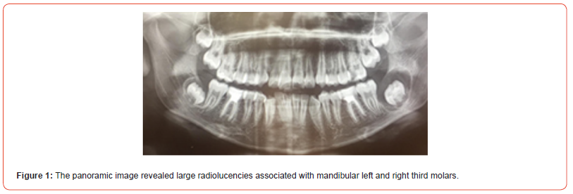
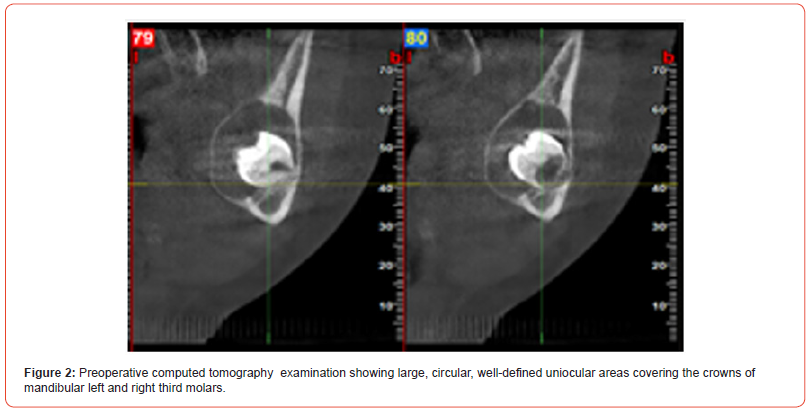
An 11-year-old boy was referred to Istanbul Kent University, Department of Pedodontics, with the complaint of dental pain related to the upper left first molar. There was no relevant medical or dental history. Intra-oral examination revealed a deep caries lesion of the upper left first molar. The panaromic radiograph and computed tomography examinations showed large, circular, welldefined uniocular areas covering the crowns of mandibular left and right third molars (Figures 1, 2).
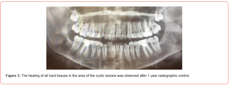
The root canal treatment was performed for the upper left first molar. The mandibular left and right third molars were extracted, and complete enucleating of the cysts was performed under general anesthesia. The specimen was submitted for histopathologic evaluation, which later confirmed the diagnosis of a dentigerous cyst. The healing of all hard tissues in the area of the cystic lesions was observed after 1 year control (Figure 3)..
Case 2
A systematically healthy 13-year-old girl was referred to Istanbul Kent University, Department of Pedodontics, for routine check-ups. The patient’s chief complaint was swelling in the lower right side of her mandible for 2 months. There was no relevant medical or dental history. The patient was moderately built and nourished. Intra-oral examination revealed palpable vestibular swelling that expanded from the mandibular right first premolar region to the mandibular right first molar region. The swelling was painless, firm, and there were no inflammatory signs in the overlying mucosa during palpation. A retained mandibular right second primary molar was present with a deep caries lesion. A bifid crown in the mandibular right lateral tooth was diagnosed as gemination according to the radiographic appearance. Lymph node examination eliminated the presence of any pathology.
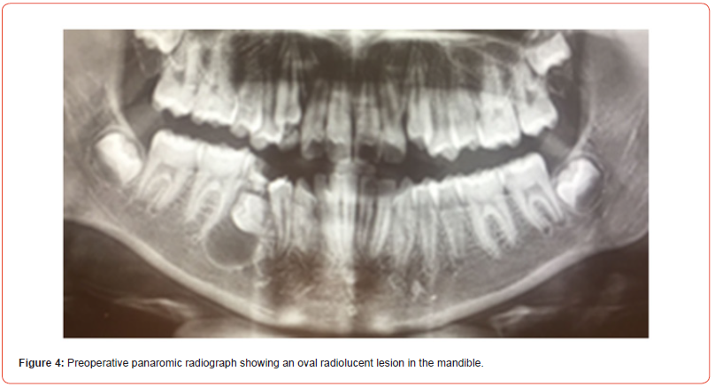
The panaromic radiograph and computed tomography examinations showed a large, circular, well-defined cyst covering the crown of the mandibular right second premolar (Figures 4, 5).
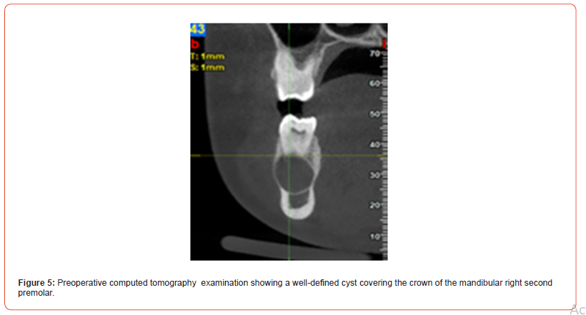
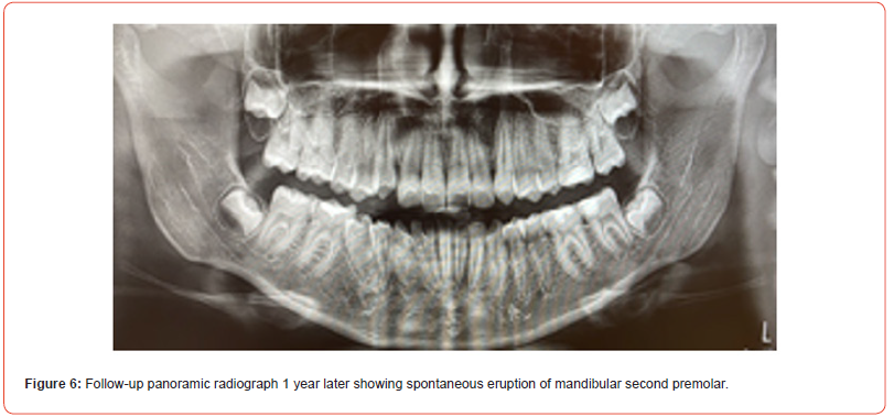
The mandibular right second primary molar was extracted, and a complete enucleation of the cysts was performed under general anesthesia. The specimen was sent for histopathological investigation, and according to the pathology result, the lesion was diagnosed as a dentigerous cyst. The spontaneous eruption of the mandibular second premolar was seen after 1 year control (Figure 6). Long-term clinical and radiographic follow-ups of the patients were planned.
Discussion
A dentigerous cyst can be inflammatory or noninflammatory [20]. Dentigerous cyst pathophysiology is still unknown, but several theories are proposed. According to the first theory, a dental follicle that has been secondarily inflamed by a non-vital tooth may be the source of the developing dentigerous cyst [4]. The second theory suggests that a dentigerous cyst of extrafollicular origin would arise from the formation of a radicular cyst at the apex of a non-vital deciduous tooth, which would be followed by the eruption of its permanent replacement into the radicular cyst [20]. Additionally, Benn and Altini [4] proposed that a dentigerous cyst could occur if a permanent successor’s follicle becomes secondary infected from other sources than the non-vital deciduous tooth [4, 21].
In this report, the first case showed a noninflammatory type of dentigerous cysts, however the second case suggested that the dentigerous cyst’s inflammation may have originated from an infection of the second primary molars.
In radiography, follicular spaces greater than 5mm are suspected of being dentigerous cysts [1, 7]. Radiographically, a dentigerous cyst is seen as a well-defined unilocular radiolucency that covers the crown of an impacted tooth and frequently has a sclerotic border [7].
Odontogenic tumors, odontogenic keratocysts, and primordial cysts must be considered in the differential diagnosis of a dentigerous cyst [22]. A histopathological examination can help an accurate diagnosis as radiography alone is unable to distinguish between the other lesions [21].
Histologically, dentigerous cysts consist of a fibrous wall that contains varying amounts of odontogenic remnants and myxoid tissue [23, 24]. Muco-sebaceous, sebaceous, and ciliated cells make up the nonkeratinized stratified squamous epithelium that lines the cyst [24]. When inflammation is present, the flattened epithelial-connective tissue interface becomes irregular [23]. Pseudoepitheliomatous hyperplasia, acute and chronic inflammatory infiltrates are frequently present [24].
Dentigerous cysts can be treated with either complete enucleation or marsupialization. Marsupialization may be recommended especially in younger patients and in cases when a large cyst increases the risk of surrounding tissue damage and a pathologic fracture of the jaw [25, 26].
Marsupialization is less complicated than enucleation with preserving of important anatomical structures and developing permanent tooth germs [21]. The pathologic tissue left in place after marsupialization is the disadvantage of marsupialization [1].
The cells in the lining of a dentigerous cyst can develop into ameloblastoma, squamous cell carcinoma, or intraosseous mucoepidermoid carcinoma; but the recurrence in dentigerous cyst is uncommon when the cyst is completely removed [26]. A radical method for removing all the cystic capsule is enucleation. Enucleation is the preferred method whenever the cyst is small, and saving the involved tooth is impossible [27].
Bilateral dentigerous cysts in the absence of a syndrome are rare, and the most common site of occurrence of the cyst is the mandibular region. The present cases were non syndromic; the lesions were in the mandibular region; and there was the presence of bilateral dentigerous cysts in our first case. In the present cases, the involved teeth were extracted, and the enucleation technique was performed. In the second case, the cyst led to a delay in the eruption of the lower right second premolar and an incorrect tooth position in the dental arch. The tooth spontaneously erupted after the surgical treatment of the cyst.
Treatment of dentigerous cysts in pediatric patients requires a multidisciplinary approach, and many factors are effective in treatment planning. The anatomic site, clinical extent, size of the cyst, and also the age and cooperation of the child must be taken into account in treatment planning. In these cases, the surgeons preferred the enucleation method according to the patients’ cooperation.
Conclusion
Asymptomatic dentigerous cysts can be diagnosed early in children during routine controls. A spontaneous eruption of a permanent tooth is possible after the surgical treatment of a dentigerous cyst. In order to prevent related complications and detect any pathological changes, periodic evaluation and long-term follow-up of the patients are necessary.
Author contributions
EO, ES, SŞ participated in designing the study. BY, ETA participated in generating the data for the study. ES, BY, ETA participated in the analysis of the data. ES, BY, ETA wrote the majority of the original draft of the paper. EO, SŞ participated in writing the paper. EO, ES, SŞ, BY, ETA have approved the final version of this paper. ES guarantee that all individuals who meet the Journal’s authorship criteria are included as authors of this paper.
Acknowledgement
This report was presented at the 29th International Congress of Turkish Society of Paediatric Dentistry, October, 12-15, 2023, Ankara, Türkiye.
Conflict of Interest
The authors had no conflict of interest to declare.
Financial Disclosure
The authors declared that this study has received no financial support.
References
- El Gaouzi R, El Harti K (2021) Dentigerous cyst: enucleation or marsupialization? (a case report). Pan Afr Med J 40(149): 1-7.
- Speight P, Fantasia FE, Neville BW (2017) Chapter 8- Odontogenic and maxillofacial bone tumours-Dentigerous cyst. In: El-Naggar AK, Chan JKC, Grandis JR, Takata T, Slootweg PJ (Eds.), WHO classification of head and neck tumours: International Agency for Research on Cancer. pp. 234-235.
- Arakeri G, Rai KK, Shivakumar HR, Khaji SI (2015) A massive dentigerous cyst of the mandible in a young patient: a case report. Plast Aesthet Res 2: 294-298.
- Benn A, Altini M (1996) Dentigerous cyst of inflammatory origin. Oral Surg Oral Med Oral Pathol Oral Radiol Endod 81: 203-209.
- Zhang LL, Yang R, Zhang L, Li W, MacDonald Jankowski D, et al. (2010) Dentigerous cyst: a retrospective clinicopathological analysis of 2082 dentigerous cysts in British Columbia, Canada. Int J Oral Maxillofac Surg 39(9): 878-882.
- Jones A, Craig G, Franklin C (2006) Range and demographics of odontogenic cysts diagnosed in a UK population over a 30-year period. J Oral Pathol Med 35: 500-507.
- Vasiapphan H, Christopher PJ, Kengasubbiah S, Shenoy V, Kumar S, et al. (2018) Bilateral dentigerous cyst in impacted mandibular third molars: a case report. Cureus 10(12): e3691.
- Devi P, Thimmarasa VB, Mehrotra V, Agarwal A (2015) Multiple dentigerous cyst: a case report and review. J Maxillofac Oral Surg 14(1): 47-51.
- Şahin O (2017) Conservative management of a dentigerous cyst associated with eruption of teeth in a 7-year-old girl: a case report. J Korean Assoc Oral Maxillofac Surg 43(1): 1-5.
- Ustuner E, Fitoz S, Atasoy C, Erden I, Akyar S (2003) Bilateral maxillary dentigerous cysts: a case report. Oral Surg Oral Med Oral Pathol Oral Radiol Endod 95(5): 632-635.
- Miller CS, Bean LR (1994) Pericoronal radiolucencies with and without radiopacities. Dent Clin North Am 38(1): 51-61.
- Shear M, Speight PM (2007) Cysts of the Oral and Maxillofacial Regions. (4th), Hoboken, New Jersey, USA: Blackwell Publishing.
- Dhupar A, Yadav S, Dhupar V, Mittal HC, Malik S, et al. (2017) Bi-maxillary dentigerous cyst in a non-syndromic child: review of literature with a case presentation. J Stomatol Oral Maxillofac Surg 118(1): 45-48.
- Alqadi S, Jambi SA, Abdullah AB, Aljuhani FM, Elsayed SA (2023) Conservative Management of Dentigerous Cysts Associated With Mixed Dentition: A Retrospective Cohort Study. Cureus 15(10): e47143.
- Sun KT, Chen MY, Chiang HH, Tsai HH (2009) Treatment of large jaw bone cysts in children. J Dent Child 76: 217-222.
- Bhardwaj S, Anand M, Altaf G, Kataria S (2019) Dentigerous cyst: a review of literatüre. Int Healthc Res J 3: 56-58.
- Tuwirqi A, Khzam N (2017) What do we know about dentigerous cysts in children, a review of literatüre. J Res Med Dent Sci 5: 67-79.
- Berti SA, Pompermayer AB, Couto Souza PH, Tanaka OM, Westphalen VP, et al. (2010) Spontaneous eruption of a canine after marsupialization of an infected dentigerous cyst. Am J Orthod Dentofacial Orthop 137: 690-693.
- Bozdogan E, Cankaya B, Gencay K, Aktoren O (2011) Conservative management of a large dentigerous cyst in a 6-year-old girl: a case report. J Dent Child 78: 163-167.
- Zerrin E, Husniye DK, Peruze C (2014) Dentigerous cysts of the jaws: clinical and radiological findings of 18 cases. J Oral Maxillofac Radiol 2(3): 77-81.
- Vinereanu A, Bratu A, Didilescu A, Munteanu A (2021) Management of large inflammatory dentigerous cysts adapted to the general condition of the patient: Two case reports. Exp Ther Med 22(1): 750.
- Duhan R, Tandon S, Vasudeva S, Sharma M (2015) Dentigerous cyst in maxillary sinus region: a rare case report and outline of clinical management for paediatric dentists. IOSR J Dent Med Sci 14(8): 84-88.
- Jain N, Gaur G, Chaturvedy V, Verma A (2018) Dentigerous cyst associated with impacted maxillary premolar: a rare site occurrence and a rare coincidence. Int J Clin Pediatr Dent 11(1): 50-52.
- Huang G, Moore L, Logan RM, Gue S (2019) Histological analysis of 41 dentigerous cysts in a paediatric population. J Oral Pathol Med 48: 74-78.
- Khandeparker RV, Khandeparker PV, Virginkar A, Savant K (2018) Bilateral maxillary dentigerous cysts in a nonsyndromic child: a rare presentation and review of the literatüre. Case Rep Dent 2018: 7583082.
- Hu YH, Chang YL, Tsai A.(2011) Conservative treatment of dentigerous cyst associated with primary teeth. Oral Surg Oral Med Oral Pathol Oral Radiol Endod 112(6): 5-7.
- Berden J, Koch G, Ullbro C (2010) Case series: treatment of large dentigerous cysts in children. Eur Arch Paediatr Dent 11(3): 140-145.
-
Emre Oğuzbal, Elif Sepet*, Sezer Şenkardeş, Burak Yazıcı and Emine Tuna Akdoğan. Treatment of Dentigerous Cyst in Pediatric Patients: Two Case Reports. On J Dent & Oral Health. 7(4): 2024. OJDOH.MS.ID.000670.
-
Dentistry, Teeth, Enamel epithelium, Dental pain, Intra oral examination, Computed tomography, Odontogenic tumors, Odontogenic keratocysts, Tooth germs
-

This work is licensed under a Creative Commons Attribution-NonCommercial 4.0 International License.


