 Research Article
The Study Result of Facial Soft Tissue Thickness of Mongolian Children
Erdenebulgan Purevjav1*, Bilguun Enkhtaivan1, Amarsaikhan Bazar2, Yerkyebulan Mukhtar3 and Badamtsetseg Mashbat4
1*Department of Dental Technology, School of Dentistry, Mongolian National University of Medical Sciences, Ulaanbaatar, Mongolia
2Department of Prosthodontics, School of Dentistry, Mongolian National University of Medical Sciences, Ulaanbaatar, Mongolia
3Department of Epidemiology and Biostatistics, School of Public Health, Mongolian National University of Medical Sciences, Ulaanbaatar, Mongolia
4Department of Dentistry, Mongolian National University of School of Medicine, Ulaanbaatar, Mongolia.
Erdenebulgan Purevjav, PhD, DDS, Department of Dental Technology, School of Dentistry, Mongolian National University of Medical Sciences, Ulaanbaatar, Mongolia.
Received Date: June 27, 2025; Published Date:July 09, 2025
Abstract
Objectives: This study created a reference dataset for facial soft tissue thickness of Mongolian children per age and sex.
Methodology: The present study was conducted on lateral cephalograms of 541 subjects (225 male and 316 females) having normal occlusion
in the age group of 6 to 15 years. All radiographs were digitized on a computer using a cephalometric software program (Winceph 11.0; Rise, Sendai,
Japan). All lateral cephalograms using a 16 landmark point. A total of eight linear measurements was measured for facial soft tissue thickness
analysis.
Results: Facial soft tissue thickness comparisons between groups per age and sex were statistically analyzed. Facial soft tissue thickness values were statistically significant concerning age and sex, except for stomion (P <0.001). There were no statistically significant differences between the sexes. (P> 0.01)
Conclusion: Statistical analysis shows that the thickness of facial soft tissue in Mongolian children increases with age. There is no significant difference in sex, but the lower part of the face is slightly thicker than in boys. It differs in most respects from the normal average for children of other nationalities and ethnic groups. There were differences between children of other nationalities and Mongolian children.
Keywords:Cephalogram, Cephalometric X-ray images, Facial soft tissue thickness, Mongolian children
Materials and Methods
Subjects
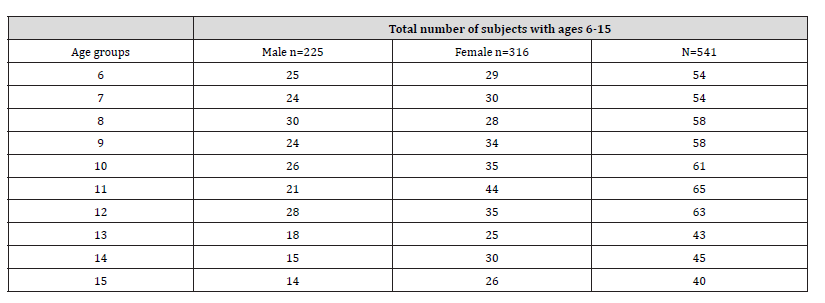
The Craniofacial Collaborative Research Project, a collaborative effort of the Tokyo Medical and Dental University and the Mongolian National University of Medical Sciences, conducted a longitudinal population-based survey of craniofacial growth of Mongolian children between 2013 and 2015. A total of 1842 students, attending the 33rd and 67th elementary and junior high schools of Ulaanbaatar participated. They were screened using a medical examination, questionnaire, cephalometric X-ray images, profile photograph, and mandibula and maxilla impressions. Based on the inclusion criteria, 541 children (225 male and 316 females) (Table 1) were enrolled to have measurements in this study and, their lateral cephalogram was performed between July 2018 and March 2019.
Table 1:Demographic characteristics of research participants.
Children were included in our study if they were 6 to 15 years of age, had normal growth and development, average body mass index, no facial asymmetry, no malocclusion or occlusal deformation, Angle’s Class I occlusion with well-aligned maxillary and mandibular dental arches, overjet and overbite scale within 2-4 mm, cephalograms of normal contrast, no previous history of orthodontic or prosthodontic treatments and no history of maxillofacial or plastic surgery.
The measurements were made on a lateral cephalometric X-ray image.
Cephalograms
Lateral cephalograms of the subjects were taken using a digital cephalometric machine (Veraviewpocs 2D CP X550, Morita, Japan). The subjects were placed in the head holder and asked to look straight ahead to establish the natural head position before adjusting the built-in nasal positioner with a millimeter scale. With teeth in centric occlusion and lips relaxed, the cephalogram was taken at a focus/object distance of 150cm and an object receptor distance of 20cm.
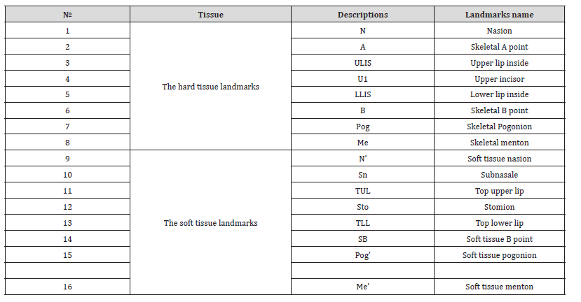
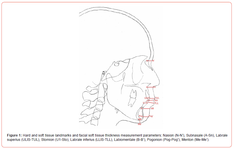
All lateral cephalograms were digitized on a computer by one radiologist with 20 years of experience doing cephalometry to eliminate inter-examiner variability. Using cephalometric software (Winceph 11.0; Rise, Sendai, Japan), eight linear measurements (Figure 1) were obtained for skeletal hard and soft tissue analysis using 16 anthropological landmark points shown in Table 2. Dentists with more than 20 years of experience with cephalometry and image manipulation have validated the landmarks and determined their reproducibility to be 95% using the ellipse method.
Table 2:Hard and soft tissue landmarks.
Landmarks
The anthropological landmarks were identified on each lateral cephalogram. All the required cephalometric landmarks were identified and marked using a cursor/mouse manually. The landmarks and measurements were taken according to the soft tissue cephalometric analysis [1-20].
Measurements
Nine measurements were selected to evaluate the changes in the soft tissue thickness and are as follows: Nasion, Subnasale, Labrale superius, Stomion, Labrale inferius, Labiomentale, Pogonion, Menton.
Statistical analysis
The results were encoded using SPSS 25.0 software, processed after whispering and error checking. The Kolmogorov - Smirnov test method was used to calculate the distribution of the numerical variable, and it was assumed that the p distribution was normal for values higher than 0.05. The Test tested the age-sex relationship for linear contrasts in One-Way ANOVA, and the Independent T-test tested the sex difference. Repeated ANOVA test was used to calculate the difference between the numerical variables of group 3 and above, after determining the distribution, and if there is a normal distribution depending on the distribution. If the p-value is less than 0.01, the difference is considered statistically significant. The reliability of the measurement method was calculated using Dahlberg’s error method [32].
Ethical statement
Ethical approval for this study was obtained from the Research Ethics Committee of the Mongolian National University of Medical Sciences on 08 June, 2018 /approval number 2018-3-10-15/. Before data collection, the parents of all children signed written, informed consent.
Results
Overall data of Mongolian children divided into ten chronological age classes (of 6 and 15 years old) are shown in table 1. General results divided between males and females are shown in Table 3, Figures 2 and 3.
Table 3 shows that facial soft tissue thickness means that standard deviations and values were compared for each age and gender. Statistically significant measurements differed with age, except for stomion (P <0.001). Statistical analysis shows that the thickness of facial soft tissue in Mongolian children increases with age.
Table 4 shows facial soft tissue thickness means, standard deviations and values were compared for gender, and there is no significant difference in gender, but the lower part of the face is slightly thicker for boys. (P <0.01).
A comparison analysis of soft tissue thickness between this study and other populations are shown in Table 5. It differs in most respects from the usual average of children of other nationalities and ethnic groups.
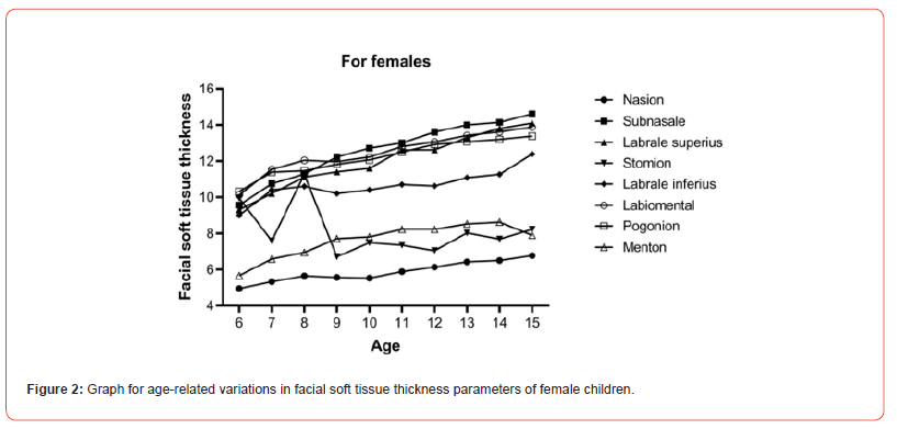
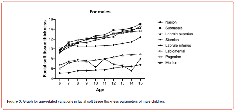
Table 3:Standard average soft tissue thickness of Mongolian children aged 6-15 years / by age and sex /
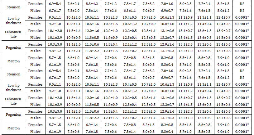
N=541, SD=Standard Deviation, Test for linear contrasts in One-Way ANOVA. NS-not significant, *p<0.001. All measurements are reported in mm.
Table 4:Standard average soft tissue thickness of Mongolian children / by sex /

N=541, SD=Standard Deviation, Independent T-test. NS-not significant, *p<0.01 All measurements are reported in mm.
Discussion
In this study, we determined the average mean and standard deviations of facial soft tissue thickness of a relatively healthy Mongolian child aged. 6-15 years (Tables 3 and 4) This is the first study of its kind in Mongolia, so we compared the results of foreign researchers’ measurements of children’s facial soft tissue thickness.
The lateral cephalograms have been shown to be a reliable method of assessing facial soft tissue, and many researchers have used this method in their research [23-25, 27, 30, 31]. Carl Stephen, a laboratory researcher at the University of Queensland, Australia International researchers estimate that there are about 1,290 studies of facial soft tissue thickness in children and 2,200 in adults [22]. Although there are many studies of facial soft tissue thickness on lateral cranial radiographs [23-25], we have clarified this because no such study has been conducted in Mongolia; The lateral cephalograms measurements were compared with American white and African black, black, European, and Asian populations. The measurement error of the study was estimated by the Dahlberg method [32].
Facial soft tissue thickness is affected by a person’s age, sex, ethnicity, lifestyle, and disease. The thickness of the facial soft tissue of a growing child varies. The study and identification of these can be used to diagnose abnormalities and distinguish between normal and abnormal growth and development; It is essential to develop a treatment plan and monitor its effectiveness [33]. The thickness of the soft tissue varies depending on the type and classification of the defect [34, 35].
According to foreign researchers, in most cases, the thickness of the soft tissue of the face of men was higher than that of women. According to Uysal, the thickness of the upper and lower lips and the thickness of the soft tissue of the anterior and inferior margins were statistically significant for the sexes [26]. Also, several studies evaluating the soft tissue thickness of different ethnic groups at different ages have shown that men outnumber women [27-29].
The thickness of the facial soft tissue of Mongolian children did not differ much by sex, but the Stomion part of a girl was thicker than that of a boy and the Menton part of a boy was thicker than that of a girl. (P <0.01) Nasion was 5.9 ± 0.9mm thinner than other parts. is similar to that of Japanese children [12, 16]; less than American [31] and European [21] children; thicker than black children [30].
The thickness of the upper (12.0 ± 1.8) and lower lip (10.7 ± 1.2) of Mongolian children is thinner than that of children of other nationalities. (P <0.01) The thickness of the soft tissue of the anterior margin is relatively thick, and the thickness of the soft tissue of the lower edge is less than that of Japanese children [23]. Facial soft tissue thickness increases at least 0.3-0.5mm per year on average in the Menton-menton area at the age of 9-12 years, and increases by a maximum of 1.2mm in the Stomion area (p <0.01). Stomion, Pogonion for female children aged 9-12 years in terms of growth trends; The Stomion and Upper lip thickness section of the male child is thicker than the other sections.
Conclusion
1. The profile and shape of the face is based in the database
of the facial soft tissue thickness.
2. It differs in most respects from the usual average of
children of other nationalities and ethnic groups.
3. It is believed that the increase in facial soft tissue
thickness varies with age and sex, in addition to the individual
characteristics of each child’s growth and development, and the
stage of sexual maturation.
4. Soft tissue thickness database of Mongolian children
aged 6-15 created and used as a diagnostic and treatment
reference in the field of orthopedics, surgery, forensic medicine,
anthropology, dermatology, and artificial intelligence base.
Acknowledgement
None.
Conflict of Interest
The authors declared no conflict of interest.
References
- Kotrashetti VS, Mallapur MD (2016) Radiographic assessment of facial soft tissue thickness in the South Indian population – an anthropologic study. J Forensic Leg Med 39(1): 161-168.
- You Soo Kim, Kang-Woo Lee (2019) The regional thickness of facial skin and superficial fat: Application to the minimally invasive procedures. Clin Anat 32(8): 1008-1018.
- Zhou Z, Peng L (2016) Virtual facial reconstruction based on accurate registration and fusion of 3D facial and MSCT scans. J Orofac Orthop 77(2): 1-8.
- Aulsebrook WA, Becker PJ (1996) Facial soft-tissue thickness in the adult male Zulu. Forensic Sci Int 79(2): 83-102.
- Philips VM, Smuts NA (1996) Facial reconstruction: utilization of computed tomography to measure facial tissue thickness in a mixed racial population. Forensic Sci Int 83(1): 51-59.
- Mevlut SK, Buyuk E (2014) Assessment of the soft tissue thickness at the lower anterior face in adult patients with different skeletal vertical patterns using cone-beam computed tomography. Angle Orthod 85(2): 211-217.
- Johari F, Esmaeili H Hamidi (2017) Facial soft tissue thickness of midline in an Iranian sample: MRI study. Open Dent J 11: 375-383.
- El-Mehallawi IH, Soliman EM (2001) Ultrasonic assessment of facial soft tissue thickness in adult Egyptians. Forensic Sci. Int 117(1-2): 99-107.
- Gungor K, Bulut O, Gurcan (2015) Variations of midline facial soft tissue thicknesses among three skeletal classes in Central Anatolian adults. Leg Med 17(6): 459-466
- Ferrario VF, Sforza (1997) Size and shape of soft-tissue facial profile: effects of age, gender and skeletal class. Cleft Palate Craniofac J 34(6): 498-504.
- De Greef S, Vandermeulen DP, Claes G, Willems G (2009) The influence of sex, age and body mass index on facial soft tissue depths. Forensic Sci Med 5(2): 60-65.
- Utsuno H, Kageyama T (2007) Facial soft tissue thickness in skeletal type I Japanese children. Forensic Sci 172(2-3): 137-143.
- Williamson MA, Nawrocki SP, Rathbun TA (2002) Variation in midfacial tissue thickness of African-American children. J Forensic Sci 47(1): 25-31
- Peckmann TR, Manhein MH, Fournier M (2013) In vivo facial tissue depth for Canadian Aboriginal children: a case study from Nova Scotia, Canada. J Forensic Sci 58(6): 1429-1437.
- Hwang HS, Kim WS, McNamara JA (2002) Ethnic differences in the soft tissue profile of Korean and European-American adults with normal occlusions and well-balanced faces. Angle Orthod 72(1): 72-80.
- Shota K, Yoshitaka M (2015) Tooth size in Chinese Oroqen ethnic minority of Inner Mongolia Autonomous Region. Odontology 103(3): 264-273.
- Zecca PA, Fastuca R (2016) Correlation assessment between three-dimensional facial soft tissue scan and lateral cephalometric radiography in orthodontic diagnosis. Int J Dent 1: 1-8.
- Wilkinson C (2010) Facial reconstruction: anatomical art or artistic anatomy?. J Anat 216(2): 235-250.
- De Greef S, Willems G (2005) Three-dimensional craniofacial reconstruction in forensic identification: Latest progress and new tendencies in the 21st century. J Forensic Sci 50(1): 12-17.
- Arnett GW, Bergman RT (1993) Facial keys to orthodontic diagnosis and treatment planning Part I. Am J Orthod Dentofacial Orthop 103(4): 299-312.
- Daniele Gibelli, Federica Collini, Chiarella Sforza (2016) Variations of midfacial soft-tissue thickness in subjects aged between 6 and 18 years for the reconstruction of the profile: a study on an Italian sample. Legal Med 22: 68-74.
- Carl N Stephan (2017) 2018 tallied facial soft tissue thickness for adults and sub-adults. Forensic Sci Int 280: 113-123.
- Utsuno H, Kageyama T, Uchida K (2010) Facial soft tissue thickness in Japanese children. Forensic Sci Int 199(1-3): 109.e1-e6.
- Gibelli D, Collini F, C. Sforza (2016) Variations of midfacial soft-tissue thickness in subjects aged between 6 and 18years for the reconstruction of the profile: a study on an Italian sample. Leg Med 22: 68-74.
- Waqar Jeelani, Mubassar Fida, Attiya Shaikh (2015) Age and sex-related variations in facial soft tissue thickness in a sample of Pakistani children. Austr J Forensic Sci 49(1): 1-14.
- Uysal T, Yagci A, Basciftci FA, Sisman Y (2009) Standards of soft tissue Arnett analysis for surgical planning in Turkish adults. Eur J Orthod 31(4): 449-456.
- Hodson G, Leiberman LS (1985) In vivo measurements of facial tissue thickness in American Caucasoid children. Forensic Sci 30(4): 1100-1112.
- Sama Hamid, Amal H (2016) Facial soft tissue thickness in a sample of Sudanese adults with different occlusions. Forensic Sci Inter 266: 209-214.
- Gyathri R, Ramesh N (2015) Facial soft tissue thickness in Forensic facial reconstruction: Is it enough if norms set? J Forensic Res 6: 5.
- Briers N, Briers TM (2015) Soft tissue thickness values for black and colored South African children aged 6-13 years. Forensic Sci Int 252: 188.e1-10.
- Smith SL, Buschang PH (2001) Midsagittal facial tissue thicknesses of children and adolescents from the Montreal growth study. J Forensic Sci 46(6): 1294-302.
- Galvão MCS, Sato JR, Coelho EC (2012) Dahlberg formula – a novel approach for its evaluation. Dental Press J Orthod 17(1): 115-124.
- Dong Y, Huang L, Feng Z, Bai S, Wu G, et al. (2012) Influence of sex and body mass index on facial soft tissue thickness measurements of the northern Chinese adult population. Forensic Sci Int 222(1-3): 396.e1-7.
- Gibelli D, Collini Zago M (2018) Modifications of midfacial soft-tissue thickness among different skeletal classes in Italian children. Open Med Imag J10(1): 1-8.
- Utsuno H, Kageyama T (2010) Pilot study of facial soft tissue thickness differences among three skeletal classes in Japanese females. Forensic Sci Int 195(1-3): 165. e1-5.
-
Erdenebulgan Purevjav*, Bilguun Enkhtaivan, Amarsaikhan Bazar, Yerkyebulan Mukhtar and Badamtsetseg Mashbat. The Study Result of Facial Soft Tissue Thickness of Mongolian Children. On J Dent & Oral Health. 9(1): 2025. OJDOH.MS.ID.000704.
-
Facial soft tissue, Maxilla impressions, Facial asymmetry, Orthodontic or prosthodontic treatments, Maxillofacial or plastic surgery, Dentists, Upper lip, Lower lips
-

This work is licensed under a Creative Commons Attribution-NonCommercial 4.0 International License.


