 Case Report
Orthodontic Treatment with the Ultima Damon System, Four Permanent Contact Points for Precise Control of Rotation, Angulation, and Torque-Case Report
Bashar Muselmani, Orthodontics department, Practice for orthodontics, Germany.
Received Date: September 07, 2023; Published Date: September 21, 2023
Summary
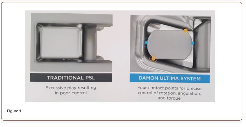
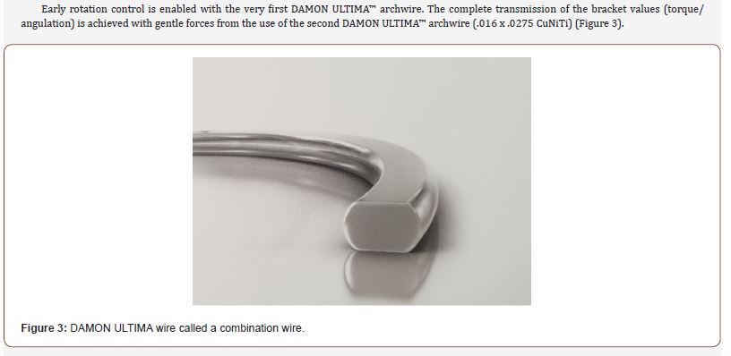
DAMON ULTIMA™ is the first full orthodontic treatment system specifically designed for faster and more accurate finishing. The DAMON ULTIMA™ system features a slot in the shape of a parallelogram. In connection with the new the patented DAMON ULTIMA™ arch (rounded square arch) enables a direct interaction at the vertical and horizontal contact points (7) (Figures 1, 2).

Keywords:Gingivitis; Dental plaques; Adjunctive therapies; Host modulation therapy; Tissue engineering; 3D printing
Clinical Case Study
Patient history
Patient: N/A Age: 17 years, 1 month.
Diagnosis
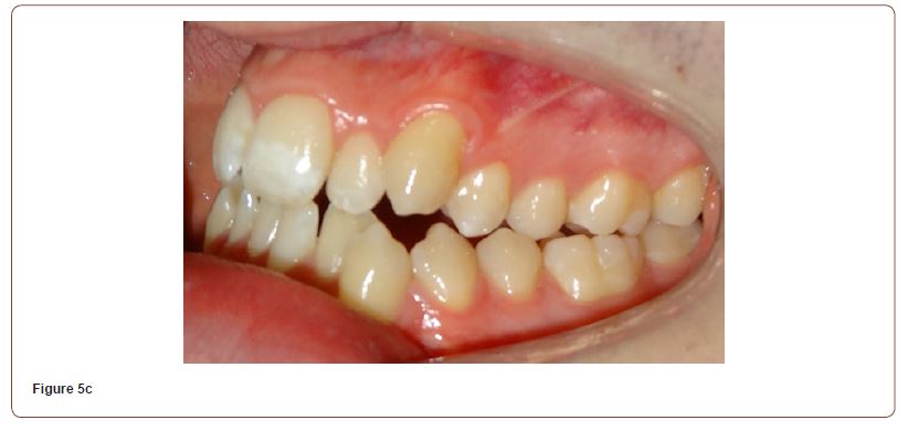
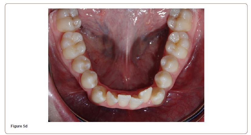
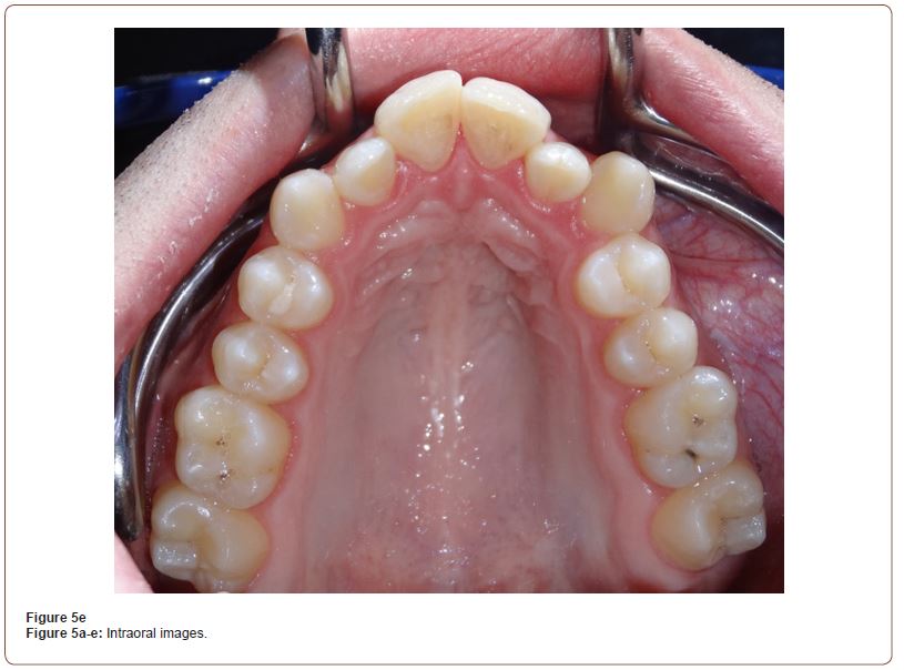
At the diagnosis appointment, the following was determined: Skeletal class III. Bilateral crossbite, narrow upper and lower jaw with crowding in the anterior and canine areas, midline shift to the left.
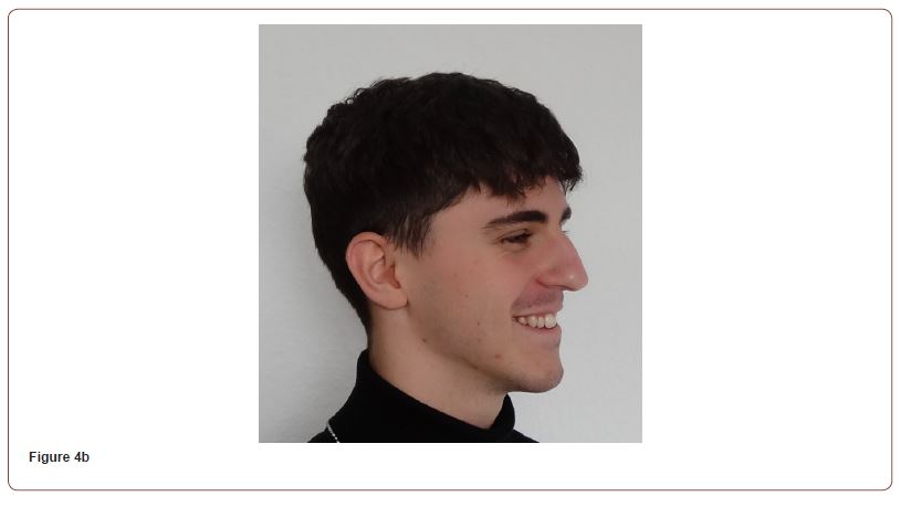
A competent lip closure with a convex mouth profile.
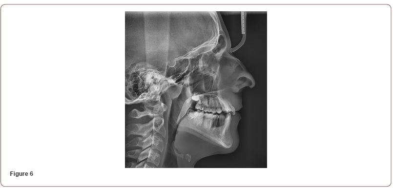
Ceph analysis:
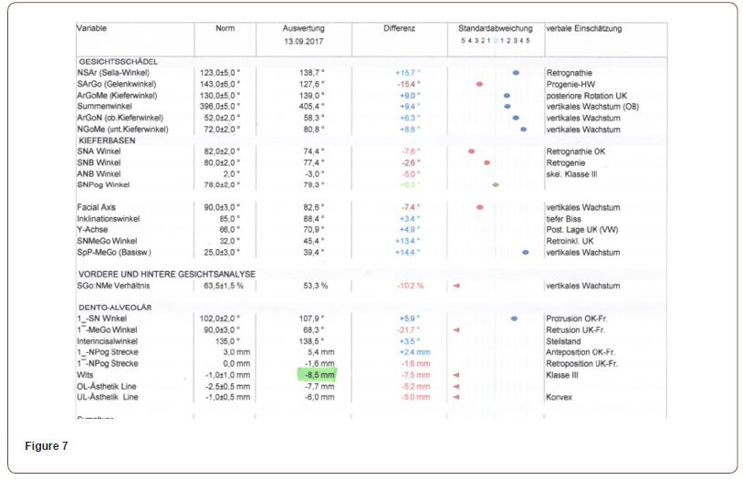
SNA angle was 74.4° (difference -7.6°, retrognacy OK), SNB angle was 77.4° (difference -2.6°, retrognacy lower), ANB angle was -3.0° (Difference -5.0° Skel. Class III), Wits was -8.5mm ((Difference -7.5mm, Skel. Class III).
The lower incisors showed a strong retrusion to the Me-Go line with -21.7°.
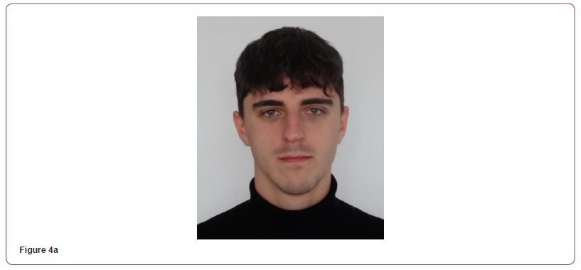
Figures 4a-d are the extraoral images and Figures 5a-e are the intraoral images.
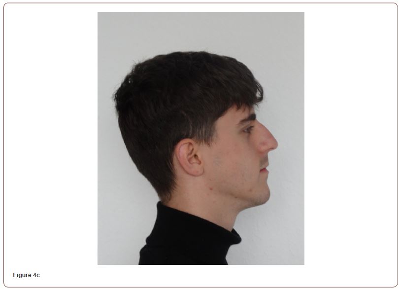
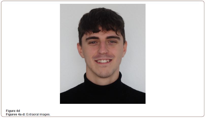
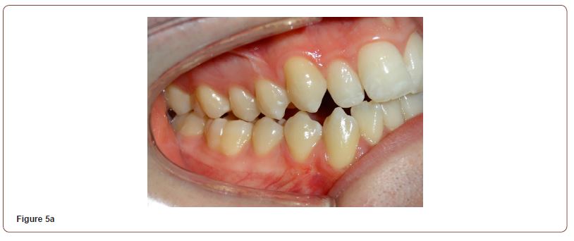
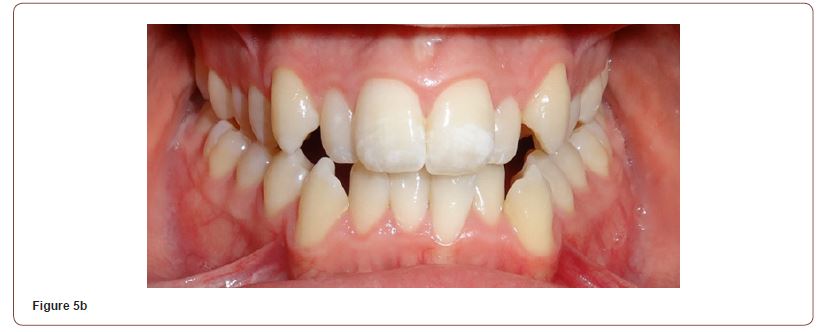
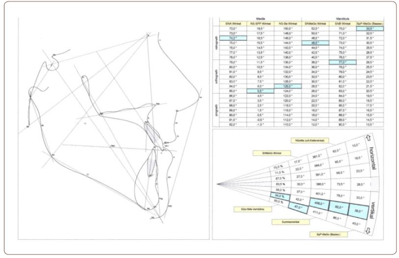
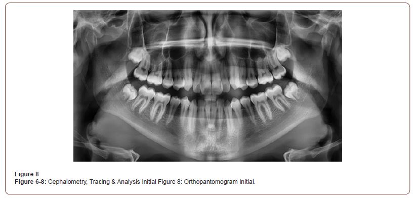
In Figures 6, 7, and 8 are, the cephalometric image with the evaluation and the orthopantomogram.
Course of treatment
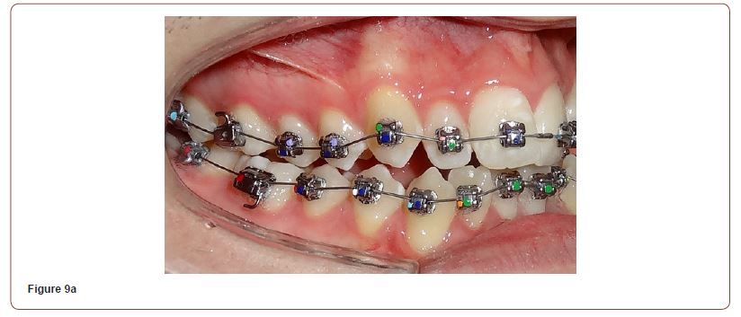
The bonding was complete in the upper and lower jaw.
Torque selection: the torque values were selected as follows: 13, 23, 33, 43,44 h.Tq, 12, 22, 31, 32, 41, 42 Low Tq, 11, 21, st.Tq.
Appointment 1: 04.05.2020
A .013” CuNiTi wire was ligated into both jaws at the start of leveling.
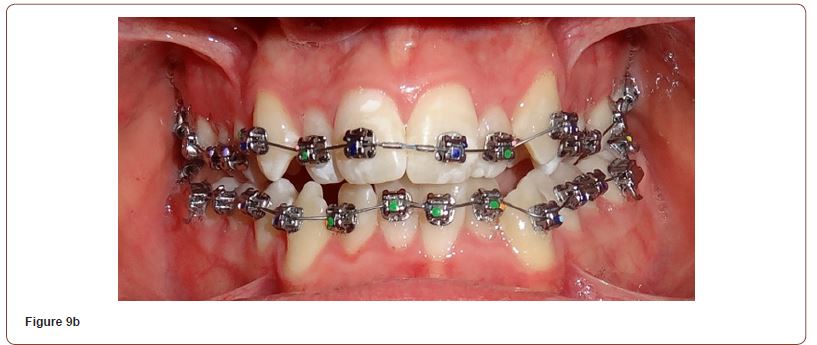
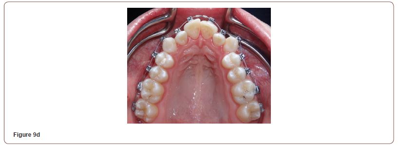
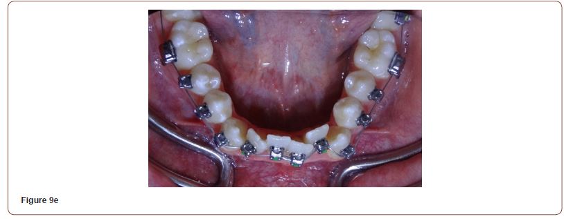
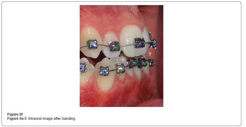
Appointment 2: 06/22/2020
Arch change in the upper jaw was a .018” CuNiTi
Appointment 3: 07/23/2020
Arch change in the upper jaw, a .0140 x .0275 CuNiTi (this is the first size of Ultima wire) in lower jaw ligated .018” CuNiTi.
Appointment 4: 09/03/2020
A cephalometry and orthopantomogram were performed at this appointment.
Appointment 5: 12.14.2020
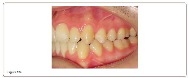
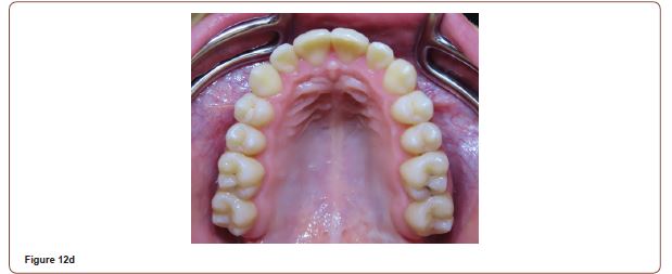
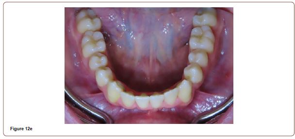
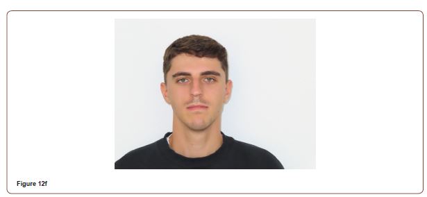
Arch change: up. 0160x.0275 SST, lwr.0140 X.0275 CuNiTi. Fig. 12a-f
Appointment 6: 04.04.2021
Arch change (up. 0.18 x .0275 CuNiTi, lwr. 0.16 x .0275 SST).
Appointment 7: 08/26/2021
Arch Change: (up. 0.18 X .275 TMA, lwr. 0.18 X .0275 CuNiTi). The patient continued to wear the vertical elastics (Figures10a-c).
Figures 10a-c
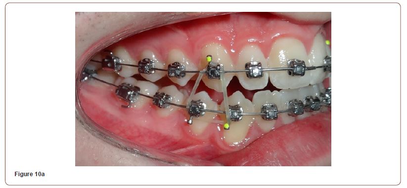
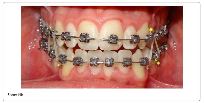
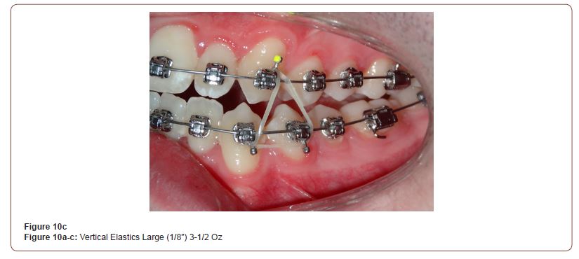
Recordings were carried out for comparison with the initial findings (Figures11a-c).
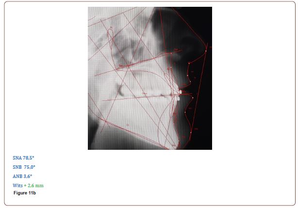
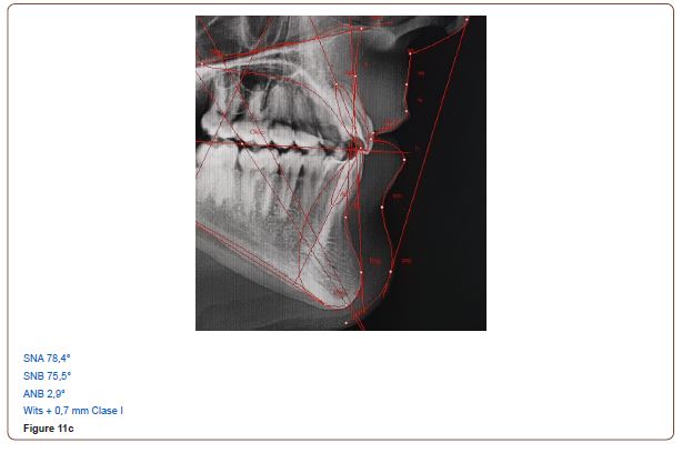
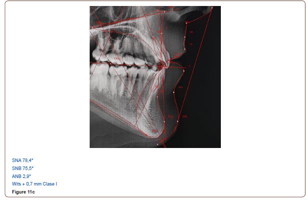
The active treatment phase is over, brackets in the upper jaw and lower jaw are removed, and a permanent lingual retainer is inserted in lower jaw 33-43. After one day, the patient wore a retention splint in the upper jaw and lower jaw.
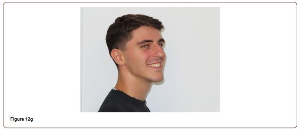
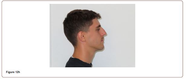
Completed documents: Models, X-rays and photos were made and evaluated. In Figures 12a-i are the results.
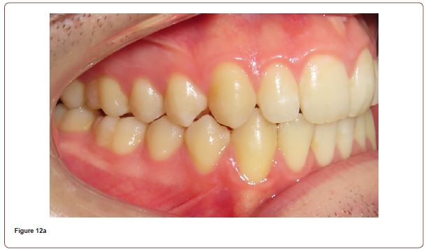
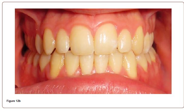

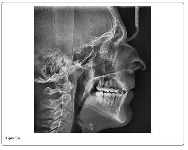
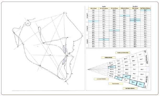
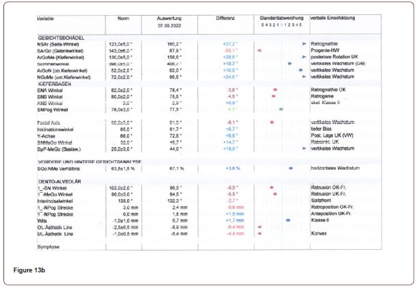
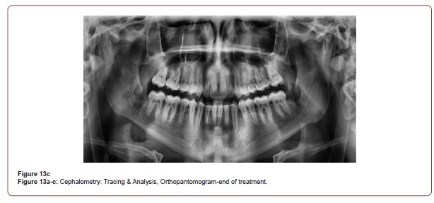
In Figure 13a-c are the cephalometric image with the evaluation and the orthopantomogram.
Discussion
Alignment, leveling, as well as Class I adjustment took about 15 months of active treatment in this case.
The clinical recordings as well as the cephalometric analyzes show a rapid and big difference between the beginning and the end of the treatment.
The cephalometric values are of greater importance, which can be well observed by comparing the three treatment phases (Figures 11a-c).
Conclusion
The presented case report shows examples of the change in size and shape of the maxillary and mandibular alveolar bone observed in adolescents treated with a passive self-ligating continuous multiband and the Damon Ultima low friction/low force treatment protocol. The Damon Ultima demonstrates precise control of rotation, angulation, and torque with shorter treatment time.
Acknowledgement
None.
Conflict of Interest
No Conflict of interest.
-
Bashar Muselmani*. Orthodontic Treatment with the Ultima Damon System, Four Permanent Contact Points for Precise Control of Rotation, Angulation, and Torque-Case Report. On J Dent & Oral Health. 7(2): 2023. OJDOH.MS.ID.000659.
-
Open access Journal of Dentistry & Oral Health, Journal of Dentistry & Oral Health, Journal of Dental, Open access journal of Oral Health, Journal of Oral Health, Dentistry & Oral Health Impact factor, High Impact factor of Dentistry & Oral Health
-

This work is licensed under a Creative Commons Attribution-NonCommercial 4.0 International License.


