




 Case Report
Pseudo-Mobitz Type II Atrioventricular Block Associated with Retrograde Ventriculoatrial Conduction Following Premature Ventricular Complexes: An Electrocardiographic Pitfall
Received Date: September 14, 2026; Published Date: September 21, 2026
Abstract
Background: Mobitz type II atrioventricular block is an important conduction abnormality that may require permanent pacing. However, electrophysiological phenomena associated with premature ventricular complexes may mimic this pattern and lead to misdiagnosis.
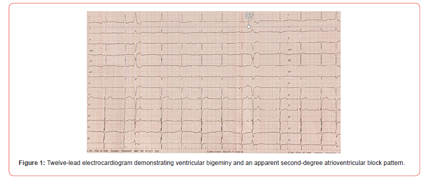
Case summary: A 58year-old man with chronic kidney disease, hypertension, and diabetes mellitus was referred for cardiological evaluation because of dizziness during hospitalization. A 12-lead electrocardiogram showed ventricular bigeminy and an apparent Mobitz type II atrioventricular block pattern. Detailed examination of the tracing revealed retrograde P waves following premature ventricular complexes, indicating ventriculoatrial conduction and favouring a pseudo-Mobitz type II pattern rather than true atrioventricular conduction disease. Twenty-four-hour ambulatory electrocardiographic monitoring was planned for further rhythm evaluation, and there was no indication for permanent pacemaker implantation at this stage.
Conclusion: Retrograde ventriculoarterial conduction following premature ventricular complexes may mimic Mobitz type II atrioventricular block. Careful assessment of the temporal relationship between premature ventricular complexes and atrial activity is important for accurate diagnosis and may prevent unnecessary permanent pacemaker implantation.
Keywords: pseudo-AV block; mobitz type ii; premature ventricular complexes; retrograde ventriculoatrial conduction; ventricular bigeminy
Introduction
Second-degree atrioventricular (AV) block is characterized by intermittent failure of atrial impulses to conduct to the ventricles. Mobitz type II AV block is particularly important because it usually reflects disease of the His-Purkinje system and may progress to advanced or complete AV block, often warranting permanent pacing [1]. Therefore, accurate recognition of this electrocardiographic pattern is essential for appropriate clinical management.
However, several electrophysiological phenomena may mimic true AV conduction abnormalities on the surface electrocardiogram. Premature ventricular complexes (PVCs) with retrograde conduction may interfere with subsequent AV conduction, potentially producing an apparent AV block pattern [2,3]. Distinguishing such pseudo-block from true conduction system disease is clinically important to avoid misdiagnosis and unnecessary interventions [2,3]. Here, we present a patient with ventricular bigeminy in whom retrograde ventriculoatrial conduction following PVCs produced an electrocardiographic appearance mimicking Mobitz type II AV block.
Case Presentation
A 58-year-old man with a history of chronic kidney disease, hypertension, and diabetes mellitus presented to the Department of Internal Medicine with fatigue and dyspnea. He was hospitalized with acute worsening of chronic kidney disease, pleural effusion, diabetes mellitus, and hypertension. A left-sided pleural effusion measuring approximately 45 mm was detected, and a pleural catheter was inserted. Laboratory evaluation revealed a hemoglobin level of 10.7 g/dL, hematocrit of 32.7%, serum creatinine of 5.32 mg/dL, blood urea nitrogen of 103 mg/dL, estimated glomerular filtration rate of 10.2 mL/min/1.73 m², and glucose level of 172 mg/dL. Serum sodium, potassium, magnesium, and calcium levels were 137 mmol/L, 4.6 mmol/L, 1.9 mg/dL, and 8.3 mg/ dL, respectively. During hospitalization, the patient experienced dizziness after getting out of bed and was referred to the cardiology department for evaluation. His blood pressure was 115/78 mmHg. The patient was not receiving any medication known to affect atrioventricular conduction. Transthoracic echocardiography showed a left ventricular ejection fraction of 55%, mild mitral regurgitation, and a 29-mm loculated pericardial effusion adjacent to the posterolateral wall of the left ventricle.
The initial 12-lead electrocardiogram demonstrated ventricular bigeminy with premature ventricular complexes, normal PR intervals, and an apparent Mobitz type II atrioventricular block pattern (Figure 1).
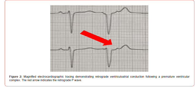
On detailed examination of the electrocardiogram, retrograde P waves associated with ventriculoatrial conduction following the premature ventricular complexes were identified, producing a pseudo-Mobitz type II atrioventricular block appearance (Figure 2).
Twenty-four-hour ambulatory electrocardiographic monitoring was planned for further rhythm evaluation. No syncope occurred during in-hospital follow-up, and there was no indication for permanent pacemaker implantation at this stage. Further evaluation was planned based on the results of ambulatory electrocardiographic monitoring, while medical treatment for the patient’s underlying clinical conditions was continued.s
Discussion
Mobitz type II atrioventricular block is an important electrocardiographic finding because it generally reflects His-Purkinje system disease and may progress to advanced atrioventricular block. [1,2] However, several electrophysiological phenomena may mimic this pattern and result in pseudo-AV block [2,3]. Premature ventricular complexes may conduct retrogradely into the atrioventricular conduction system and interfere with subsequent antegrade conduction. Concealed retrograde penetration may render the atrioventricular node refractory to the following atrial impulse, producing an apparent Mobitz type II pattern despite the absence of intrinsic conduction system disease [2-4]. In our patient, careful examination of the electrocardiogram revealed retrograde P waves following the premature ventricular complexes. The temporal relationship between the premature ventricular complexes, retrograde atrial activation, and apparent blocked beats favored a pseudo-Mobitz type II pattern rather than true Mobitz type II atrioventricular block. Distinguishing pseudo-AV block from true Mobitz type II atrioventricular block is clinically important because the latter may require permanent pacing, whereas pseudo-AV block related to premature beats does not necessarily indicate intrinsic conduction system disease [1-3]. Careful assessment of P-wave timing and the relationship between premature ventricular complexes and subsequent atrioventricular conduction may prevent misdiagnosis and unnecessary pacemaker implantation [2-4].
Conclusion
Retrograde ventriculoatrial conduction following premature ventricular complexes may produce an electrocardiographic pattern mimicking Mobitz type II atrioventricular block. Careful evaluation of P-wave timing and its relationship with premature ventricular complexes is essential to distinguish pseudo-AV block from true conduction system disease and may prevent unnecessary permanent pacemaker implantation.
References
- Fernández-Sanlés A, Sayols-Baixeras S, Subirana I, Sentí M, Pérez-Fernández S, et al. (2021) DNA methylation biomarkers of myocardial infarction and cardiovascular disease. Clin Epigenetics 13(1): 86.
- Rask-Andersen M, Martinsson D, Ahsan M, Enroth S, Ek WE, et al. (2016) Epigenome-wide association study reveals differential DNA methylation in individuals with a history of myocardial infarction. Human Molecular Genetics 25(21): 4739-4748.
- Peixoto P, Cartron PF, Serandour AA, Hervouet E (2020) From 1957 to Nowadays: A Brief History of Epigenetics. Int J Mol Sci 21(20): 7571.
- Ospelt C (2022) A brief history of epigenetics. Immunology Letters 249: 1-4.
- Jarrell DK, Lennon ML, Jacot JG (2019) Epigenetics and Mechanobiology in Heart Development and Congenital Heart Disease. Diseases 7(3): 52.
- Kelsey G (2020) Imprints in the history of epigenetics. Nat Rev Mol Cell Biol 21(10): 566-567.
- Wang G, Wang B, Yang P (2022) Epigenetics in Congenital Heart Disease. J Am Heart Assoc 11(7): e025163.
- Yan L, Geng Q, Cao Z, Liu B, Li L, et al. (2023) Insights into DNMT1 and programmed cell death in diseases. Biomedicine & Pharmacotherapy 168: 115753.
- Zhang L, Xia C, Yang Y, Sun F, Zhang Y, et al. (2023) DNA methylation and histone post-translational modifications in atherosclerosis and a novel perspective for epigenetic therapy. Cell Commun Signal 21(1): 344.
- Zhu L, Jia L, Liu N, Wu R, Guan G, et al. (2022) DNA Methyltransferase 3b Accelerates the Process of Atherosclerosis. Oxid Med Cell Longev 2022: 5249367.
- Liu Y, Tian X, Liu S, Liu D, Li Y, et al. (2020) DNA hypermethylation: A novel mechanism of CREG gene suppression and atherosclerogenic endothelial dysfunction. Redox Biol 32: 101444.
- Han W, Wang W, Wang Q, Maduray K, Hao L, et al. (2024) A review on regulation of DNA methylation during post-myocardial infarction. Front Pharmacol 15: 1267585.
- Boovarahan SR, AlAsmari AF, Ali N, Khan R, Kurian GA (2022) Targeting DNA methylation can reduce cardiac injury associated with ischemia reperfusion: One step closer to clinical translation with blood-borne assessment. Front Cardiovasc Med 9: 10231909.
- He K, Cao X, Deng X (2021) Histone methylation in epigenetic regulation and temperature responses. Curr Opin Plant Biol 61: 102001.
- Zhang Y, Sun Z, Jia J, Du T, Zhang N, et al. (2021) Overview of Histone Modification. Adv Exp Med Biol 1283: 1-16.
- Wang Y, Yuan Q, Xie L (2018) Histone Modifications in Aging: The Underlying Mechanisms and Implications. Curr Stem Cell Res Ther 13(2):125-135.
- Wei X, Yi X, Zhu XH, Jiang DS (2020) Histone methylation and vascular biology. Clin Epigenetics 12(1): 30.
- Neele AE, Chen HJ, Gijbels MJJ, van der Velden S, Hoeksema MA, et al. (2021) Myeloid Ezh2 Deficiency Limits Atherosclerosis Development. Front Immunol 11: 594603.
- Gao R, Liu M, Yang H, Shen Y, Xia N (2025) Epigenetic regulation in coronary artery disease: from mechanisms to emerging therapies. Front Mol Biosci 12: 1548355.
-

This work is licensed under a Creative Commons Attribution-NonCommercial 4.0 International License.




