 Research Article
Application of Infrared Thermography in “Color Diagnosis” of Traditional Chinese Medicine
Chih-Sheng Chen1,2,6,7, Zhi-Ren Tsai4,5, Hung-Jen Lin6,7, Tung-Ti Chang6,7, Yung-Ming Chang8,9, and Rungsheng Chen3*
1Assistant Professor, Department of Food Nutrition and Health Biotechnology, Asia University, Taichung 413, Taiwan
2Director of Chinese Medicine, Asia University Hospital, Taichung 413, Taiwan
3Associate Professor, Department of Optometry, Asia University, Taichung 413, Taiwan
4Associate Professor, Department of Computer Science and Information Engineering, Asia University, Taichung 413, Taiwan
5Researcher, Department of Medical Research, China Medical University Hospital, Taichung, Taichung 404, Taiwan
6Department of Chinese Medicine, China Medical University Hospital, Taichung, 404, Taiwan
7School of Post-Baccalaureate Chinese Medicine, China Medical University, Taichung 404, Taiwan
8School of Chinese Medicine for Post-Baccalaureate, I-Shou University, Kaohsiung 840, Taiwan.
9Chinese Medicine Department, E-DA Hospital, Kaohsiung, Taiwan
Rung-sheng Chen, Associate Professor, Department of Optometry, Asia University, Taichung 413, Taiwan.
Received Date:August 14, 2021; Published Date:October 25, 2021
Abstract
The color diagnosis is to observe the changes of the patient’s facial features and facial features can be a diagnostic method to understand the condition. The theory and technology of TCM color diagnosis are self-contained and have accumulated rich experience in about 2000 years. However, it is still in a visual description of visual inspection and expression in language. It is influenced by the amount of doctor’s experience and language expression ability. It has strong subjectivity and poor clarity.
Using infrared thermal imaging technology to carry out objective research on color diagnosis of Traditional Chinese Medicine (TCM), it can establish the model for color diagnosis of TCM.
Introduction
Color diagnosis is a diagnostic method to understand the patient’s condition by observing the changes of facial features and complexion. “Nan Jing”: looking and knowing is called God. looking and knowing is to see its five colors to know its disease. “Qianjin Yifang”: the supreme doctor is to observe the color, the moderate doctor is to listen to the sound, and the inferior doctor is to follow the pulse. Therefore, a good doctor must be named in the five colors, which can determine life and death, and determine suspicious. It can be seen both of the above statements are highly respected color diagnosis is a very smart diagnosis.
Current Situation of Color Diagnostics
The theory and technology of TCM color diagnosis are selfcontained and have accumulated rich experience in the past 2000 years. However, it is still in the intuitive description of visual observation and language expression. It is influenced by the doctor’s experience and language expression ability. It is highly subjective and unclear, and it is difficult to master.
In the electromagnetic spectrum, the wavelength range of visible light is very narrow, which greatly limits the research and application of color diagnosis.
Week eight registered similar results as nutritional intake was further adjusted to reduce, but not eliminate caffeine and processed foods. The journal entries reflected average daily caloric intake between 1500-1700 calories per day, 3 liters of water, and a regular meditation and exercise regimen. Weight was recorded as 297 pounds.
Principle and Method
Principle
Any object above absolute zero (- 273 ℃) in nature, its molecular thermal motion will continuously radiate infrared to the external space. Human body temperature is about 36 ~ 37 ℃, which is a natural biological infrared light source, and it will continuously emit infrared radiation. This radiation will change with physiological activities or pathological changes in the body, and these changes can be sensed and received by the infrared thermal imager. The infrared thermal radiation will be converted into electronic signals, and then processed into color images. Moreover, it does not need to contact the patient and will not change any state of the object, so this is a non-invasive test method. It can also continuously measure the temperature change on the human body surface and detect the temperature difference in different parts of patient. The color image will show temperature change, display the location and nature of the lesion, and accurately and indicate the temperature point by point.
Method
Instrument: Infrared thermal imaging camera (Figure 1), with a temperature resolution of 0.04°C at 30°C.

Testees: The thermal images measurement was proved by Institutional Review Board (IRB), and there are 30 cases collected images from Chinese Medicine Clinic in Asia University Hospital.
Test environment: Room temperature is 21.5~23.5℃, and the relative humidity 30.2% ~56.5%.
Measure process: The testee sat still in the test room for 30 minutes to adapt to the environment, and then tested the subject. At 1.5 meters away from the infrared thermal imager, the testee was photographed three times according to sitting, sitting on the left and right. Collecting the thermal image, the testee was required to open his eyes and hold his breath for several seconds, and then the three thermal images taken were stored in the file.
Measurement
After the completion of thermography in the traditional Chinese medicine clinic of Asian University Hospital, the central point temperatures of the color areas of the five internal organs (lungs, heart, liver, spleen, and kidneys) and their counterparts of hollow organs (large intestine, large intestine, gallbladder, stomach, and bladder) of 30 testees were collected for statistical analysis.
The physiological and pathological state of five internal and hollow organs is reflected in the specific part of the face, which is called the “color part” [1,2]. The color part is of great significance for the positioning of understanding the disease. Tradition Chinese medicine often uses the Mingtang color part to gain insight into the disease. The Mingtang color part comes from the “Lingshufive colors chapter”. The patient’s complexion can be shown in the Mingtang color part. The corresponding “central coordinate fixed point method” is described as:
According to the observation of clinical cases, if the disease color appears according to the color part of Mingtang, although the size of its range changes, it always takes a fixed point as the center. The appearance or aggregation or dispersion of the disease color of each viscera will always surround the center of a specific color part. The drawing method is based on (as shown in Figure 2), it shows the front width of a person’s face is about five eye widths [3,4].

Therefore, in terms of longitudinal coordinates, the surface can be divided into approximately equal parts symmetrical through the longitudinal lines of the anterior 1) median line, 2) inner canthus vertical line, 3) pupil vertical line, 4) outer canthus vertical line and 5) Temple vertical line. In addition, in terms of transverse coordinates, the transverse coordinates are composed of 1) line at the medial end of eyebrow, 2) line at the inner canthus of two eyes, 3) line at the highest end of zygomatic bone, 4) line 1 / 3 above the center of nasal wing and 5) horizontal line at the base of nasal wing; Such a coordinate system can position the viscera at the intersection of these vertical and horizontal coordinates. The specific positions are shown in Tables 1 and 2.
Table 1: The position of the internal organs at the central coordinate.

Table 2: The position of the hollow organs at the central coordinate.

Results and Discussion
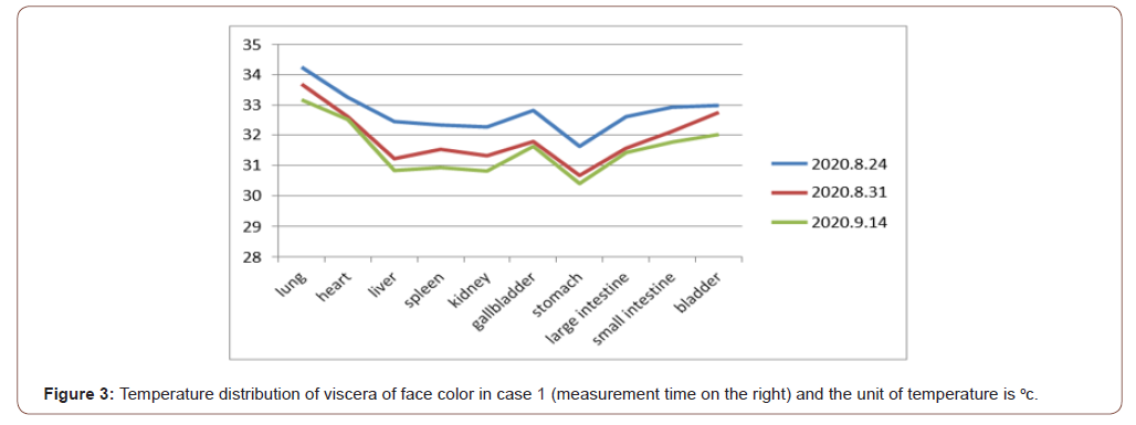
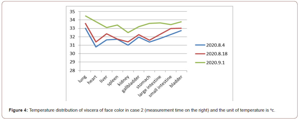
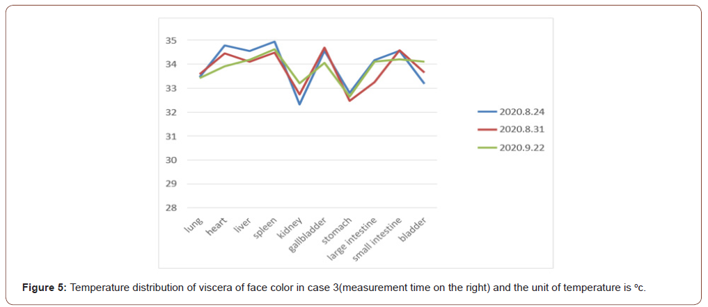
According to measurements given by sec.4, we can analyze the temperature change of the central point of the viscera color part of the received cases. A total of 30 cases will be collected, and each case will measure 1-3 infrared thermal images; After the thermal image was converted to temperature data, the following phenomena were found. Each case has its unique temperature distribution map of viscera and face color, as shown in Figures 3, 4 and 5.


From the above temperature distribution corresponding to the internal and hollow organs, we can get the following results:
1) Each case has its own unique color temperature pattern, just like a fingerprint.
2) The graph pattern of Case 1 moves down with time.
3) The graph pattern of Case 2 moves up with time.
4) The graph pattern of Case 3 has no significant change over time.
Therefore, the inference is as follows:
1) by TCM hall color portion theory, infrared thermal imaging observed cases facial temperature, each has its temperature profile pattern (TFP, temperature feature pattern) [5,6],
2) Whether the diseases of viscera and viscera are relative to their corresponding TFP, such as whether patients with specific disease have their special TFP. If so, measuring TFP can be used as an auxiliary method or tool for medical diagnosis.
3) With the progress of the treatment, if the rise and decline of TFP are relative to the health changes of the case. Then It is worth further research with the integrated method of big data analysis and computer vision.
Acknowledgement
I would like to thank Asia University in Taiwan for providing a suitable research environment and supporting this research granted by No. ASIA-108-CMUH-17. And thanks also go to Ministry of Science and Technology by No. MOST 110-2321-B-468-001.
Conflict of Interest
Author declare no conflict of interest.
References
- Paul U Unschuld (2003) Huang Di nei jing su wen. University of California Press.
- M Vollmer, KP Möllmann (2018) Infrared Thermal Imaging: Fundamentals, Research and Applications, 2nd
- Thermal Imaging to Diagnose Disease.
- Thermography in Medicine.
- Kurt Ammer, EFJ Ring (2012) Standard Procedures for Infrared Imaging in Medicine. Chapter 32. Medical Infrared Imaging. Principles and Practice, CRC Press, Taylor & Francis Group.
- Robert Koprowski (2018) Processing Medical Thermal Images Using Matlab.
-
Chih-Sheng Chen, Zhi-Ren Tsai, Hung-Jen Lin, Tung-Ti Chang, Yung-Ming Chang, and Rungsheng Chen*. Application of Infrared Thermography in “Color Diagnosis” of Traditional Chinese Medicine. On J Complement & Alt Med. 7(1): 2021. OJCAM. MS.ID.000653.
-
Nutrition, Exercise, Diabetic Control, Diabetes, Weight Reduction, Reduced Pharmaceuticals, Blood Sugar, Nutritional Control, T2D Diabetic Condition, Decreased weight, decreased dosing, Pharmaceutical interventions
-

This work is licensed under a Creative Commons Attribution-NonCommercial 4.0 International License.


