 Research Article
Effects of Resting in Different Postures and Ankle Pump Movement on Hemodynamics of Deep Venous Blood Flow of Lower Limb
Gao Ruijiao1, Cao Pengkai1, Liu Xiangdong1, Zhang Yanrong1, Li Tianhua1 and Ding Junqing2*
1Vascular Surgery Department, The Third Hospital of Hebei Medical University, China
2Nursing Department, The Third Hospital of Hebei Medical University, China
Junqing Ding, Nursing Department, The Third Hospital of Hebei Medical University Shijiazhuang 050051, China.
Received Date:February 22, 2022; Published Date:May 17, 2022
Abstract
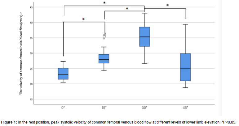
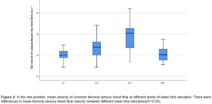
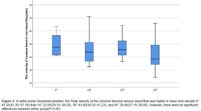
Purpose: The aim of this study was to provide evidence of individualized prevention of deep venous thrombosis (DVT) through evaluating venous hemodynamics of lower limb in response to ankle pump movement in different positions. Methods: Peak systolic velocity and mean flow velocity of common femoral vein were measured in 10 health volunteers (20 limbs) using color Doppler ultrasound in different positions, including at rest and when ankle pump movement were performed at lower limb elevation of 0, 15, 30, 45 degrees. Results: In the rest position, there were differences in the peak and mean femoral venous blood velocity among 0°, 15°, 30° and 45° (P=0.00, P=0.00); The Peak velocity of the common femoral venous blood flow was higher in lower limb elevate 30° (35.36±4.97) than 0° (23.38±2.23), 15° (28.45±3.01) and 45° (25.89±5.51). (P<0.05). The mean velocity of the common femoral venous blood flow was higher in lower limb elevate 30° (29.14±7.23) than 0° (20.20±2.51), 15° (23.67±4.52) and 45° (20.37±3.39). (P<0.05). In ankle pump movement position, the Peak velocity of the common femoral venous blood flow was higher in lower limb elevate 0° 47.35(41.35~57.18) than 15° 32.05(28.13~39.20), 30° 43.60(36.33~51.23), and 45° 29.80(27.15~36.08). However, there were no significant differences between either group(P>0.05). Conclusion: A better individualized prevention of DVT could be achieved through appropriate selection of position and ankle pump movement.
Keywords:Ankle pump movement; Position; Lower extremity; Venae profundae; Hemodynamics
Introduction
Deep venous thrombosis (DVT) is a common complication of trauma, operation and immobility. The incidence of DVT is 80 cases per 100,000, with a prevalence of lower limb DVT of 1case per 1000 population. DVT may cause pulmonary thromboembolism (PTE), even death [1]. The pathophysiological mechanisms of DVT involves damage of the vessel wall, blood flow turbulence and hypercoagulability. Immobility may reduce blood flow, which allows the accumulation of procoagulant factors that may induce thrombosis. The primary goal of pharmacologic DVT prophylaxis is to prevent fatal PE. There are three protocols for the prevention of DVT, which including basic prevention, physical prevention and drug prevention. Early clinical studies in DVT prevention focused on the potential benefit of prophylactic anticoagulation in populations at high risk of DVT. And, anticoagulation is the mainstay of treatment for DVT. It aims to reduce mortality, thrombus extension, recurrence, the risk of post thrombotic syndrome (PTS) and chronic thromboembolic pulmonary hypertension. However, effective anticoagulant also increases the risk of bleeding, and anticoagulant may prevent DVT results in a 50-70% reduction in the rate of DVT [2], there are still about 30-50% patients would suffered DVT. Physical prevention of DVT includes ankle pump exercise and lower limb elevation, which may promote the venous blood return [3].
Physical prevention is one of measures of prevention recommended by guideline and has been used in clinical application. However, there are few studies focus on how much lower limb elevation will promote lower limb return is the best. In this study, we measure the flow the velocity of femoral vein using color Doppler ultrasound in different degrees of lower limb elevation, analysis the effect of elevation of lower limb on blood return. Comparing the velocity of femoral venous blood flow aims to form a suitable plan which provide a theoretical basis for clinical application.
Materials and Methods
Participants
In this study, twenty participants were enrolled form May 2019 to September 2019. Among them, there were 10 females, aged from 20-32 years. Both sides of femoral veins were detected.
Exclusion criteria:
I. Deep venous thrombosis
II. Iliac venous compression syndrome
III. Chronic venous insufficiency
IV. With heart disease.
V. With a history of vascular disease
VI. Operational history or trauma of lower limb
VII. Coagulation disorders
Methods
The participant lies on examination bed, the femoral vein was located inside the femoral artery above the bilateral ilioinguinal ligament. Femoral venous blood flow was detected at 1cm upon the saphenous femoral vein valve. Duplex sonographic measurements were made with a PHILIPS CX50, fitted with a 5.0MHz sonar head. The angle between the probe and the femoral vein was kept at 60°. The trapezoidal position was selected for lower limb elevation. The line between the top of the greater trochanter of femur and the lateral malleolus of femur in the same side was taken, and then the horizontal line between the top of the greater trochanter of femur and the bed surface was taken. The angel of the two lines was the angle of lower limb elevation.
Quiescent condition: the ankle actively dorsiflexes 20° and then plantarflexes 45°, the frequency was 30-40 times/min. The velocities of femoral venous blood flow were detected at horizontal position, lower limb elevation 15, 30 and 45° (Figure 1). All detections were operated by same ultrasonologist, each position would detect 3 times. The participant would rest 10min before detection.

Statistical Analysis
SPSS 22.0 software was used for the statistical analysis. The date presented is regarded as being normal distribution and the results are presented as mean ± SD (χ±s). The date presented is regarded as being abnormal distribution and the results are presented as medians and interquartile range. A paired t-test was used to analyze the both side of femoral venous blood flow. The variance analysis of repeated measurement was performed for different groups. Significance was set at P<0.05.
Results
The flow velocity of both femoral venous blood flow at rest
In the rest position, there was no difference between right and left femoral venous blood flow velocity at different limb elevation degrees(P>0.05) (Table 1).
Table 1: The peak and mean flow of right and left femoral venous blood flow.

Effects of lower limb elevation on the velocity of common femoral venous blood flow at rest
In the rest position, there were differences in the peak and mean femoral venous blood velocity among 0°, 15°, 30° and 45° (P=0.00, P=0.00); The Peak velocity of the common femoral venous blood flow was higher in lower limb elevate 30° (35.36±4.97) than 0° (23.38±2.23), 15° (28.45±3.01) and 45° (25.89±5.51). (P<0.05). The mean velocity of the common femoral venous blood flow was higher in lower limb elevate 30° (29.14±7.23) than 0° (20.20±2.51), 15° (23.67±4.52) and 45° (20.37±3.39). (P<0.05) (Figures 1&2).

The flow velocity of both femoral venous blood flow at ankle pump movement
In ankle pump movement position, there was no difference between right and left femoral venous flow velocity at different limb elevation degrees(P>0.05) (Table 2).
Effects of lower limb elevation on the velocity of common femoral venous blood flow at ankle pump movement.
Table 1:The peak and mean flow of right and left femoral venous blood flow.

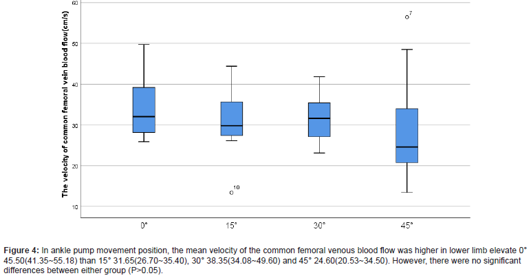
In ankle pump movement position, there were differences in the peak and mean femoral venous blood velocity among 0°, 15°, 30° and 45° (P=0.02), however, there were no differences in the mean femoral venous blood velocity among 0°, 15°, 30° and 45° (P=0.10); The Peak velocity of the common femoral venous blood flow was higher in lower limb elevate 0° 47.35(41.35~57.18) than 15° 32.05(28.13~39.20), 30° 43.60(36.33~51.23), and 45° 29.80(27.15~36.08). However, there were no significant differences between either group(P>0.05). The mean velocity of the common femoral venous blood flow was higher in lower limb elevate 0° 45.50(41.35~55.18) than 15° 31.65(26.70~35.40), 30° 38.35(34.08~49.60) and 45° 24.60(20.53~34.50). However, there were no significant differences between either group (P>0.05) (Figures 3&4).

Discussion
Venous stasis is one of the major components of Virchow’s triad [4]. Improving the blood return of lower limb is an effective method to prevent DVT [5]. Both lower limb elevation and ankle pump movement are use full for blood return of lower limb [3,6-8].
In this study, by analyzing the effects of lower limb elevation on the femoral venous blood flow. In the rest position, the femoral venous blood flow with lower limb elevation of 30° had the faster blood flow. The reason may be that: the venous return is affected by negative pressure which produced by diastolic heart and inspiratory movement [9]. In additional, hydrostatic pressure of lower limb vein may also affect the venous return. Elevate lower limb may increase the hydrostatic pressure, which may promote the venous return. However, when the lower limb is raised more than 45°, the peak velocity and mean velocity of femoral venous blood flow were not significantly increased compared with the rest position. The reasons may be related to the following factors:\ (1) with elevation of lower limb, the curvature of the femoral and popliteal vein increased, which causing to swirl of blood flow. Swirly status will reduce the velocity of blood flow. (2) Popliteal vein may be compressed by knee flexion, which lead to reduction of venous blood flow. Similarly, the return of femoral vein will reduce when the hip is bent [7,10]. In this study, we verified that the peak and mean velocity of femoral vein was the highest at 30° of lower limb elevation in rest position. In ankle pump movement position, the femoral vein velocity was the highest in 0°of lower limb elevation. This result provides scientific basis of the suitable potion for venous return in the resting state.

According to our research, we have provided scientific guidance for the prevention of DVT in clinical work for patients with different diseased. For the patients with normal lower limb function, elevate 30° of lower limb, when occurred fatigue or discomfort, changed to ankle pump movement at horizontal position, which would be the best scheme for prevention of DVT. For patients with dysfunction of lower limb, elevate 30° of lower limb, intermittently lower elevation angle, which may be the best scheme.
Conclusion
In conclusion, the effects of rest and ankle pump movement on deep venous blood flow dynamics of lower limb are different at different position of lower limb elevation. Personalized prevention scheme is needed for prevention of DVT on different patients.
Acknowledgment
Medical Science Research Project of Hebei Province: 20201028.
Conflict of Interest
The authors have no conflict of interest to disclose.
- Fernandez-Capitan C, Rodriguez Cobo A, Jimenez D, Madridano O, Ciammaichella M, et al. (2021) Symptomatic subsegmental versus more central pulmonary embolism: Clinical outcomes during anticoagulation. Res Pract Thromb Haemost 5(1): 168-178.
- Nicholson M, Chan N, Bhagirath V, Ginsberg J (2020) Prevention of Venous Thromboembolism in 2020 and Beyond. J Clin Med 9(8): 2467.
- Li Y, Guan XH, Wang R, Li B, Ning B, et al. (2016) Active Ankle Movements Prevent Formation of Lower-Extremity Deep Venous Thrombosis After Orthopedic Surgery. Med Sci Monit 22: 3169-3176.
- Kumar DR, Hanlin E, Glurich I, Mazza JJ, Yale SH (2010) Virchow's contribution to the understanding of thrombosis and cellular biology. Clin Med Res 8(3-4): 168-172.
- Stein PD, Yaekoub AY, Ahsan ST, Matta F, Lala MM, et al. (2009) Ankle exercise and venous blood velocity. Thromb Haemost 101(6): 1100-1103.
- Hickey BA, Cleves A, Alikhan R, Pugh N, Nokes L, et al. (2017) The effect of active toe movement (AToM) on calf pump function and deep vein thrombosis in patients with acute foot and ankle trauma treated with cast - A prospective randomized study. Foot Ankle Surg 23(3): 183-188.
- Kropp AT, Meiss AL, Guthoff AE, Vettorazzi E, Guth S, et al. (2018) The efficacy of forceful ankle and toe exercises to increase venous return: A comprehensive Doppler ultrasound study. Phlebology 33(5): 330-337.
- Lattimer CR, Franceschi C, Kalodiki E (2018) Optimizing calf muscle pump function. Phlebology 33(5): 353-360.
- Li T, Yang S, Hu F, Geng Q, Lu Q, et al. (2020) Effects of ankle pump exercise frequency on venous hemodynamics of the lower limb. Clin Hemorheol Microcirc 76(1): 111-120.
- Kim JT, Park CS, Kim HJ, Lee JM, Kim HS, et al. (2009) The effect of inguinal compression, Valsalva maneuver, and reverse Trendelenburg position on the cross-sectional area of the femoral vein in children. Anesth Analg 108(5): 1493-1496.
-
Gao Ruijiao, Cao Pengkai, Liu Xiangdong, Zhang Yanrong, Li Tianhua and Ding Junqing. Effects of Resting in Different Postures and Ankle Pump Movement on Hemodynamics of Deep Venous Blood Flow of Lower Limb. On J Complement & Alt Med. 7(3): 2022. OJCAM.MS.ID.000664.
-
Ankle pump movement; Position; Lower extremity; Venae profundae; Hemodynamics; Deep Venous; Different Postures; Blood Flow; Lower Limb; Trauma; Prophylactic anticoagulation; Hypercoagulability; Procoagulant factors; Thrombosis
-

This work is licensed under a Creative Commons Attribution-NonCommercial 4.0 International License.


