


 Research Article
Constant Membrane Surface Charge Density and Membrane Potential Generation Mechanism Backed by Association-Induction Hypothesis
Received Date: March 21, 2024; Published Date: April 08, 2024
Abstract
The author built a simple experimental living cell model consisting of two electrolytic solutions separated by a charged membrane. The author found that the membrane surface charge density looked almost constant regardless of the electrolytic solution ion concentrations. Then, the author derived a membrane potential formula using only the common physical chemistry concepts instead of the conventionally accepted physiological concept of membrane theory. Consequently, the derived formula turns out to be the sum of two terms: One is identical to Goldman-Hodgkin-Katz equation (GHK eq.) and the other one is a function of membrane surface charge density. But the magnitude of the latter term is often cases negligible compared with the former term, that is, the derived membrane potential formula (denoted by reGHK eq.) virtually identical to the GHK eq., and at the same time the membrane surface charge density was found to be often cases constant irrespective of the solution ion concentration as expected. This constant membrane surface charge density appears to be a key for the derivation the reGHK eq. The reGHK eq. is built on the premise that the membrane potential is generated by the spatially heterogeneous ion distribution caused by the ion adsorption. This concept is equivalent to the principle of the unconventional physiological theory called Association-Induction Hypothesis (AIH). Although the AIH has been long overlooked, it could replace the long-accepted membrane theory.
Keywords: Membrane theory; Membrane potential; Goldman-Hodgkin-Katz equation; Association-Induction Hypothesis; Membrane surface charge densitys
Introduction
Membrane potential and AIH
The membrane potential generation mechanism was elucidated especially by the biophysicists in the last century [1, 2]. They say that the membrane potential is generated owing to the occurrence of transmembrane ion transport. This mechanism is one of the consequences of the broadly-accepted physiological concept called membrane theory. However, this mechanism is still controversial among a small number of physiology research groups [1- 17]. It is not appropriate to summarize that their works have been performed for the purpose of fully denying the current physiological views. It must be more appropriate to say that they accept the experimental observations the past scientists have done. However, they think that it is worth rescrutinizing those observations from more realistic view in a constructive way, and they also think that it is scientifically meaningful to find more appropriate explanation to the physiological phenomena such as a membrane potential generation mechanism which the author of this paper deals within this work.
The author would like to show some reports by the researchers who have not accepted the current physiological concepts. Ling suggested the membrane potential is generated by the spatial heterogeneous ion distribution caused by the ion adsorption in the living cell [1, 2]. Consequently, Ling deny the ion transporters (channels and pumps). He did not deny the existence of molecules called channel and pump as far as the author knows. But he denies their ion transport functionalities that the most of physiologists have believed up until today. Edelman performed some experiments decades ago, and his works support the Ling’s theory [3]. Pollack views the cell water not as free water but as the highly-motion-restricted molecule [4]. As a matter of fact, the existence of such a motion- restricted water molecule has been accepted even in our daily life as the surface tension. Pollack suggested that there exists the significant influence of the motion-restricted water molecules on the ion transport across the plasma membranes. He states that the water molecules in the living cell are highly structured, and it influences on the ion level disparity between the cell inside and the cell outside. Therefore, the water state must be involved in the membrane potential generation. Pollack’s study of water originates from Ling’s work of the water state in the living cell [1, 2]. But Pollack’s water study goes beyond the physiology and reaches the discoveries of new aspects of water molecules [5]. Wnek has worked on the biomimicking materials. The living cell excitability looks the sign of life. However, the synthetic polylelctrolytes hold the excitability as well. His work may require us to change the view to bioelectricity, namely, bioelectricity may not be the biological activity origin. Matveev touches upon the bioelectricity of nonliving microsphere [6]. Microsphere exhibits the action potential, and its profile is indistinguishable from the action potential profile of real living cell. It makes us ponder if the action potential generation is truly the sign of life and further makes us think that the current physiological theory may be at least incomplete if not wrong.
Bagatolli et.al. suggests that the realistic view to the living cell is the key to understand the cell activity [8, 9]. The realistic view means, for example, that the water in the living cell is not in the free state but in the motion-restricted state like Pollack suggests. Thermodynamics suggests that the substances such as a water molecule and ions are not in the ideal state at all. Therefore, Bagatolli et.al. suggest the fundamental importance of viewing the living cell in more realistically. Schneider also suggests the importance of more physics-rooted view to the living cell [10]. He thinks that the cell characteristics cannot be represented by so simple manner. Principle may be based on the simple thermodynamics. But the emerging cell characteristics involve the various aspects of many substances. Lee describes that the membrane potential generation is due to the capacitor formation on the membrane surface [11, 12]. The capacitor formation means that the charge separation. Lee’s work is similar to the ion selective electrode (ISE) mechanism advocated by Cheng in the electrochemistry field [13, 14]. Cheng sates that the ISE selectively detects the ions by their adsorption on the electrode surface. The adsorbed substance forms a sort of capacitor on the ISE surface, then the capacitor potential is detected by the ISE. So, Lee’s theory in physiology is quite consistent with the Cheng’s theory in electrochemistry.
Funk and Scholkmann published a review on the current studies of bioelectricity [15]. They don’t say which mechanism is right for the membrane potential generation. The just report the recent progress in bioelectricity study. Their review suggests that the membrane potential is not determined only by the cell chemical composition but even the cell geometry is involved in the determination of cell characteristics. This is equivalent to the Schneider’s thought to the effect that the cell characteristics are not determined only by a single factor.
The author has repeatedly observed firsthand the potentials equivalent to the membrane potential in the experimental artificial cell models in which the transmembrane ion transport never takes place [16, 17]. The author detail it below.
Goldman-Hodgkin-Katz equation (GHK eq.) is a formula in the purview of the membrane theory and used for numerically predicting the membrane potential [1, 2, 18-20]. The GHK eq. contains the physical quantity Pi which represents the membrane permeability to the mobile ions. Eq. 1 is the typical example of GHK eq. If Pi = 0, namely, the membrane is impermeable, Eq. 1 collapses.

But Tamagawa and Ikeda found that the potential across an impermeable silver oxide-coated silver wire which separates two electrolytic solutions exhibits the potential characteristics in line with the reinterpreted GHK eq. prediction [16, 17]. What is the reinterpreted GHK eq.? Tamagawa and Ikeda suggested that the typical reinterpreted GHK eq. expression is given by Eq. 2. Pi of Eq. 2 is replaced by Ki , resulting in Eq. 2. Ki is the association constant between the mobile ion and the adsorption site of membrane surface.

Eq. 2 is derived employing the long-forgotten physiological theory called Association-Induction Hypothesis (AIH) put forth by Ling [1, 2]. The AIH was proposed as the alternative theory to the membrane theory. Hence, its emphasis is totally in conflict with the membrane theory. The typical difference between them regarding the membrane potential generation mechanism is that AIH states that the membrane potential is generated by the spatial heterogeneous intracellular and extracellular distribution of mobile and immobile ion charges due to the ion adsorption while membrane theory states that the transmembrane ion transport is essential for the membrane potential generation.
The membrane theory does not agree with the experimental fact. It has faced an even more problematic issue. The membrane theory states that the ion passage through the plasma membrane of a living cell is governed by the ion channel and pumps. However, Ling suggested that the living cell does not have enough energy to activate the pumps [1, 2]. It violates the foundation of the law of the conservation of energy. One may say that Ling’s energy estimation for the activation of the pumps is not reliable. But Ling reported a quite intriguing experimental result regarding the intracellular and the extracellular ion distribution as described below [1, 2, 22].
It is well-known that there exists a disparity between the intra and extracellular ion level in the living cell. Physiologists agree that such an ion level disparity is due to the function of ion transporter (ion channels and pumps) embedded in the plasma membrane. However, he found that K+ accumulates even in the dead cell and Na+ hardly enters the same dead cell. The dead cell Ling used in his experiment does not contain any functioning sodium-potassium pump. Without the sodium-potassium pumps, K+ cannot accumulate in the dead cell or Na+ cannot be prohibited to enter the same dead cell. Nevertheless, accumulation of K+ in the dead cell and the expel of Na+ from the same dead cell was observed. The membrane theory appears to be short of something physiologically fundamental. On the other hand, the AIH can explain such an issue. The AIH states that the ion level disparity is regulated by the activity of water and the cell potential characteristics are governed by the ion adsorption [1-5]. Water molecules in the cell is in the highly structured state and its activity is low. The degree of the affinity of K+ and Na+ to the structured water molecule is different from that to the free water. On top of that the immobile ion adsorption sites of cell have the different degree of affinity to the individual ions. Hence, these effects cause the ion level disparity. Consequently, the nonzero membrane potential is generated even without the pumps. Therefore, no energy is in need for the nonzero potential generation in the purview of the AIH states.
Reinterpreted GHK eq. and AIH
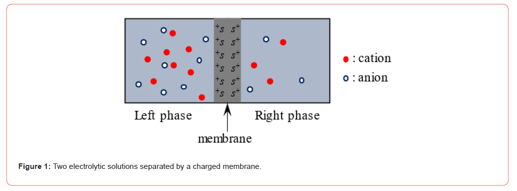
It is possible to theorize the membrane potential generation mechanism by employing the ion adsorption phenomenon rather than the transmembrane ion transport. Imagine that two electrolytic solutions separated by a membrane and the surface of this membrane bears the positively charged ion adsorption site s+’s, and s+ associates with mobile anions (see the illustration Figure 1).
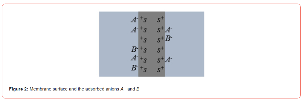
Assuming that there exist two species of mobile anions, A− and B− in the two electrolytic solutions, and the mobile anions can adsorb on the membrane surface sites s+’s as given by Eq. 3 (see Figure 2). Consequently, Eq. 4 is given where KJ (J − = A− , B− ) is the association constant (see the notation table Table 1), and this is the Langmuir isotherm [23] where the coordinate systems are set to the system of Figure 2 as illustrated in Figure 3.

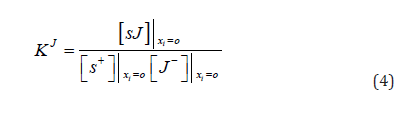
( i = L (Left phase), R (Right phase); J − = A− , B− )
Focusing on the individual phase of the solutions illustrated in Figure 3, the formula of the membrane surface charge density is derived by following the procedure given in the ref. [16, 17].
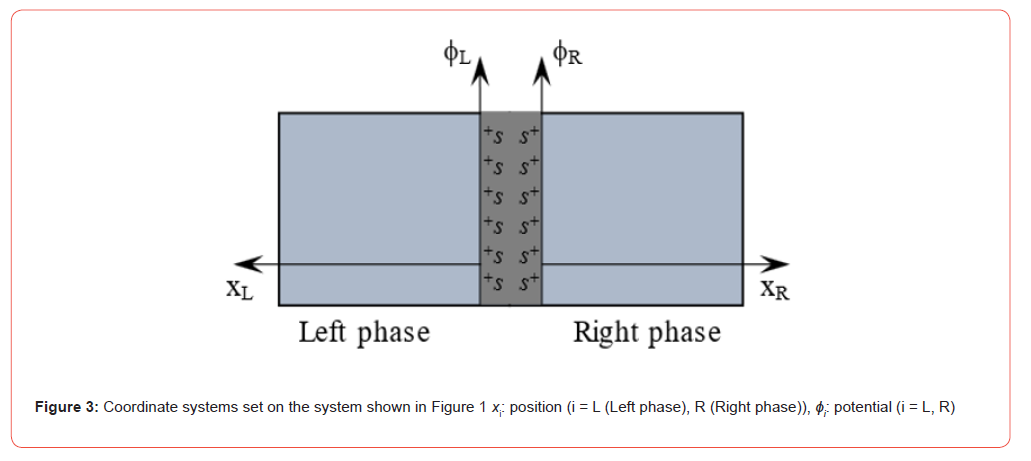
Table 1:Notation

† :  is computed by plugging the experimentally measured
 into Eq. 16.
Ion distribution is given by Eq. 5.

Poisson-Boltzmann equation (P.-B. eq.) is given by Eq. 6. Charge density ρi in the i phase is given by Eq. 7 where C+ A and C+B represent the counter cations of A− and B− , respectively.
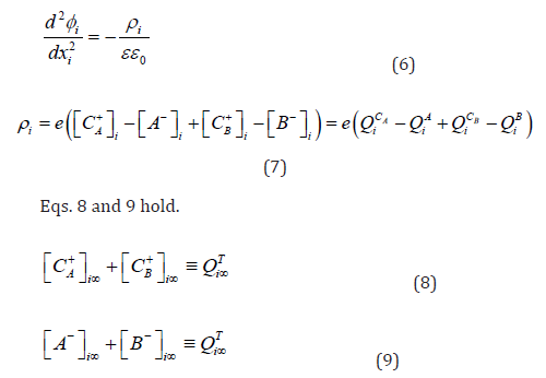
Arranging the P.-B. eq. of Eq. 6 using Eqs. 7, 8, 9 and the boundary conditions of Eqs. 10 and 11, Eq. 12 is derived.
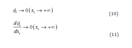
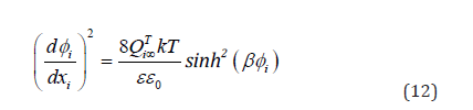
Since the membrane surface is positively charged in this case, Eq 13 holds.

Eq. 12 results in Eq. 14.
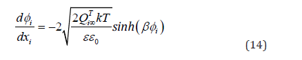
The membrane surface charge density is given by Eq. 15.
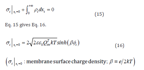
The membrane surface charge density  can be given by
another formula as detailed below [7, 8].

Eqs 18 and 19 are derived using Eqs. 4 and 17.
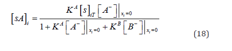
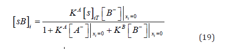
Therefore,  can be given by Eq. 20 as well as Eq. 16.


RHS of Eq. 20 can be arranged into Eq. 22 using Eqs. (18) and (19).

Tamagawa and Ikeda performed an experiment and found that
Eq. 23 holds regardless of [16, 17]. So, the membrane surface
charge density is quite indifferent to the solution ion concentration.
Hence, Eq. 22 can be arranged into Eq. 24 using Eq. 23.


The membrane potential, ΔΦAds , can be given by Eq. 25. It can be arranged into Eq. 26 by plugging Eq. 24 into Eq. 25. Eq. 26 rests on the concept that the membrane potential is generated by the ion adsorption, and quite importantly the ion adsorption-based membrane potential generation mechanism is virtually equivalent to the AIH-based mechanism [1, 2]. On the other hand, the GHK eq. rests on the premise that the transmembrane ion transport is responsible for the membrane potential generation. Nevertheless, the last term of RHS of Eq. 26 is identical to the GHK eq. for this system Eq. 27. Therefore, the GHK eq. can be reinterpreted in view of the ion adsorption mechanism. From now on, we call this equation reGHK eq. (reinterpreted GHK equation), and the last term of RHS of Eq. 26 is again explicitly shown as Eq. 28 along with introducing the new notation ΔΦRE which represents the reGHK eq. In the derivation process of Eq. 28, the author employed the mass action law, the Langmuir isotherm and the P.-B. eq. only [23], and all of them are within the range of ordinary and broadly accepted physical chemistry, and this derivation process is never in conflict with the AIH.
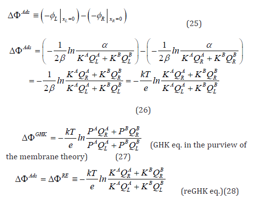
The fundamentally important starting equation for reaching Eq. 28 is Eq. 23. Eq. 23 means that the membrane surface charge density is constant irrespective of solution ion concentration. Eq. 23 is an empirical condition. Therefore, we don’t know if it is universally right. Hence, the author tested if Eq. 23 is valid more broadly.
Verification of the membrane surface charge density constancy
The author performed the membrane surface potential measurement in order to confirm the broad applicability of Eq. 23. The experimental procedure is described firs here.
Membrane preparation
Four kinds of ion exchange membranes were used for measuring the membrane surface potential, and they are Selemion AMV, CMV, AMVN and CMVN (Asahi Glass. Co., Ltd. (Tokyo)). From now on, these membranes are to becalled, AMV, CMV, AMVN and CMVN, respectively, for short. AMV and AMVN contain atomic functional groups which can dissociate into immobile cations and mobile anions, while CMV and CMVN contain atomic functional groups which can dissociate into immobile anions and mobile cations. Tiny pieces of these membranes were prepared by cutting the original sheets of AMV, CMV, AMVN and CMVN.
Solutions for bathing the membranes
Two types of electrolytic solutions were prepared. One is the mixture of KCl and KBr. The other one is the mixture of NaCl and LiCl. The procedure of preparing the KCl-KBr mixture is given as follows: 0.1 M KCl solution was prepared by dissolving KCl into the deionized water. 0.1 M KBr solution was prepared by the same procedure. The same quantity of resulting 0.1 MKCl solution and the 0.1 M KBr solution were mixed together. The resulting KCl-KBr mixture contains 0.05 M KCl and 0.05 M KBr. This mixture is to be called 0.05_KCl-KBr. KCl-KBr mixture whose ion concentration is 1/10 of 0.05_KCl-KBr was prepared by diluting 0.05_KCl-KBr by 10-fold with the deionized water. The resulting KCl-KBr mixture is denoted by 0.005_KCl-KBr. By the same procedure, the further diluted KCl-KBr mixtures were prepared, resulting in two KCl-KBr mixtures. One contains 0.0005 M KCl and 0.0005 M KBr, and it was denoted by 0.0005_KCl-KBr. The other one contains 0.00005M KCl and 0.00005 M KBr, and it is denoted by 0.00005_KCl-KBr. The mixtures of NaCl solution and LiCl solution were prepared in the similar manner. The resulting mixtures are denoted in the same manner as the KCl-KBr solutions. Therefore, the following four solutions were prepared: 0.05_NaCl-LiCl, 0.005_NaCl-LiCl, 0.0005_NaCl-LiCl, 0.00005_NaCl-LiCl.
Prior treatment to the membranes
The tiny pieces of AMV prepared earlier were stored in 0.00005_KCl-KBr ∼ 0.05_KCl-KBr at least five days in a row. During this storage period, the bathing solution for these tiny AMV pieces were exchanged with the new solutions once a day as illustrated in Figure 4. AMVN had undergone exactly the same treatment. The tiny pieces of CMV had undergone the same treatment using 0.00005 NaCl-LiCl ∼ 0.05 NaCl-LiCl instead of 0.00005_KCl-KBr ∼ 0.05_KCl-KBr. CMVN had undergone exactly the same treatment as the CMV had undergone.
Surface potential measurement
The membrane surface potential was measured as illustrated
in Figure 4 using a pair of Ag/AgCl electrodes. One electrode was
placed in the bathing solution of membrane and the other one was
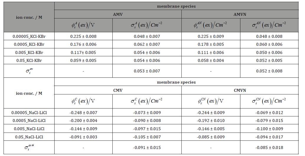
placed on the surface of membrane. The experimentally measured
surface potentials represented by φs M (ex )
are summarized in Table 2 along with the species of membrane in the electrolytic solutions
and their concentration where M of φs M (ex ) represents the
species of membrane used, M = A (AMV), C (CMV), AN (AMVN), CN
(CMVN). Data of  in Table 2 is shown in Figure. 5. Horizontal
axis represents the logarithm of ion concentration of the bathing
solution of the membranes. Irrespective of the bathing solution ion
concentration, it is not inappropriate to say that Eq.23 is basically
valid for any species of membrane. Therefore, Eq. 26 can be derived.
Consequently, it is possible to derive the reGHK eq. of Eq. 28, which
is identical to the GHK eq. Furthermore, the surface charge density
appears to be constant in various experimental systems. Therefore,
ΔΦRE is expected to be valid in various systems.
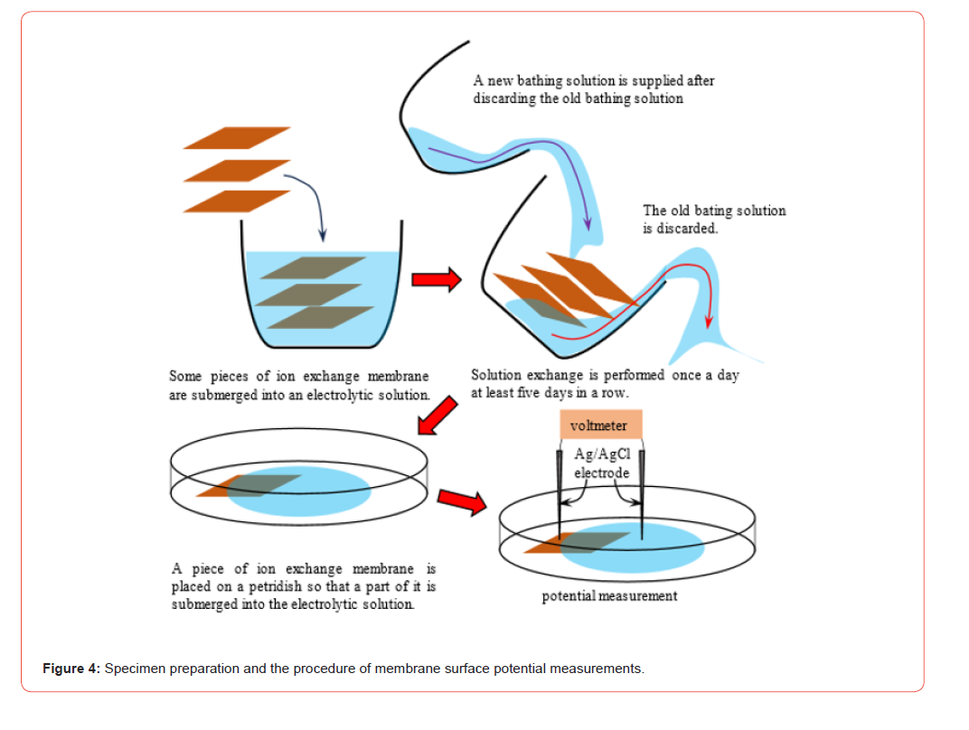
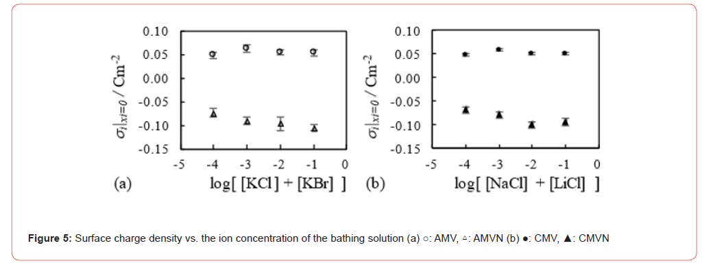
Table 2:Experimentally measured membrane surface potential and the computed membrane surface charge density ,†
*: M = A, AN, C, CN A, AN, C and CN represent AMV, AMVN, CMV and CMVN, respectively.
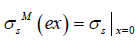 is computed by plugging the experimentally measured
 into Eq. 16.
#: averaged membrane surface charge density.
Results and Discussion
Theorization of membrane potential without the assumption of membrane surface charge density constancy According to what has been described up until here, Eq. 22 can be arranged into Eq. 28 which is identical to the GHK eq. as long as Eq. 23 is valid. That discussion is within the range of ordinary physical chemistry, and it is also in line with the AIH. Now, what if Eq. 23 is not employed? The author will derive the membrane potential formula by employing the ion adsorption mechanism without Eq. 23 as below.
Assuming that the surface charge density  depends on
 and it can be represented by
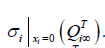 For example, if we
have four data of surface charge density
 M and 10−1 M, the average surface charge density,
 , can be given by Eq. 29.
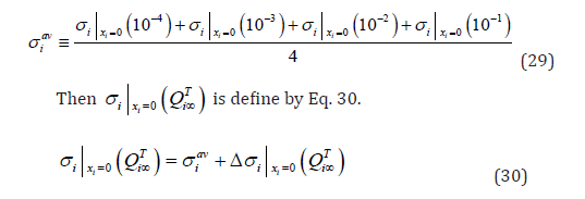
Eq. 22 is arranged into Eq. 31 and can be further arranged into Eq. 32 using Eq. 30.
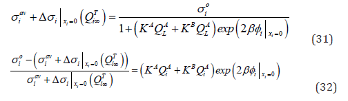
Introducing Eqs. 33 and 34, Eq. 32 can be arranged into Eq. 35.
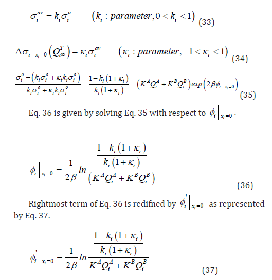
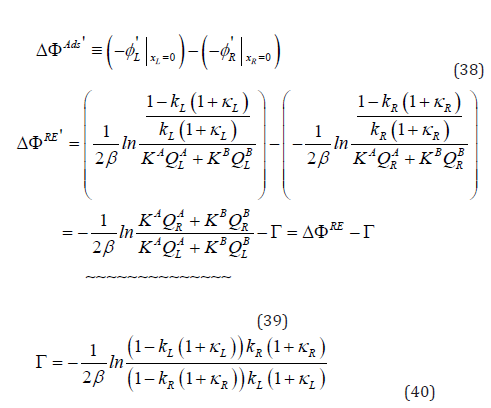
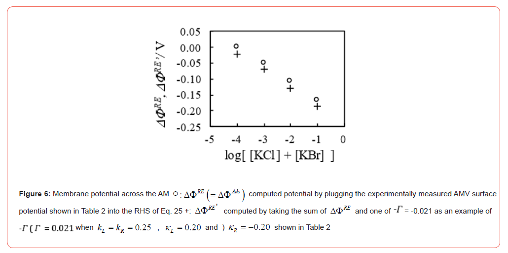
Figure 1 is the electrolytic solution system when the two electrolytic solutions are separated by a membrane. What is the membrane potential formula (ΔΦAds ' )like when Eq. 37 is used? According to Eq. 25, ΔΦAds ' is given by Eq. 38. RHS of Eq. 38 can be transformed into Eq. 39 using Eq. 37 where LHS of Eq. 39 is re-denoted by ΔΦRE' . So, even though the assumption Eq. 23 is not employed, the potential formula Eq. 39 quite similar to the GHK eq. is derived. ~~~~ term in Eq. 39 is the same as ΔΦRE of Eq. 28. ΔΦRE' of Eq. 39 is different from ΔΦRE' merely by −Γ (Γ is define by Eq. 40).
The author computed the numerical values of ΔΦRE' using the
last equation of Eq. 39 when the membrane AMV was used as an
example since the deviation of its surface charge density  from its average
 is the most significant among four membranes
cases as clearly shown in Table 2.
Assuming that the Right phase solution is 0.0005_KCl-
KBr and the Left phase solution varies from 0.00005_KCl-KBr
through 0.05_KCl-KBr, the experimentally measured potential
 summarized in Table 2 are plugged into the RHS of Eq.
25. For example, when the Left phase solution is 0.005_KCl-
KBr,
 . Hence,
 where the error term
is neglected in this computation for simplicity. The computed
ΔΦAds can be regarded as ΔΦRE owing to Eq. 28. But ΔΦRE' deviates
from ΔΦRE = (ΔΦAds ) by −Γ . Therefore, the author computed
−Γ . In order to compute −Γ , the author had to determine the
numerical values of kR and kL . The deviation of membrane surface
charge
 from the average
 is basically in the range of
Eq. 41 as shown in Table 2 regardless of the membrane species.

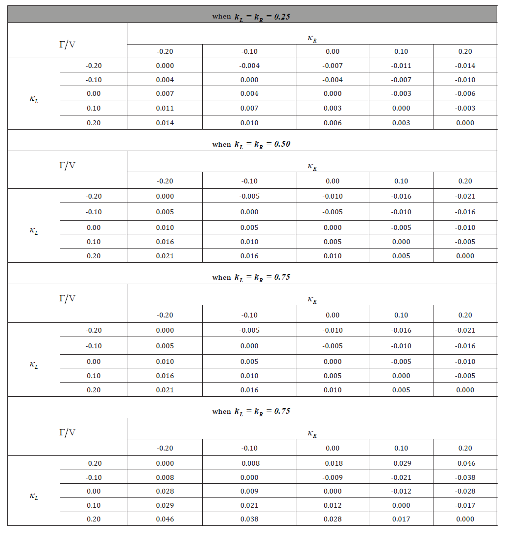
The author computed Γ under three conditions Eq. 42 ∼ Eq. 44 where kL = kR takes 0.25, 0.50 and 0.75, and κL and κR take -0.2, -0.1, 0.0, +0.1 and +0.2. The computational result is given in Table 3. Table 3 says that as long as kL = (kR) is 0.5 or lower than 0.5, the magnitude of −Γ is not so large. Therefore, ΔΦRE' ~ ΔΦRE holds (Eq. 45). Eq. 45 is valid under a certain condition. Therefore, the ~~~~ term of Eq. 39 which is identical to the GHK eq. is basically valid within the range of experimental error. This means that the AIH can provide us the potential formula which is identical to the GHK eq. such as Eq. 27.
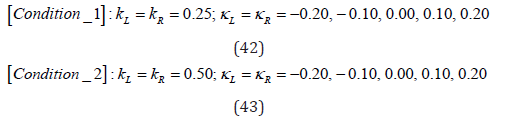
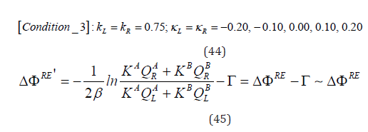
Figure 6 is an example. ΔΦRE in this diagram is computed by plugging the experimentally measured membrane surface potential in Table 2 into the RHS of Eq. 25. ΔΦRE' is computed by taking the sum of ΔΦRE and −Γ following Eq. 45. The author employed one of −Γ ' s, −Γ = -0.021 (which is obtained when 0.25 kL = kR , κL =0.20 and κR −0.20 shown in Table 2). ΔΦRE' is different from ΔΦRE by −Γ = -0.021 V. This is not so small difference as a membrane potential, but the entire potential data curves of ΔΦRE and ΔΦRE' in Fig. 6 look quite similar each other, and as mentioned earlier, the data in Table 3 suggest that the magnitude of −Γ is mostly small enough as long as kL = (kR) is 0.5 or lower than 0.5. But of course, ΔΦRE' largely deviates from ΔΦRE when kL = kR=0.75 (see Table 3). As a matter of fact, it is well-known that even the broadly employed GHK eq. does not necessarily for explaining all the membrane characteristics [24, 25]. Therefore, the validity limitation of the ΔΦRE' ∼ ΔΦRE (Eq. 45) is quite understandable. The potential formula ΔΦRE' under a certain condition is almost same as ΔΦRE whose mathematical expression is virtually indistinguishable from the GHK eq. This outcome is largely owing to the constant membrane surfacecharge density (though not perfectly constant) and is also owing to the experimental fact that −Γ is not so sensitive to the deviation of the membrane surface charge density from the average value.
The formulas so far obtained are in line with the AIH model but its mathematical expression such as ΔΦRE and ΔΦRE' are often the case indistinguishable from the mathematical expression of GHK eq. of Eq. 27. Therefore, we have not been aware of the shortcomings of the GHKeq. such that the GHKeq. does not take into consideration the ion adsorption effect on the membrane potential generation.
Table 3:
Directly measured membrane potential
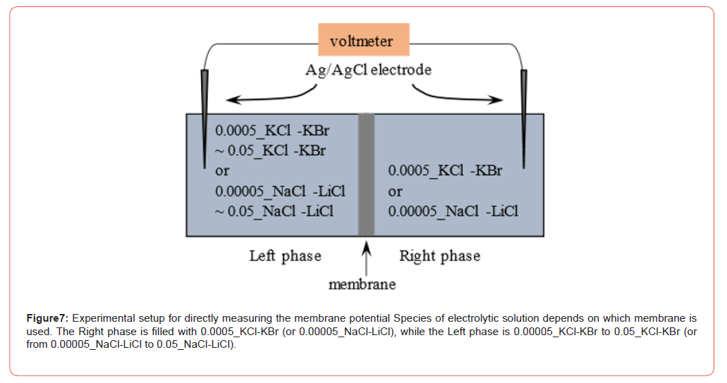
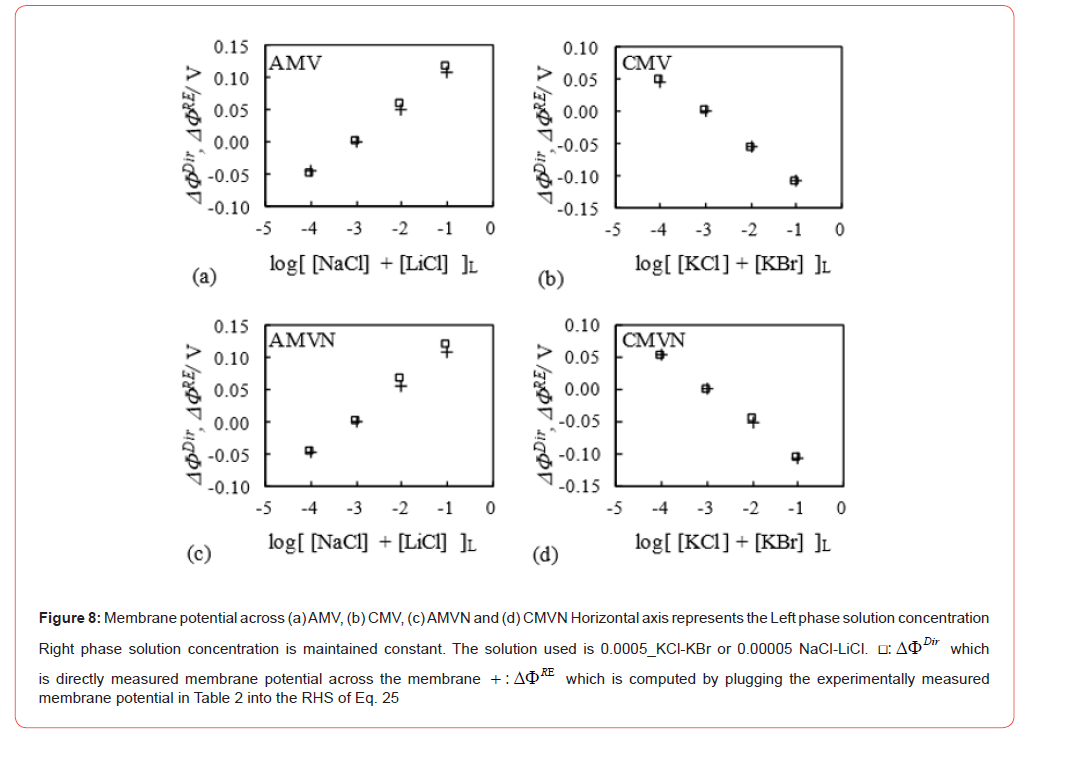
The author also performed the direct measurement of membrane potential, ΔΦDir , across the AMV, AMVN, CMV and CMVN. The experimental setup is illustrated in Figure 7. When the membrane potential was measured, 0.0005_KCl-KBr (or 0.00005_Na- Cl-LiCl) was always used as the Right phase electrolytic solution while the Left phase solution varied from 0.00005_KCl-KBr to 0.05_ KCl-KBr (or from 0.00005_NaCl-LiCl to 0.05_NaCl-LiCl). ΔΦDir and ΔΦRE earlier computed using Eq. 25 are shown in the diagrams in Figure 8. ΔΦRE perfectly reproduces the experimental membrane potential ΔΦDir .
Summary of the sections 3.1 and 3.2
The sections 3.1 and 3.2 deal discuss the membrane potential generation from the view of ion adsorption mechanism. For the theorization, merely Boltzmann distribution, mass action law and Langmuir isotherm are used [23] along with and without the assumption of membrane surface charge constancy in the section 3.1. Then the author reached the conclusion that it is natural to reach the membrane potential formula identical to the GHK eq. in purview of the ion adsorption mechanism. Therefore, the ion channel and pumps are not necessary for the membrane potential generation. In the section 3.2, it was found that the membrane potential can be quantitatively reproduced as in Fig.8 merely using the membrane surface potential data. The membrane surface potential is governed by the ion adsorption. Therefore, this physical chemistry- based theorization and its successful data reproducibility shown in Figure 8 serve as the supportive evidence to the AIH.
Behind the success of the ion adsorption-based membrane potential generation mechanism, the membrane surface charge density constancy appears to play a fundamental role. If the membrane surface charge density is constant, the reGHK eq. can be derived. If not, the reGHK eq., which is identical to the GHK eq., cannot be derived but the potential deviates by “ −Γ ” from there GHK eq. As touched upon earlier, it is not so uncommon that the membrane potential does not obey the potential predicted by the GHK eq. Therefore, even the deviation of the actual potential from the potential predicted by the reGHK eq. is not so problematic. The reGHK eq. involves the membrane surface charge density constancy. It appears to hold under the relatively broad range of experimental conditions. This must be one of the reasons that the GHK eq. works fine though the foundation of the GHK eq. may be wrong. Namely, the right membrane potential formula is the reGHK eq., but its expression is identical to the expression of the GHK eq. Therefore, they are indistiguishable each other. But why does the membrane surface charge density is constant under so broad conditions? It is still incomprehensible, but it must be the important theme to solve.
Conclusion
Membrane potential formula identical to the GHK eq. was found to be derivable using merely the basic physical chemistry concepts as long as the membrane surface charge density is constant regardless of the ion concentration of solutions. This concept was built on the premise that the membrane potential is governed by the ion adsorption and this principle is in harmony with the long-forgotten physiological theory called Association-Induction Hypothesis (AIH). The ion adsorption mechanism-based membrane potential formula worked quite fine but such a fineness of the formula appears to rests on the membrane surface charge density constancy under the relatively broad conditions. At this moment, the author has no clues regarding why such a condition well establish but should address it as a next theme.
Funding
No financial supports.
Acknowledgment
None.
Conflict of Interest
The authors declare that they have no conflict of interest.
References
- Ling G N (1992) A Revolution in the Physiology of the Living Cell, Krieger Pub Co. Malabar, Florida.
- Ling G N (2001) Life at the Cell and Below-Cell Level: The Hidden History of a Fundamental Revolution in Biology. Pacific Press, New York.
- Edelman L (2005) DOUBTS ABOUT THE SODIUM-POTASSIUM PUMP ARE NOT PERMISSIBLE IN MODERN BIOSCIENCE. Cellular and Molecular Biology 51(8): 725-729.
- Pollack G H (2001) Cells, Gels and the Engines of Life: A New, Unifying Approach to Cell Function, Ebner & Sons, Seattle, USA.
- Pollack G H (2013) The Fourth Phase of Water: Beyond Solid, Liquid, and Vapor, Ebner & Sons, Seattle, USA.
- Matveev V V (2017) Comparison of fundamental physical properties of the model cells (protocells) and the living cells reveals the need in protophysiology. Int. J. Astrobiol 16: 97-104.
- Wnek G E, Costa A C S, Kozawa S K (2022) Bio-Mimicking, Electrical Excitability Phenomena Associated With Synthetic Macromolecular Systems: A Brief Review With Connections to the Cytoskeleton and Membraneless Organelles. Frontiers in Molecular Neuroscience 15: 830892.
- Bagatolli L A, Mangiarotti A, Stock R P (2020) Cellular metabolism and colloids: Realistically linking physiology and biological physical chemistry. Prog. Biophys. Mol. Biol 162: 79-88.
- Bagatolli L A, Stock R P (2021) Lipids, membranes, colloids and cells: a long view. BBA Biomembranes 1863(10): 183684.
- Schneider M F (2020) Living systems approached from physical principles. Prog. Biophys. Mol. Biol. 162: 2-25.
- Lee J W (2019) Electrostatically localized proton bioenergetics: better understanding membrane potential. Heliyon 5(7): e01961.
- Lee J W (2020) Protonic conductor: better understanding neural resting and action potential. J. Neurophysiol 124(4): 1029-1044.
- Cheng K L (1990) Capacitor Theory for Nonfaradaic Potentiometry. MICROCHEMICAL JOURNAL 42: 5-24.
- Cheng K L (1990) Explanation of Misleading Nernst Slope by Boltzmann Equation. MICROCHEMICAL JOURNAL 42: 5-24.
- Funk R H W, Scholkmann F (2023) The significance of bioelectricity on all levels of organization of an organism. Part 1: From the subcellular level to cells. Progress in Biophysics and Molecular Biology 177: 185-201.
- H Tamagawa, K Ikeda (2018) Another interpretation of Goldman-Hodgkin-Katz equation based on the Ling’s adsorption theory. Euro. Biophys. J 47(8): 869-879.
- Tamagawa H (2019) Mathematical expression of membrane potential based on Ling’s adsorption theory is approximately the same as the Goldman–Hodgkin–Katz equation. J. Biol. Phys 45(1): 13-30.
- Cronin J (1987) Mathematical aspects of Hodgkin-Huxley neural theory. Cambridge University Press, New York.
- Keener J, Sneyd J (2008) Mathematical physiology: I: cellular physiology. Interdisciplinary applied mathematics. Springer, New York.
- Ermentrout G B, Terman T H (2010) Mathematical foundations of neuroscience, vol 35. Interdisciplinary applied mathematics book. Springer, New York.
- Ling G N (1997) Debunking the alleged resurrection of the sodium pump hypothesis. Physiol. Chem. Phys. Med. NMR 29(2): 123-198.
- Ling G N (1978) MAINTENANCE OF LOW SODIUM AND HIGH POTASSIUM LEVELS IN RESTING MUSCLE CELL. J. Physiol 280: 105-123.
- Barrow G M (1979) Physical chemistry, McGraw-Hill, New York.
- Ohki S, Aono O (1979) Membrane Potential of Squid Axons: Effect of Internal and External Ion Concentration. Japanese Journal of Physiology 29(4): 373-382.
- Chang D (1983) DEPENDENCE OF CELLULAR POTENTIAL ON IONIC CONCENTRATION Data Supporting a Modification of the Constant Field Equation. BIOPHYS. J 43(2): 149-156.
-

This work is licensed under a Creative Commons Attribution-NonCommercial 4.0 International License.