 Research Article
An Interesting Limitation on Application of THz Spectroscopy to Characterization of DNA
Daniel Shreiber*1, Sarah Stranieri2 and Molleshree Karna3
1WMRD, CCDC Army Research Laboratory, USA
2University of Illinois at Urbana-Champaign, USA
3Columbia University, USA
Daniel Shreiber, WMRD, CCDC Army Research Laboratory, Aberdeen, MD, USA.
Received Date: September 22, 2021; Published Date: October 27, 2021
Abstract
THz (terahertz) spectroscopy has been proposed as a novel method to characterize DNA molecules when applied on plasmonic surfaces. This approach can be used as a less expensive alternative for a DNA characterization method. It has been successfully demonstrated that a significant shift of the resonant frequency in THz spectrum between TRIS buffer and TRIS buffer containing the DNA can be observed. The sensitivity of the method can be enhanced when the resonant frequency of the DNA molecule under consideration matches the plasmon frequency of the plasmon surface yielding a non-linear effect of overlapping resonances. It is shown here that this method only works when the THz radiation is applied above certain energy threshold.
Keywords: THz spectroscopy; Plasmon surfaces; DNA characterization
Abbreviations: THz: Terahertz; TDS: Time Domain Spectroscopy; DNA: Deoxyribonucleic acid; FFT: Fast Fourier Transform
Introduction
The ultimate goal of the proposed approach is to establish conditions and limitations of application of THz spectroscopy to characterize DNA. If successful, this approach will yield a novel method that will be able to analyze base pair sequences in full strands that will in turn improve forensic analysis, genetic testing, and DNA production [1]. This is due to the proposed capability to identify short oligonucleotides. The proposed method will be cheaper than the existing ones.
THz spectroscopy is a very good candidate to characterize molecules like DNA because in this spectrum (wavelength of 50 μm- 1mm) a resonance can be observed with vibrational modes of these large molecules. Through this approach it is possible to monitor different processes occurring in the DNA molecules such as weak hydrogen bonding of the molecule base pairs, genetic material replication [2] and transcription, movements of the entire double helix [3].
Experiment and Methods: In order to achieve the stated goal, a plasmon surface has been utilized [4]. In fact, in the THz spectrum, due to the fact that the surface plasmon is not tightly bound to the surface, the condition is of a metamaterial with modes where the resonant frequency can be calculated from the same formalism (1) as a regular plasmon (spoof plasmon) [5]. The plasmon surface consists of a stainless steel 100 μm thick foil perforated with an array of periodic holes with diameter of 200 μm and pitch (L) of 500 μm (Figure 1).

The Drude Model Approximation is valid for the spoof plasmon surface in THz spectrum where the resonant wavelength, λ can be determined from the following formalism:

Where m and n are surface “plasmon” orders and ε is a real part of the dielectric constant o the dielectric surrounding the surface.
The surface of the mesh was coated with 450 nm thin film of gold for better binding of the DNA to the surface.
It is important to state again that the approach of utilizing a plasmon surface (the mesh) was elected because the overall effect can be significantly enhanced if the geometrical parameters of the plasmon surface will yield the resonant frequency similar to the one of the DNA to benefit from the overlapping resonance effect.
The solution containing 200 μL of DNA in TRIS buffer was spin coated onto the meshes at 203 rpm and dried at this rotational speed for 10 minutes. The DNA oligomer sequence used was 5’-CATTAACGAGTTACTCAATGAGT5CTTTCTG-3’.
The mesh was inserted between emitting and receiving antennae in transmission mode of THz TDS (time domain) spectrometer TeraKit15 (Menlo Systems) (Figure 2). Each reciprocal side also contained a collimating lens and a focusing lens which could be taken out. The focused beam had diameter of 3 mm whereas a collimated one (when the focusing lens is out) was 30 mm.

The results were normalized by the free space when no mesh is inserted between the antennae and the scans were converted to frequency domain via Fast Furrier Transform (FFT) and analyzed.
Results and Discussion
It has been previously shown [6] that both TRIS buffer and TRIS buffer with DNA have distinct resonance peaks in the THz spectrum yielding a blue shift of about 0.1 THz (from 1.5 THz to 1.4 THz when DNA is added). This provides a substantial support to the idea that THz TDS Spectrometry can be used as a method to characterize DNA. As a reminder, the resonant peaks in current experimental configuration were not coinciding with the plasmon peaks of the plasmon surface which from (1) were calculated (and experimentally confirmed) to be at λ=0.58 THz and λ=0.82 THz. Again, coinciding the resonant peak with the plasmon peak would result in much more pronounced effect due to the overlapping resonances.
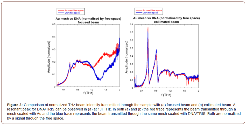
In this work, a very important limitation for the proposed method is discussed. As it was previously mentioned, the THz TDS Spectroscope can operate in transmission mode in two configurations. Namely with or without focusing lenses between the antenna/ collimating lens and the sample and, reciprocally, on the other side of the sample. Using the focusing lens would reduce the focus spot diameter at maximum wavelength from about 30 mm when the beam is collimated to only 3 mm. This would result in 100 times increase in the energy density of the beam for the focused beam. Interestingly enough, as it is obvious from Figure 3, the resonant peak for the DNA with TRIS buffer can be observed when the mesh is illuminated with a focused beam and is not observed at all when the sample is illuminated with the collimated beam which suggests a certain threshold of energy required to trigger the resonant response in the sample.

Conclusion
THz TDS Spectroscopy presents as an interesting approach to characterize DNA molecules. DNA molecules are resonant in this spectrum of radiation but, as it was shown here, the resonant peaks can be observed only when the sample is illuminated with a beam that has energy density above certain threshold. We speculate that this is due to the non-linear nature of the effect. This approach merits additional investigation in order to determine fully all its benefits and limitations. We believe that this work is a significant step forward in this process.
Acknowledgment
None.
Conflict of Interest
No conflict of interest.
References
- AL Chernev, NT Bagraev, LE Klyachkin, AK Emelyanov, MV Dubina (2014) DNA detection by THz pumping. Condensed Matter.
- DL Woolard, TR Globus, BL Gelmont, M Bykhovskaia, AC Samuels, et al. (2001) Submillimeter-wave Phonon Modes in DNA Macromolecules. Phys Rev E Stat Nonlin Soft Matter Phys 65(5 Pt 1): 051903.
- Globus T, Khromova T, Woolard D, Gelmont B (2004) Terahertz Fourier transform characterization of biological materials in solid and liquid phases. Chemical and Biological Standoff Detection 5268: 1-9.
- M Tanaka, F Miyamaru, M Hangyo, T Tanaka, M Akazawa, et al. (2005) Effect of a thin dielectric layer on terahertz transmission characteristics for metal hole arrays. Opt Lett 30(10): 1210.
- JB Pendry, L Martın-Moreno, FJ Garcia-Vidal (2004) Mimicking Surface Plasmons with Structured Surfaces. Science 305(5685): 847-848.
- S Stranieri, M Karna, D Shreiber (2015) Terahertz Characterization of DNA: Enabling a Novel Approach. ARL-CR-0788, US Army Research Lab Tech Report.
-
Daniel Shreiber, Sarah Stranieri, Molleshree Karna. An Interesting Limitation on Application of THz Spectroscopy to Characterization of DNA. Mod Concept Material Sci. 4(4): 2021. MCMS. MS.ID.000590. DOI: 10.33552/MCMS.2021.04.000590.
-
THz spectroscopy, Plasmon surfaces, DNA characterization, Molecules, Frequency, Material, THz spectrum, Wavelength, Yielding, Focused beam, Threshold
-

This work is licensed under a Creative Commons Attribution-NonCommercial 4.0 International License.


