 Case Report
Resin Infiltration Applications for Aesthetic Improvement
Hatice Kizilhan, Melike Tandogan, Deniz Ozkuyucu, Elif Sepet*
Department of Pediatric Dentistry, İstanbul Kent University, Faculty of Dentistry, Türkiye
Elif Sepet, Department of Pediatric Dentistry, İstanbul Kent University, Faculty of Dentistry, Turkey
Received Date:December 01, 2023; Published Date:March 21, 2024
Abstract
White spot lesions (WSLs) occur due to hypomineralization of the enamel. Conditions causing enamel hypomineralization are fluorosis, trauma, molar-incisor hypomineralization, genetic defects, and environmental factors. Resin infiltration is a minimally invasive technique that aims to stabilize WSLs, prevent the progression of caries, and enhance the aesthetic appearance of affected teeth. In this report, the treatment of WSLs in four pediatric patients is presented. The present study aims to discuss the aesthetic results of resin infiltration treatment based on lesion variety in pediatric patients. A mild fluorosis case was treated only with the resin infiltration technique and a satisfactory outcome was achieved in the presented study. The efficacy of resin infiltration in the presented MIH cases was evaluated, and it was concluded that superficial and small white lesions presented favorable outcomes in terms of patient and practitioner satisfaction. Compared to white lesions, the deeper and yellowish-brown lesions required multiple etching applications. It has been determined that the aesthetic results do not provide sufficient satisfaction and reveal the need for additional composite restoration in deep WSLs. To determine the success of the treatment, it is important to select the cases carefully and assess the density of lesions before the treatment. In conclusion, standardized pretreatment, treatment, and posttreatment procedures would be required for the resin infiltration technique in pediatric patients to achieve better results.
Keywords:Resin infiltration technique; White spot lesions; Enamel hypomineralization
Abbreviations:WSL: White spot lesion; CPP-ACP: Casein phosphopeptide-amorphous calcium phosphate; MIH: Molar incisor hypomineralisation
Introduction
White spot lesions (WSLs), defined as ‘white opacities’, occur as an initial manifestation of enamel demineralization, frequently indicating the early stages of caries development. [1,2] Key etiological factors contributing to the formation of white spot lesions are poor oral hygiene, the accumulation of bacterial biofilm, and dietary habits. [3] Additional factors may include orthodontic treatment, decreased saliva flow, and systemic conditions. [4] These lesions are characterized by observable changes in the tooth’s appearance and texture, often appearing as chalky or opaque white spots, indicating a loss of mineral content in the enamel. [5,6] The reason for the white appearance is the changes in light-scattering optical properties of the decalcified enamel. [2] Non-invasive methods like remineralization cures, topical fluoride applications, and casein-phosphopeptide-amorphous calcium phosphate (CPP-ACP) products have shown promise in reversing enamel demineralization. In more advanced cases, minimally invasive restorative techniques, such as resin infiltration and microabrasion, may restore the enamel’s integrity [2,6].
WSLs occur due to the hypomineralization of the enamel. Enamel formation is a complicated process that is influenced by both environmental and genetic factors. [7] The diagnosis should take into account conditions that cause hypomineralization, such as fluorosis, trauma, molar-incisor hypomineralization (MIH), environmental factors, and genetic defects causing enamel hypoplasia. [6,7] Developmental enamel defects are associated with hypoplasia, hypomineralization, or hypomaturation. Enamel hypoplasia occurs if the matrix formation is affected and results in pits or grooves, or thin and missing enamel. [8] Hypomineralization, or decreased mineralization as a result of maturation disruption, typically presents as soft enamel. Hypomaturation is defined by altered translucency that occurs as a result of a decrease in the deposition of minerals at the final stage of mineralization [8-10].
Excessive incorporation of fluorides during enamel formation results in fluorosis. [9] Excessive fluoride uptake and the use of fluoride-containing substances occur after symmetrical interaction of the homologous teeth and influence different tooth groups. In the early stage, convergent horizontal white lines cause a parchmentlike appearance accompanied by irregular chalky areas. [10] Histopathologically, hypermineralization occurs in the superficial layer of teeth with dental fluorosis, and hypomineralization occurs in the subsurface of the external third of enamel. Then, the presence of exogenous chromophoric proteins causes a color shift to brown [9,10].
MIH is a prevalent developmental dental condition characterized by qualitative enamel defects in the first permanent molars and incisors. [11, 12] The etiology of MIH is multifactorial, involving a combination of genetic and environmental influences during tooth development. Genetic predisposition, complications during prenatal and perinatal periods, childhood illnesses, and exposure to environmental toxins are identified as potential factors contributing to the development of MIH. [13] MIH is clinically characterized by white to yellow to brown demarcated enamel opacities of different colors, occasionally undergoing posteruptive breakdown due to soft and porous enamel in affected molars and incisors. [11] The effective management of MIH necessitates a comprehensive, multidisciplinary approach that addresses both the functional and aesthetic aspects of the affected teeth [11-13].
Non-invasive and minimally invasive treatments aim to preserve enamel, given that demineralization initiates beneath the enamel surface through clinically evident, non-cavitated white spot lesions, which may evolve into cavities if left untreated. [14- 16] Compared to surrounding healthy enamel, demineralized and hypomineralized enamel have different refractive indices owing to larger pore sizes. This results in light dispersion at the subsurface level visibly alters colors and creates aesthetic issues [16-20].
The therapeutic strategy varies depending on the specific types of lesions. In the first phase, the use of preventative therapies proves to be advantageous [21].
The infiltration of a low viscosity, hydrophilic resin substance in the porous lesion has been presented as a new treatment for WSLs. [16] Resin infiltration has several benefits, like the absence of tooth structure loss, allowing stability for white spot lesions, preventing caries progress, plugging of the micropore forms in the body of the lesion, delaying the necessity for a restoration, decreasing recurrent decay, and better aesthetic outcomes in covering demineralized enamel [22].
The present study aimed to assess the effectiveness of treating WSLs with resin infiltration technique and present the clinical and aesthetic outcomes in four cases.
Case Presentation
Written informed consent was obtained from the patient’s parents to publish anonymized information in this article. No ethical committee was required for this study because routinely approved procedures were carried out.
In all cases presented in the article, the standard ICON resin infiltration procedure was applied according to the manufacturer’s instructions described below:
To remove debris and plaque, tooth surfaces were cleaned with a prophylaxis paste without fluoride (Cleanic Prophy Paste, Kerr, Rastatt, Germany). The isolation is provided by a light-cured gingival barrier (Fläsh Gingiva Protector LC Syringe, Birkenau, Germany) and cotton rolls. 15% hydrochloric acid (Icon-Etch, DMG, Hamburg, Germany) was applied to the dried affected tooth surfaces for 2 minutes. The acid was completely cleaned by suctioning from the tooth surface, washing for 30 seconds, and drying with water-free air. 99% ethanol (Icon-Dry, DMG, Hamburg, Germany) was applied for 30 seconds and dried with water-free air. With the application of Icon-Dry, whitish, opaque lesion color changes are expected to diminish significantly. The dental operating light was switched off, and infiltrating resin (Icon Infiltrant, DMG, Hamburg, Germany) was applied to the tooth surface for 3 minutes. Material on the tooth surface was dispersed with air and photopolymerized with a UV lamp (WoodPecker C, 1000 mW/cm2-1200 mW/cm2, China) for the 40s. Resin infiltration (Icon Infiltrant, DMG, Hamburg, Germany) was applied a second time for 1 minute. Dispersing and lightning were applied the same as in the previous step. The contact surfaces were checked, and the surfaces were polished. Finishing was done with polishing discs (Kerr OptiDisc, Rastatt, Germany).
The etching procedure was performed 2–5 times in the deeper and yellowish-brown lesions.
Case 1
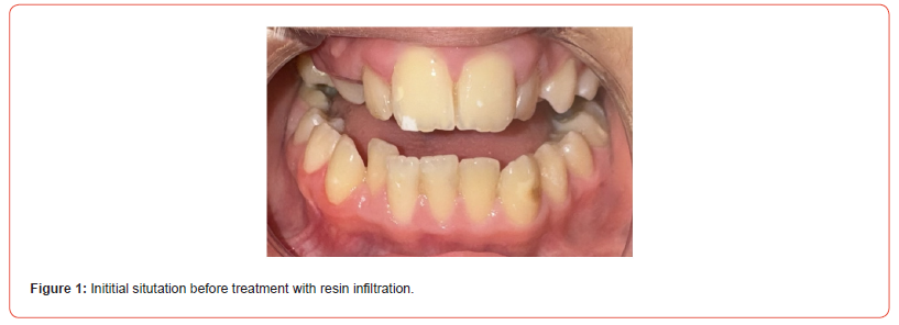
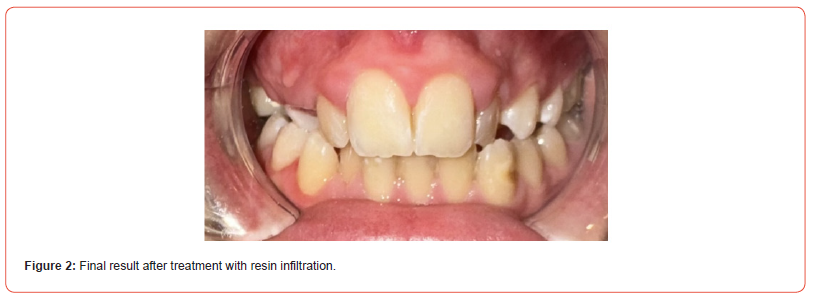
A 12-year-old girl was referred to Istanbul Kent University, Department of Pedodontics, with a complaint of aesthetic concerns on her anterior teeth. The clinical examination showed WSLs on teeth numbers 11 and 21 and a yellowish-brown spot lesion on tooth number 33, and mild opacities were detected in permanent first molars. MIH is diagnosed based on radiographic and clinical examinations. (Figure 1). The resin infiltration was performed for teeth numbers 11, 21, and 33. As a result of the application, a satisfactory aesthetic result was achieved for maxillary incisors, but the technique was found unsatisfactory for a deep-seated lesion on tooth number 33. (Figure 2)
Case 2
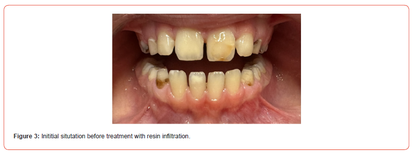
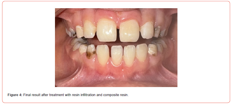
An 8-year-old girl was referred to Istanbul Kent University, Department of Pedodontics, with the complaint of aesthetic concerns on anterior teeth and pain in lower permanent molars. MIH is diagnosed based on radiographic and clinical examinations. (Figure 3) Dental treatments were performed on permanent molars. In tooth number 21, a thin layer of composite filling on the vestibule surface, which had previously been applied in another clinic, was removed. The white spot lesions on the anterior teeth, 21,41,42 were treated by resin infiltration. As a result of the resin infiltration application, an aesthetically satisfactory result was obtained only tooth number 42. The technique was found unsatisfactory for masking the deep lesion on teeth number 21,41 For this reason, upon the request of the family, aesthetic composite restorations were applied to tooth number 21. (Figure 4)
Case 3
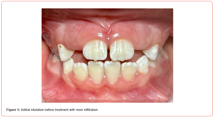
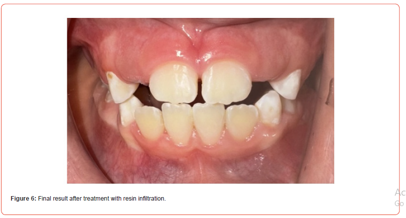
An 8-year-old girl was referred to Istanbul Kent University, Department of Pedodontics, with complaints of dental pain related to the maxillary molar teeth and an unusual appearance of permanent teeth that have recently erupted. Radiographic and clinical examinations revealed a deep caries lesion of the upper left deciduous first molar. Examination of the oral cavity revealed the presence of white spots on all the permanent incisors, to a greater extent on the vestibular and a lesser extent on the oral surfaces. The patient was diagnosed with fluorosis, according to the classification of Thylstrup and Fejerskov [23]. The case was assessed on a scale of 3-4 points, with a higher score of damage to the upper incisors. (Figure 5) The treatment plan consisted of resin infiltration of the crown surfaces of all permanent incisors that erupted. As a result of the application, a satisfactory aesthetic result was achieved for the patient. (Figure 6)
Case 4
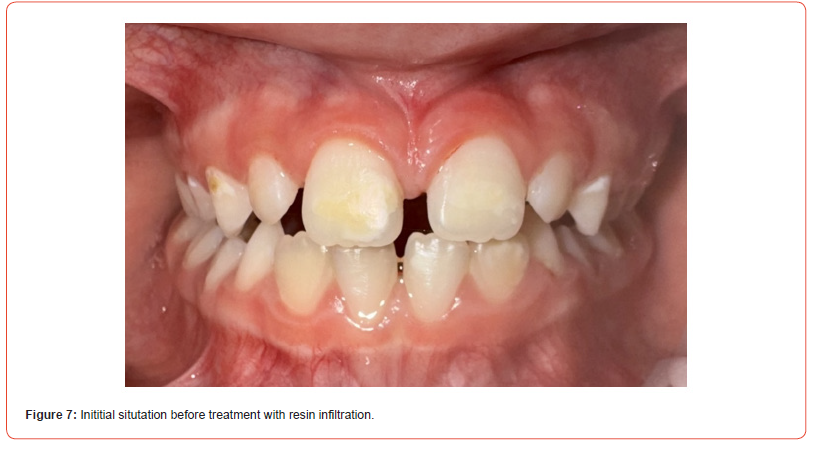
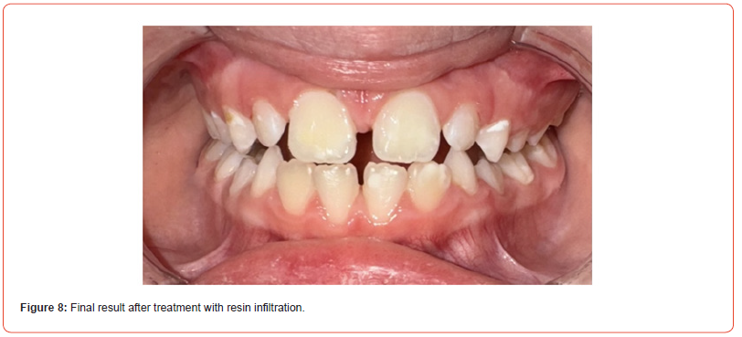
An 8-year-old girl was referred to Istanbul Kent University, Department of Pedodontics, with esthetic concern on anterior teeth due to yellowish-white lesions. The clinical examination revealed deep caries lesions of the permanent first molars and hypomineralization of the permanent incisors. (Figure 7) MIH is diagnosed based on radiographic and clinical examinations. Dental treatments were performed on permanent molars. As a result of the resin infiltration application on 11-21, an aesthetically satisfactory result was obtained from tooth number 21. The color masking was more satisfactory on tooth 21. (Figure 8)
Discussion
Hypoplastic or hypomineralized enamel defects are common in pediatric patients and cause serious problems in the permanent and primary dentitions. To improve children’s quality of life, pediatric dentists must be aware of the risk factors and provide conservative treatments that enhance the aesthetic appearance of such deformities [24, 25].
A correct diagnosis must be established in WSLs with a comprehensive evaluation of the patient’s medical and dental history, as well as a clinical examination that assesses the lesion’s location, symmetry, outline form, depth, and opacity [8].
There are several alternatives available for treating hypoplastic or hypomineralized enamel defects, ranging from less-invasive to more-invasive treatments. Choosing the appropriate treatment option depends on the severity of the lesion [26].
Effective management of cases can be achieved with various conservative approaches and minimally invasive methods developed for lesions detected at the initial stage. Resin infiltration is one of these methods. This concept is less invasive than microabrasion, macro-abrasion, or restorations and is, therefore, a more conservative approach [24].
The resin infiltration technique was developed in the 1970s, and since then, low-viscosity resin materials have been used to restore demineralized enamel. [17,27,28] Since the late 2000s, the content of the materials used in resin infiltration has improved, and more successful clinical and laboratory results have been obtained. The outcome of these developments is that resin infiltration has begun to be used frequently in daily clinical practice. [17,29,30] Resin infiltration methods utilize 15% hydrochloric acid to open the pores within the lesions due to hypomineralized enamel’s resistance to conventional etching. This promotes penetration of the resin infiltrant and can mask minor, white developmental defects by penetrating into the pores. Resin infiltration allows masking even in the more in-depth regions of the lesion due to the change in the refractive light index. The resin infiltration method reduces white spots instantly compared to remineralizing agents [30]. Enamel also gets mechanically strengthened by blocking the diffusion of acid into the enamel which helps to arrest lesions at earlier stages. Therefore, the risk of secondary caries can be minimized. It is preferred as an alternative to invasive restorations because there is no risk of postoperative sensitivity and pulpal inflammation; it reduces the risk of gingivitis and periodontitis; it provides immediate improved aesthetic results when used as a masking resin for WSLs; and it offers high patient satisfaction [31].
In this report, the management of four pediatric cases with WSLs was presented. All the lesions were diagnosed as hypomineralization.
The mild fluorosis case was treated only with the resin infiltration technique and a satisfactory outcome was achieved in the presented study.
Patients with varying degrees of dental fluorosis will require different treatments depending on their expectations and aesthetic concerns, which are subjective and vary from person to person. When treating dental fluorosis, combination treatment modalities like microabrasion, bleaching, and resin infiltration are appropriate to maintain the desired results [32].
Even though the resin infiltration technique has several advantages for minimally invasive treatment of WSLs, it is important to discuss the factors that could compromise the treatment’s efficacy. Inefficient isolation, incomplete resin polymerization and the depth of the lesion are the most important factors that may affect the success of the treatment [33, 34]. This technique requires an extremely dry field and works on the infiltration concept. In addition to maintaining a moisture-free environment, the lesion needs to be dried several times for application steps. The application procedure must be applied according to the manufacturer’s instructions to enhance the process of infiltration [33]. Resin infiltration effectively masks post-orthodontic initial carious lesions and stabilizes the optical improvement for at least 12 months [35].
A smaller WSL is considerably easier to treat with the resin infiltration technique than a larger lesion. Multiple applications may be necessary for medium-to-large-sized lesions. The probability of achieving a complete infiltration will decrease with the increasing depth of the WSLs. Large lesions are also related to increased polymerization shrinkage. Considering the weak capillary activity of the resin in these lesions, the infiltration of deep lesions does not produce satisfactory results. [33,36] Resin infiltration is a minimally invasive technique that may be relied upon in MIH opacities to produce satisfactory outcomes [37].
The opacities in MIH syndrome are not the same as those in fluoride spots or caries etiology, hence treatment results may vary. For a deep resin penetration, the authors recommend a longer infiltration time, a modification to the etching application, a longer etching duration, and preliminary tooth structure preparation [38].
The high level of protein in hypomineralized enamel might be a cause of a weak penetration of the resin [39] In hypomineralized lesions, the use of HCI demineralizes the disordered hydroxyapatites, resulting in a more ordered structure. The excess protein and peptides can be removed with NaOCl and H2O2 application. [39,40]
It is critical to take the thickness of the surface layer into account during treatment [40]. The repeated etching procedure (2-4 times) may cause an average erosion depth of 77 μm [41].
There may be unwanted side effects following the resin infiltration treatment, including teeth and gum ache, soft tissue damage, and sour aftertaste [38]. Besides, a perfect aesthetic result cannot be guaranteed, so it is necessary to inform patients before the treatment [42].
The efficacy of resin infiltration in the presented MIH cases was evaluated, and it was concluded that superficial and small white lesions presented favorable outcomes in terms of patient and practitioner satisfaction. Compared to white lesions, the deeper and yellowish-brown lesions required multiple etching applications. The etching procedure was performed 2–5 times in the deeper lesions. It has been determined that the aesthetic results do not provide sufficient satisfaction and reveal the need for additional composite restoration in deep WSLs.
To determine the success of the treatment, it is important to select the cases carefully and assess the density of lesions before the treatment. There are several opinions such as the CIELAB system, light-induced fluorescence, and magnifying glasses, over the most reliable diagnostic method.
In conclusion, standardized pretreatment, treatment, and posttreatment procedures would be required for the resin infiltration technique in pediatric patients to achieve better results.
Acknowledgement
None.
Conflict of Interest
The authors has no conflict of interest to declare.
References
- Lopes PC, Carvalho T, Gomes ATPC, Veiga N, Blanco L, et al. (2024) White spot lesions: diagnosis and treatment–a systematic review. BMC Oral Health 24(1): 58.
- Ando M, Shaikh S, Eckert G (2018) Determination of Caries Lesion Activity: Reflection and Roughness for Characterization of Caries Progression. Oper Dent 43(3): 301-306.
- Pitts NB, Zero DT, Marsh PD, Ekstrand K, Weintraub JA, et al. (2017) Dental caries. Nat Rev Dis Primers 25(3): 17030.
- Srivastava K, Tikku T, Khanna R, Sachan K (2013) Risk factors and management of white spot lesions in orthodontics. J Orthod Sci 2(2): 43-49.
- Zandoná AF, Zero DT (2006) Diagnostic tools for early caries detection. J Am Dent Assoc 137(12): 1675-1684.
- Sadıkoğlu İS (2020) White Spot Lesions: Recent Detection and Treatment Methods. Cyprus J Med Sci 5(3): 260-266.
- Smith CE (1998) Cellular and chemical events during enamel maturation. Crit Rev Oral Biol Med 9(2): 128-161.
- Clarkson J (1989) Review of terminology, classification, and indices of developmental defects of enamel. Adv Dent Res 3(2): 104-109.
- Deveci C, Çınar Ç, Tirali RE (2018) Chapter 6/Management of White Spot Lesions. In: Akarslan Z, Ge hrke SA (Editors), Dental Caries - Diagnosis, Prevention and Management. (1st ed). IntechOpen Ltd, London, UK.
- Denis M, Atlan A, Vennat E, Tirlet G, Attal JP (2013) White defects on enamel: Diagnosis and anatomopathology: Two essential factors for proper treatment (Part 1). Int Orthod 11(2): 139-165.
- Padavala S, Sukumaran G (2018) Molar Incisor Hypomineralization and Its Prevalence. Contemp Clin Dent 9 (Suppl 2): S246-S250.
- Yannam SD, Amarlal D, Rekha CV (2016) Prevalence of molar incisor hypomineralization in school children aged 8-12 years in Chennai. J Indian Soc Pedod Prev 34(2): 134-138.
- Koruyucu M, Özel S, Tuna EB (2018) Prevalence and etiology of molar-incisor hypomineralization (MIH) in the city of Istanbul. J Dent Sci 13(4): 318-328.
- Prasada K, Penta P, Ramya K (2018) Spectrophotometric evaluation of white spot lesion treatment using novel resin infiltration material (ICON®). J Conserv Dent 21(5): 531-535.
- Perdigão J (2020) Resin infiltration of enamel white spot lesions: An ultramorphological analysis. J Esthet Restor Dent 32(3): 317-324.
- Çehreli ZC (2023) Resin Infiltration: Ultraconservative Treatment Options for Carious and Non-carious Enamel Lesions. Curr Oral Health Rep 10(2): 23-7.
- Borges AB, Caneppele TMF, Masterson D, Maia LC (2017) Is resin infiltration an effective esthetic treatment for enamel development defects and white spot lesions?A systematic review. J Dent 56: 11-18.
- Hallgren K, Akyalcin S, English J, Tufekci E, Paravina RD, et al. (2016) Color Properties of Demineralized Enamel Surfaces Treated with a Resin Infiltration System. J Esthet Restor Dent 28(5): 339-346.
- Gençer MDG, Kirzioğlu Z (2019) A comparison of the effectiveness of resin infiltration and microabrasion treatments applied to developmental enamel defects in color masking. Dent Mater J 38(2): 295-302.
- Oliveira A, Felinto L, Francisconi-dos-Rios L, Moi G, Nahsan F (2020) Dental Bleaching, Microabrasion, and Resin Infiltration: Case Report of Minimally Invasive Treatment of Enamel Hypoplasia. Int J Prosthodont 33(1): 105-110.
- Stoica AM, Stoica OE, Dako T, Kovàcs-Ivàcson AC, Monea M, et al. (2023) Evaluation of the therapeutic performance of ICON infiltration resin in the treatment of White Spot Lesions in esthetic dental areas. Clinical Report. Acta Stomatologica Marisiensis Journal 6(2): 25-32.
- Bergstrand F, Twetman S (2011) A Review on Prevention and Treatment of Post-Orthodontic White Spot Lesions – Evidence-Based Methods and Emerging Technologies. Open Dent J 5: 158-162.
- Thylstrup A, Fejerskov O (1978) Clinical appearance of dental fluorosis in permanent teeth in relation to histological changes. Community Dent Oral Epidemiol 6(6): 315-328.
- Acosta MG, Natera A (2021) Level of knowledge concerning enamel defects and their treatment among pediatric dentists. ALOP 7: 25-34.
- Casaña-Ruiz MD, Marqués Martínez L, García Miralles E (2023) Management of Hypoplastic or Hypomineralized Defects with Resin Infiltration at Pediatric Ages: Systematic Review. Int J Environ Res Public Health 20(6): 5201.
- Meyer-Lueckel H, Paris S (2008) Improved resin infiltration of natural caries lesions. J Dent Res 87(12): 1112-1116.
- Robinson C, Hallsworth AS, Weatherell JA, Künzel W (1976) Arrest and control of carious lesions: a study based on preliminary experiments with resorcinol-formaldehyde resin. J Dent Res 55(5): 812-818.
- Paris S, Meyer-Lueckel H, Kielbassa AM (2007) Resin infiltration of natural caries lesions. J Dent Res 86(7): 662-666.
- Meyer-Lueckel H, Paris S, Kielbassa AM (2007) Surface layer erosion of natural caries lesions with phosphoric and hydrochloric acid gels in preparation for resin infiltration. Caries Res 41(3): 223-230.
- Saluja I, Pradeep S, Shetty N (2022) Minimally invasive management of white spot lesion using resin infiltration technique: A case report. Gülhane Tıp Dergisi 64(1): 120-122.
- Paris S, Meyer-Lueckel H, Cölfen H, Kielbassa AM (2007) Penetration coefficients of commercially available and experimental composites intended to infiltrate enamel carious lesions. Dent Mater 23(6): 742-748.
- Nicholas LS, Yew Christopher QE, Fei Frank LK (2023) Conservative esthetic management of brown enamel fluorosis using combination therapy: A clinical report. J Conserv Dent 26(3): 349-354.
- Manoharan V, Arun Kumar S, Arumugam SB, Anand V, Krishnamoorthy S, et al. (2019) Is Resin Infiltration a Microinvasive Approach to White Lesions of Calcified Tooth Structures? A Systemic Review. Int J Clin Pediatr Dent 12(1): 53-58.
- Taher NM, Alkhamis HA, Dowaidi SM (2012) The influence of resin infiltration system on enamel microhardness and surface roughness: an in vitro study. Saudi Dent J 24(2): 79-84.
- Wierichs RJ, Abou-Ayash B, Kobbe C, Esteves-Oliveira M, Wolf M, et al (2023) Evaluation of the masking efficacy of caries infiltration in post-orthodontic initial caries lesions: 1-year follow-up. Clin Oral Invest 27(5): 1945-1952.
- Meyer-Lueckel H, Chatzidakis A, Naumann M, Dörfer CE, Paris S (2011) Influence of application time on penetration of an infiltrant into natural enamel caries. J Dent 39(7): 465-469.
- Alghawe S, Raslan N (2024) Management of permanent incisors affected by Molar-Incisor-Hypomineralisation (MIH) using resin infiltration: a pilot study. Eur Arch Paediatr Dent.
- Athayde G, Reis P, Jorge R, Americano G, Fidalgo T, et al. (2022) Impact of masking hypomineralization opacities in anterior teeth on the aesthetic perception of children and parents: A randomized controlled clinical trial. J Dent 123: 104168.
- Natarajan AK, Fraser SJ, Swain MV, Drummond BK, Gordon KC (2015) Raman spectroscopic characterisation of resin-infiltrated hypomineralised enamel. Anal Bioanal Chem 407(19): 5661-5671.
- Bulanda S, Ilczuk-Rypuła D, Dybek A, Pietraszewska D, Skucha-Nowak M, et al. (2022) Management of Teeth Affected by Molar Incisor Hypomineralization Using a Resin Infiltration Technique-A Systematic Review. Coatings 12: 964.
- Tam CP, Manton DJ (2021) Esthetic management of incisors affected with molar incisor hypomineralisation. Clin Dent Rev 5: 6.
- Marouane O, Douki N, Chtioui F (2020) A combined approach for the aesthetic management of stained enamel opacities: External bleaching followed by resin infiltration. J Esthet Restor Dent 32: 451-456.
-
Hatice Kizilhan, Melike Tandogan, Deniz Ozkuyucu, Elif Sepet*. Resin Infiltration Applications for Aesthetic Improvement. Mod Concept Material Sci. 6(1): 2024. MCMS. MS.ID.000626.
-
Resin infiltration technique, White spot lesions, Enamel hypomineralization, White opacities, Genetic factors, Hydrophilic, Yellowish-white lesions, Micro-abrasion, Hydroxyapatites
-

This work is licensed under a Creative Commons Attribution-NonCommercial 4.0 International License.


