

 Case Report
Vein of Galen Aneurysmal Malformation: Neonatal Management Challenges
1 Department of Neonatology, Centro Materno Infantil do Norte (CMIN), Unidade Local de Saúde de Santo António (ULSSA), Porto, Portugal
2 Department of Obstetrics, CMIN, ULSSA, Porto Portugal
3 Department of Pediatric Cardiology, CMIN, ULSSA, Porto, Portugal
4 Department of Neuroradiology, ULSSA, Porto, Portugal
Received Date:January 24, 2026; Published Date:February 23, 2026
Abstract
Introduction: Vein of Galen aneurysmal malformation (VGAM) is a rare congenital vascular anomaly involving arteriovenous shunting in the
median prosencephalic vein. Prenatal diagnosis is key for optimized perinatal care.
Case Presentation: We report a case of VGAM diagnosed by ultrasound at 29 weeks and confirmed by fetal MRI. Progressive fetal cardiac
dysfunction led to cesarean delivery at 38 weeks. The newborn developed high-output heart failure and pulmonary hypertension. With a Bicêtre
score of nine, anticongestive therapy was initiated, followed by transarterial embolizations on days one and eight. The patient was discharged on day
44. Definitive treatment was planned at 5–6 months, but worsening hydrocephalus during a viral respiratory infection required urgent intervention.
Despite technically successful embolizations, the infant died at six months from pneumonia and multiorgan failure.
Conclusion: This case highlights the clinical complexity of VGAM and the need for early referral to centers with neonatal intensive care and
interventional neuroradiology expertise.
Keywords: Vein of Galen malformation; Prenatal diagnosis; High-output heart failure; Congenital vascular anomalies; Endovascular treatment; Perinatal care
Abbreviations: ARDS: acute respiratory distress syndrome; CE-MRI: contrast-enhanced brain magnetic resonance imaging; CSF: cerebrospinal fluid; HOHF: high-output cardiac failure; VGAM: Vein of Galen aneurysmal malformation
Introduction
Vein of Galen aneurysmal malformation (VGAM) is a rare congenital vascular malformation, occurring in fewer than one in 25,000 deliveries. However, it remains the most common congenital cerebral vascular malformation, accounting for approximately 30% of all pediatric vascular anomalies [1,2]. VGAM results from the formation of arteriovenous fistulae between the primitive choroidal circulation and the median prosencephalic vein of Markowski-an embryonic precursor of the vein of Galen that normally involutes between eight and 11 weeks of gestation. Persistence of Markowski’s vein leads to marked venous dilation with multiple arterial feeders, creating a high-flow, low-resistance arteriovenous shunt through which up to 70% of cardiac output may be diverted to the cerebralcirculation [2,3].
During intrauterine life, the low vascular resistance of the placenta effectively competes with the VGAM shunt, limiting diversion of blood flow through the malformation. After birth, abrupt interruption of placental circulation results in a sudden increase in flow across the shunt, frequently triggering rapid clinical deterioration characterized by severe respiratory distress and high-output cardiac failure (HOHF), observed in more than 50% of affected neonates. Without timely intervention, this hemodynamic instability may progress to refractory pulmonary hypertension and multisystem organ failure [2,4,5]. In less critical presentations, pharmacologic stabilization may allow elective endovascular embolization to be delayed until approximately five to six months of age [6,7].
Prenatal identification of VGAM is essential for early diagnosis and coordinated perinatal management through a multidisciplinary approach [2].
We present a case in which VGAM was suspected prenatally on obstetric ultrasonography and subsequently confirmed by fetal brain Magnetic Resonance Imaging (MRI). The case was notable for ultrasonographic findings suggestive of intrauterine cardiac decompensation, consistent with congestive heart failure. These features indicated a high-flow vascular malformation unlikely to respond adequately to immediate postnatal medical stabilization. The patient underwent endovascular embolization of the feeding vessels with initial clinical improvement.
Case Presentation
A male neonate, born to non-consanguineous parents with no relevant family medical history, was diagnosed prenatally with VGAM at 29 weeks of gestation via obstetric ultrasonography, subsequently confirmed by fetal MRI (Figure 1). Fetal echocardiography demonstrated findings consistent with moderate HOHF. Given the severity of the condition, medical termination of pregnancy was proposed but declined by the parents. The pregnancy was thereafter monitored with twice-weekly ultrasonography. Due to progressive worsening of the fetal cardiac status, an elective cesarean delivery was performed at 38 weeks of gestation, with a plan for early postnatal endovascular treatment. For this purpose, the umbilical artery was cannulated immediately after birth to allow vascular access.

At birth, Apgar scores were seven, eight, and eight at one, five, and 10 minutes, respectively. The neonate required immediate resuscitation, including endotracheal intubation. Anthropometric measurements were appropriate for gestational age. Physical examination revealed a cranial bruit audible over the anterior fontanelle. Postnatal cranial ultrasonography and contrastenhanced brain MRI (CE-MRI) demonstrated a large choroidal-type aneurysmal dilation predominantly supplied by the left posterior cerebral artery and bilateral choroidal arteries (Figure 2).

On day one, the newborn exhibited signs of HOHF, including right heart chamber dilation, increased flow through the superior vena cava, features of pulmonary hypertension, and diastolic retrograde flow in the descending aorta (Figure 3).

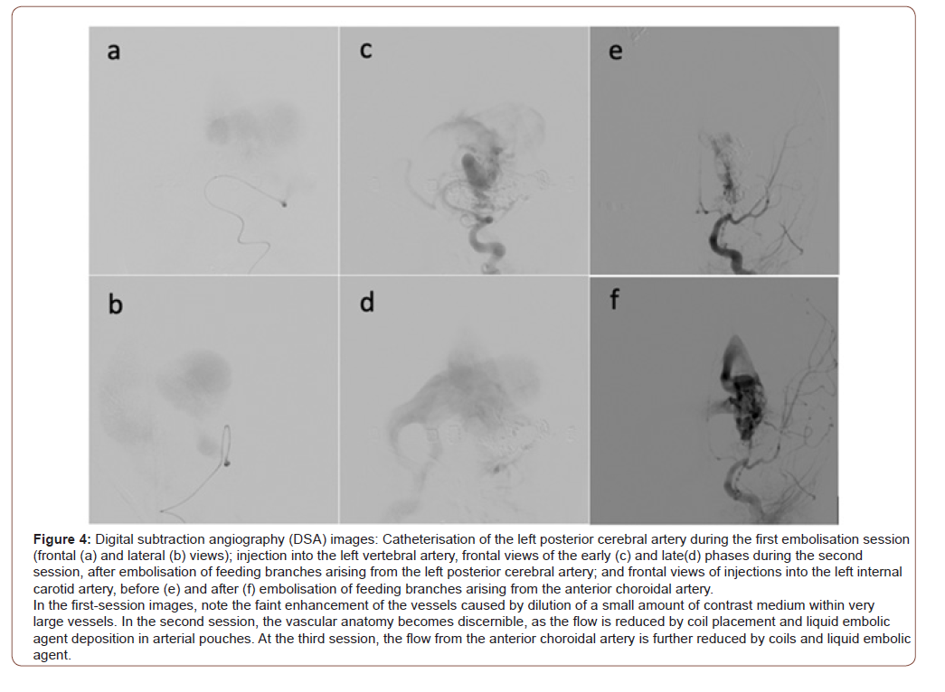
He required invasive mechanical ventilation and was initiated on analgesia and inotropic support with dopamine. The Bicêtre score was nine, indicating the need for urgent endovascular embolization. Embolization of the left posterior cerebral artery was performed via umbilical arterial catheterization using both coil deployment and Onyx injection (Figure 4).
The Bicêtre score is a clinical assessment tool developed to guide the management of neonates with VGAM. It evaluates the functional status of multiple organ systems, including cardiac, respiratory, hepatic, neurological, and general condition, with scores ranging from 0 to 21. Higher scores indicate greater clinical stability and eligibility for endovascular treatment (Table 1) [8].
Despite the procedure, echocardiography on day three showed persistent right-sided cardiac overload, prompting initiation of anti-congestive therapy. A repeat CE-MRI that day demonstrated reduced arterial inflow to the malformation, though complete occlusion had not been achieved. A second embolization was therefore performed on day eight (Figure 4).
Following the second procedure, gradual clinical and echocardiographic improvement in right heart overload was observed, consistent with decreased arteriovenous shunting. On day 11, CE-MRI showed reduced aneurysmal dilation but revealed areas of acute ischemia involving the left thalamus and posterior, predominantly left-sided, corpus callosum, attributed to procedural complications. The infant was discharged on day 44 on furosemide and spironolactone, with a final Bicêtre score of 16. At three months of age, the patient exhibited appropriate psychomotor development, though weight gain remained insufficient. Echocardiography continued to show cardiomegaly, but ventricular function was preserved. CE-MRI demonstrated further reduction in aneurysmal dilation; however, the arteriovenous shunt persisted, accompanied by progressive hydrocephalus.
At five months of age, during evaluation for an intercurrent viral respiratory infection, markedly prominent scalp veins and worsening hydrocephalus were noted on cranial ultrasonography. Repeat CE-MRI confirmed persistent aneurysmal dilation, now predominantly supplied by the right posterior cerebral artery and the anterior choroidal artery, along with hydrocephalus and left cerebellar tonsillar herniation (Figure 2). The patient underwent urgent re-embolization of the main feeding arteries using Onyx and coils, following selective catheterization of the left vertebral and posterior cerebral arteries (Figure 4). Postoperative recovery was complicated by prolonged mechanical ventilation requirements. Despite hemodynamic stability without vasopressor support, the infant developed hospital-acquired pneumonia progressing to Acute Respiratory Distress Syndrome (ARDS) and sepsis, culminating in multiorgan failure and death one month after the procedure, at six months of age.
Discussion
Lasjaunias’ classification describes two primary types of VGAM: the choroidal type-by far the most common (56–76% of cases)-and the mural type. The choroidal type typically affects male neonates, features multiple arterial feeders forming a complex vascular network, and produces high-flow shunts that frequently lead to HOHF, as seen in our case. In contrast, the mural type is less common, has a more favorable prognosis, and typically presents later in infancy with milder symptoms. Heart failure, when present in mural VGAM, is generally mild and asymptomatic [1-4,8,9]. According to the literature, if left untreated, this malformation is associated with near 100% morbidity and mortality, particularly in newborns [3]. Clinical presentation is predominantly agedependent, with prognosis closely linked to both the age of symptom onset and the severity of heart failure. The manifestations encompass a wide clinical spectrum, ranging from minimal signs to overt cardiogenic shock. Notably, earlier clinical manifestation is consistently associated with a less favorable outcome [2].
In the presented case, we were faced with an early manifestation of prenatal heart failure which, despite the absence of brain lesions, may be associated with a mortality rate of approximately 80%, leading some authors to consider pregnancy termination as an acceptable option [1,2]. Prenatal ultrasonography, complemented by fetal MRI, is critical for early diagnosis and delivery planning at a specialized center equipped with neonatal intensive care and interventional neuroradiology resources. [6,9,10] Despite advancements in prenatal imaging, VGAM should remain a differential diagnosis in any neonate presenting with unexplained HOHF refractory to medical therapy or with unexplained hydrocephalus.
Early medical management of congestive heart failure and fluid status (diuretics, vasodilators and inotropic agents) is essential for stabilizing neonates prior to definitive intervention and for addressing multiorgan dysfunction [3,11]. Once admitted to the Neonatal Intensive Care Unit, the newborn should undergo continuous monitoring and evaluation with echocardiography, cranial ultrasound, CE-MRI to identify any previously undetected brain lesions, and MR angiography to delineate the anatomy of the VGAM and guide therapeutic planning. Although the immediate clinical priority is the management of HOHF, VGAM is also associated with significant neurological comorbidities, including encephalopathy, hydrocephalus, seizures, and developmental delay [1].
Ventriculoperitoneal shunting is not typically performed in cases of VGAM because it carries a high risk of serious complications and does not address the underlying pathophysiology of the malformation. VAGM-associated hydrocephalus is primarily due to hydrovenous congestion from the high-flow arteriovenous shunt, not a primary CSF circulation problem. Direct CSF diversion can precipitate acute neurological deterioration, including status epilepticus, intraventricular hemorrhage, subdural hematoma, venous infarction, and worsening developmental delay, as well as increased mortality, as demonstrated in pediatric case series and literature reviews [8,12,13].
Endovascular embolization (selective occlusion of feeder vessels using coils or liquid embolic agents) remains the treatment of choice and urgent intervention is performed in cases of cardiac heart failure, developmental delay or regression, and ischemic signs on imagery. Otherwise, the treatment should be delayed to 5 or 6 months of age for better results [14,15]. The use of the Bicêtre score, developed by Lasjaunias and colleagues, supports appropriate patient selection and helps guide the timing of intervention (Table 1) [8]. According to this scoring system, our patient achieved a score of nine out of 21, placing him in the category requiring urgent endovascular intervention. A Bicêtre score ≤12, and especially a value as low as nine, is strongly associated with unfavorable outcomes, including early neonatal mortality, as demonstrated in contemporary cohorts from the United Kingdom and European tertiary centers [5,16]. A meta-analysis of outcomes following embolization of VGAM demonstrated that studies employing the Bicêtre neonatal evaluation score for patient selection reported higher rates of favorable neurological outcomes compared to those that did not. Although this is a validated tool, it presents certain limitations and, therefore, should not be used as the sole basis for decision-making but rather in conjunction with clinical assessment and neuroimaging findings [11,17,18].
Table 1:Bicêtre Score for the evaluation of neonatal arteriovenous malformations.
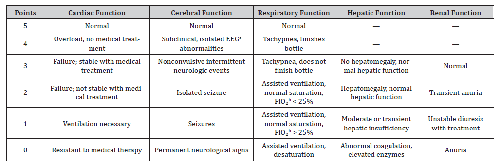
a EEG: Eletroencephalogram, bFiO2: Fraccional inspired oxygen; Maximal score: 5 (cardiac) + 5 (cerebral) + 5 (respiratory) + 3 (hepatic) + 3 (renal) = 21
The presence of HOHF, often accompanied by pulmonary hypertension and the need for inotropic support, is an additional negative prognostic marker [19-22]. Optimal management involves aggressive hemodynamic stabilization and, when possible, early endovascular embolization, which can improve survival but does not eliminate the high risk of mortality and severe neurological morbidity. For the profile described (prenatal diagnosis, high output heart failure, Bicêtre score nine), the risk of neonatal mortality is extremely high, often above 70% in the best contemporary series [5,15,16,19,22,23].
In the context of VGAM, endovascular embolization often necessitates multiple sessions to attain satisfactory results. Importantly, the primary objective of embolization is not complete occlusion of the malformation, but rather achieving a sufficient reduction in arteriovenous shunting to enable hemodynamic stabilization and mitigate the risk of progressive neurological deterioration [18,24-27].
Early postnatal care must include a multidisciplinary team involving neonatologists, obstetricians, pediatric cardiologists, neurologists, and interventional neuroradiologists. In this case, timely prenatal diagnosis enabled coordinated perinatal planning, allowing for prompt supportive care and eventual successful intervention.
This case underscores the importance of prenatal identification of VGAM and the complexities of neonatal management. VGAM is a rare but severe cerebral arteriovenous malformation associated with high neonatal mortality rates in the absence of treatment. It predominantly presents in neonates with high-output cardiac failure, often complicated by pulmonary hypertension and multiorgan dysfunction. Prenatal detection of VGAM is critical for early diagnosis and strategic perinatal management through a multidisciplinary approach. Endovascular embolization of the arterial feeders using coils or liquid agents is the preferred therapeutic approach for VGAM, frequently necessitating multiple sessions to achieve adequate occlusion. Advances in neonatal intensive care and endovascular therapy have improved survival outcomes and reduced morbidity among affected patients.
Acknowledgment
All authors have reviewed the final version to be published and agreed to be accountable for all aspects of the work.
Concept and design:
Ricardo Liz Almeida, Elisa Proença
Acquisition, analysis, or interpretation of data: Ricardo Liz
Almeida, João Xavier, Cláudio Henriques, Luís Guedes-Martins, Elisa
Proença
Drafting of the manuscript:
Ricardo Liz Almeida
Critical review of the manuscript for important intellectual
content: João Xavier, Cláudio Henriques, Luís Guedes-Martins, Elisa
Proença
Supervision: Elisa Proença
Conflicts of Interests
The authors declare no potential conflicts of interest with respect to the research, authorship, and/or publication of this article.
References
- Recinos PF, Rahmathulla G, Pearl M, Recinos VR, Jallo GI, et al. (2012) Vein of Galen Malformations: Epidemiology, Clinical Presentations, Management. Neurosurg Clin N Am 23(1): 165-177.
- Gillet de Thorey A, Ozanne A, Melki J, Dumery G, Benachi A, et al. (2022) State of the art of antenatal diagnosis and management of vein of Galen aneurysmal malformations. Prenat Diagn 42(9): 1073-1080.
- Frawley GP, Dargaville PA, Mitchell PJ, Tress BM, Loughnan P (2002) Clinical course and medical management of neonates with severe cardiac failure related to vein of Galen malformation. Arch Dis Child Fetal Neonatal Ed 87(2): F144-9.
- Alvarez H, Garcia Monaco R, Rodesch G, Sachet M, Krings T, et al. (2007) Vein of Galen Aneurysmal Malformations. Neuroimaging Clin N Am 17: 189-206.
- Giorgi L, Durand P, Morin L, Miatello J, Merchaoui Z, et al. (2020) Management and Outcomes of Neonatal Arteriovenous Brain Malformations with Cardiac Failure: A 17 Years’ Experience in a Tertiary Referral Center. Journal of Pediatrics 218: 85-91.e2.
- Arko L, Lambrych M, Montaser A, Zurakowski D, Orbach DB (2020) Fetal and Neonatal MRI Predictors of Aggressive Early Clinical Course in Vein of Galen Malformation. American Journal of Neuroradiology 41(6): 1105-1111.
- Patel N, Mills JF, Cheung MMH, Loughnan PM (2007) Systemic haemodynamics in infants with vein of Galen malformation: assessment and basis for therapy. Journal of Perinatology 27(7): 460-3.
- Lasjaunias PL, Chng SM, Sachet M, Alvarez H, Rodesch G, et al. (2006) The management of vein of Galen aneurysmal malformations. Neurosurgery 59: S184-94.
- Pimenta M, Araújo C, Clode N (2020) Vein of Galen Aneurysm: Prenatal Ultrasound Diagnosis. Acta Med Port 33: 521-521.
- Sepulveda W, Platt CC, Fisk NM (1995) Prenatal diagnosis of cerebral arteriovenous malformation using color Doppler ultrasonography: case report and review of the literature. Ultrasound in Obstetrics and Gynecology 6(4): 282-6.
- Cory MJ, Durand P, Sillero R, Morin L, Savani R, et al. (2023) Vein of Galen aneurysmal malformation: rationalizing medical management of neonatal heart failure. Pediatr Res 93(1): 39-48.
- Giorgi L, Durand P, Morin L, Miatello J, Merchaoui Z, et al. (2020) Management and Outcomes of Neonatal Arteriovenous Brain Malformations with Cardiac Failure: A 17 Years’ Experience in a Tertiary Referral Center. J Pediatr 218: 85-91.e2.
- Bhatia K, Mendes Pereira V, Krings T, Ter Brugge K, Kortman H, et al. (2020) Factors Contributing to Major Neurological Complications from Vein of Galen Malformation Embolization. JAMA Neurol 77(8): 992-999.
- Yan J, Wen J, Gopaul R, Zhang CY, Xiao SW (2015) Outcome and complications of endovascular embolization for vein of Galen malformations: A systematic review and meta-analysis. J Neurosurg 123(4): 872-90.
- Berenstein A, Paramasivam S, Sorscher M, Molofsky W, Meila D, et al. (2019) Vein of Galen Aneurysmal Malformation: Advances in Management and Endovascular treatment. Clin Neurosurg 84(2): 469-478.
- Lecce F, Robertson F, Rennie A, Heuchan AM, Lister P, et al. (2018) Cross-sectional study of a United Kingdom cohort of neonatal vein of galen malformation. Ann Neurol 84(4): 547-555.
- Cordova EG, Levy P, Kheir JN, Orbach DB, Barnewolt C, et al. (2020) Vein of galen malformation. Neoreviews 21: e678-86.
- Brinjikji W, Krings T, Murad MH, Rouchaud A, Meila D (2017) Endovascular treatment of vein of Galen malformations: A systematic review and meta-Analysis. American Journal of Neuroradiology 38(12): 2308-2314.
- Pereira C, Gouveia R, Rebelo M, Abrantes M, Costa J (2017) Aneurisma da Veia de Galeno: Descrição de Quatro Casos Clínicos. Portuguese Journal of Pediatrics 48: 337-41.
- De Rosa G, De Carolis MP, Tempera A, Pedicelli A, Rollo M, et al. (2019) Outcome of Neonates with Vein of Galen Malformation Presenting with Severe Heart Failure: A Case Series. Am J Perinatol 36(2): 169-175.
- Khurana J, Orbach DB, Gauvreau K, Collins SL, Tella JB, et al. (2023) Pulmonary Hypertension in Infants and Children with Vein of Galen Malformation and Association with Clinical Outcomes. Journal of Pediatrics 258: 113404.
- Cornet MC, Li Y, Simmons RL, Baker A, Fullerton HJ, et al. (2022) Outcome of Neonates Presenting with Severe Cardiac Failure due to Cerebral Arteriovenous Fistula. Pediatr Neurol 131: 25-29.
- Berenstein A, Fifi JT, Niimi Y, Presti S, Ortiz R, et al. (2012) Vein of galen malformations in neonates: New management paradigms for improving outcomes. Neurosurgery 70(5): 1207-13.
- Lasjaunias P, Garcia-Monaco R, Rodesch G, Ter Brugge K, Zerah M, et al. (1991) Vein of Galen malformation - Endovascular management of 43 cases. Child’s Nervous System 7(7): 360-7.
- Nurimanov C, Makhambetov Y, Menlibayeva K, Nurakay N, Makhambetov N, et al. (2023) Long-Term Outcomes of Endovascular Embolization in a Vein of Galen Aneurysmal Malformation: A Single-Center Experience. Diagnostics 13(16): 2704.
- Matsoukas S, Shigematsu T, Bazil MJ, Fifi J, Berenstein A (2022) Transvenous embolization of vein of galen aneurysmal malformations with coils as a final procedure for cure: A single-institution experience of 18 years. Interventional Neuroradiology 30(4): 463-469.
- Habiba MAE-D, Saleh SM (2023) Endovascular management of vein of Galen aneurysmal malformations: case series and review of the literature. Journal of Medicine in Scientific Research pp. 6.
-

This work is licensed under a Creative Commons Attribution-NonCommercial 4.0 International License.