

 Case Report
Right Ventricular Hypertrophy in a Newborn Related to Paracetamol Use During Pregnancy
1 Neonatal Intensive Care Unit - Neonatology Department, Centro Materno Infantil do Norte, Unidade Local de Saúde Santo António, Portugal
2 Fetal Medicine Center - Obstetrics Department, Centro Materno Infantil do Norte, Unidade Local de Saúde Santo António, Portugal
3Pediatric Cardiology Department, Centro Materno Infantil do Norte, Unidade Local de Saúde Santo António, Portugal
Received Date:January 14, 2026; Published Date:January 23, 2026
Abstract
Right ventricular hypertrophy (RVH) in neonates is a rare form of hypertrophic cardiomyopathy and may result from congenital anomalies, perinatal stress, maternal diabetes or drug-induced premature ductal arteriosus (DA) closure. Emerging evidence suggests that high-dose paracetamol in late pregnancy may cause DA constriction through cyclooxygenase inhibition. We report the case of a neonate born to a 34-year-old woman with well-controlled gestational diabetes, who reported self-medication with paracetamol (up to 3 grams per day) throughout the second and third trimesters for persistent sciatica. Prenatal ultrasounds and fetal echocardiography at 26 weeks were normal. Despite clinical stability, screening for critical congenital heart disease revealed pre-ductal hypoxemia. Echocardiography at 48 hours showed RVH with interventricular septal thickening, mild pulmonary hypertension, closed DA and a bidirectional shunt through a patent foramen ovale. Serial echocardiograms demonstrated progressive improvement in RVH and shunt dynamics without specific therapy. The newborn was discharged on day 10 and followup at one month confirmed complete RVH resolution. This case supports a possible association between high-dose maternal paracetamol use in late pregnancy and transient neonatal RVH, highlighting the need for caution and further pharmacovigilance studies regarding analgesic strategies during pregnancy.
Keywords: Cardiomyopathy; Right ventricular hypertrophy; Paracetamol
Abbreviations: DA: Ductus arteriosus; NB: Newborn; NSAIDS: Nonsteroidal anti-inflammatory drugs; PFO: Patent foramen ovale; RVH: Right ventricular hypertrophy
Case Presentation
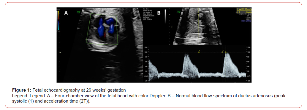
A 34-year-old woman, gravida 3, para 2, with well-controlled asthma, was followed during her third pregnancy. Gestational diabetes was diagnosed in the second trimester and effectively managed through dietary modifications. At 17 weeks’ gestation, the patient was evaluated in the Emergency Department for sciatica pain and discharged with paracetamol. First-trimester biochemical screening indicated an increased risk for trisomy 21; however, the couple declined further invasive diagnostic testing. Other routine prenatal laboratory tests, including the infection screening, and ultrasound examinations were normal. A fetal echocardiography performed at 26 weeks of gestation showed no abnormalities (Figure 1).
Persistent sciatica led to frequent self-medication with paracetamol, with reported doses of up to 3 grams per day, during the second and third trimesters. No alternative analgesics, including nonsteroidal anti-inflammatory drugs (NSAIDs), were used.
Labor was induced at 40 weeks’ gestation, resulting in a spontaneous vaginal delivery in cephalic presentation. Membrane rupture occurred five hours prior to delivery and meconiumstained amniotic fluid was noted during the expulsive phase. The postpartum course was uneventful and the mother was discharged in stable condition.
A liveborn female newborn (NB) required brief nasopharyngeal aspiration due to meconium-stained secretions. Apgar scores were 7, 9 and 10 at 1, 5 and 10 minutes, respectively. Physical examination was unremarkable and birth anthropometry was appropriate for gestational age according to the World Health Organization growth charts.
Despite appearing clinically stable, the screening for critical congenital heart diseases detected hypoxemia, with pre-ductal oxygen saturation levels of 90-92%. The NB maintained good vitality and exhibited no signs of respiratory distress. Cardiovascular assessment revealed no abnormalities, including normal cardiac auscultation, symmetric femoral pulses and no differential blood pressure between the limbs. The NB was subsequently transferred to the Neonatal Intensive Care Unit (NICU) for further evaluation.
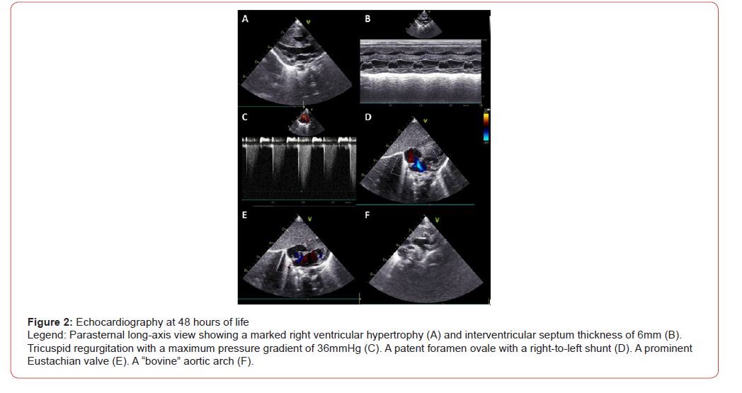
Initial assessments, including chest X-rays and blood tests with a septic screening, showed no abnormalities. Echocardiography at 48 hours of life showed right ventricular hypertrophy (RVH), interventricular septal thickening of 6mm, signs of pulmonary hypertension, a bidirectional shunt through a patent foramen ovale (PFO) with a right-to-left predominance and a closed ductus arteriosus (DA) (Figure 2 A-D). Additional findings included a prominent Eustachian valve and a “bovine” aortic arch (Figure 2 E-F). During the stay at NICU, serial echocardiograms demonstrated improvement in RVH with preserved biventricular systolic function, a progressive reduction of tricuspid regurgitation and a bidirectional flow through the PFO, without the need for specific medications. The NB required supplemental oxygen (maximum FiO2 of 0.28) until the 6th day of life and was discharged on the 10th day with marked improvement in RVH, low-grade tricuspid regurgitation (maximum gradient pressure of 15 mmHg) and a PFO with a predominantly left-to-right shunt.
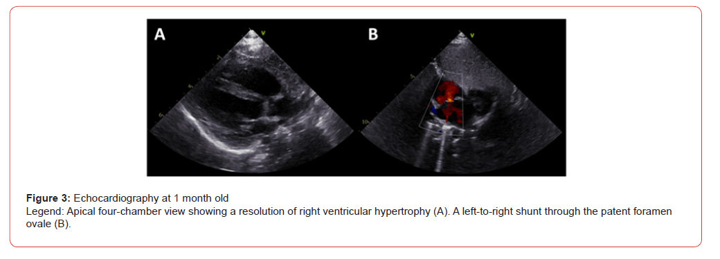
At one month of age, during a follow-up appointment at the Pediatric Cardiology clinic, the infant was asymptomatic, with a normal growth and development. The Guthrie testing excluded inborn errors of metabolism. Echocardiographic evaluation demonstrated a complete resolution of RVH, with a residual leftto- right shunt through the PFO (Figure 3). An electrocardiogram showed sinus rhythm (heart rate of 132 beats per minute), consistent with normal age-related findings.
Discussion
Right ventricular hypertrophy (RVH) is an uncommon form of hypertrophic cardiomyopathy in neonates. It can be associated with several factors, including perinatal stress, congenital heart anomalies, maternal diabetes, the use of drugs leading to premature ductal constriction, maternal consumption of polyphenol-rich foods and metabolic diseases [1,2].
The initial echocardiographic findings were crucial in ruling out other structural heart defects that are typically associated with a closed DA and RVH, such as tetralogy of Fallot, truncus arteriosus and pulmonary stenosis [3]. Furthermore, the absence of a family history of cardiomyopathy or other hereditary conditions further reduced the likelihood of a genetic origin for the observed RVH. Although the patient had a history of gestational diabetes, its impact on the neonatal outcome appears limited in this case due to the good glycemic control maintained throughout the pregnancy and the normal findings on the prenatal echocardiography performed at 26 weeks of gestation. Additionally, no other complications typically associated with gestational diabetes, such as macrosomia or neonatal hypoglycemia, were observed.
A notable aspect of the maternal history, revealed during indepth postnatal questioning, was the frequent use of paracetamol for sciatica pain, with a self-reported dose of up to 3 grams per day, during the second and third trimesters. Although NSAIDs are more commonly implicated in premature DA closure, recent clinical evidence suggests that high-dose paracetamol during the third trimester can also cross the placenta freely and inhibit cyclooxygenase-1/2 and subsequent prostaglandin signaling, leading to DA constriction in late pregnancy [4,5]. Such constriction poses serious risks, including fetal loss or life-threatening cardiac failure in the neonate [6]. This emerging evidence strengthens the hypothesized link between maternal paracetamol intake and fetal DA closure, providing a plausible explanation for the transient RVH observed in this neonate.
It was also important to exclude other antenatal factors that could contribute to DA constriction, such as the maternal consumption of polyphenol-rich foods, which are known to inhibit prostaglandin synthesis and could lead to premature closure of the DA, similar to NSAIDs [7]. In this case, the mother denied consumption of foods and beverages rich in polyphenols, such as herbal teas, fruit juices and chocolate, reducing the likelihood of this contributing to the RVH. Additionally, the negative Guthrie screening for inborn errors of metabolism, combined with the transient nature of the isolated RVH, allowed for the exclusion of metabolic disorders as a contributing factor.
To conclude, this case highlights the importance of a cautious use of paracetamol during pregnancy, given emerging evidence of potential adverse effects on fetal health. Notably, the possibility that paracetamol may contribute to premature closure of the DA could provide an explanation for some idiopathic cases of transient RVH. These findings underscore the necessity for robust pharmacovigilance studies and the development of analgesic strategies that prioritize maternal and fetal safety throughout pregnancy.
Conflict of Interest
The authors have no conflicts of interest to disclose.
References
- Tyagi V, Chopra A, Jain A (2019) Premature Ductal Constriction and Reversible Early Isolated Right Ventricular Cardiomyopathy in a Neonate at Birth. Journal of Fetal Medicine 06(01): 45-46.
- Alp H, Karataş Z, Baysal T (2014) Transient severe isolated right ventricular hypertrophy in neonates. J Clin Neonatol 3(3): 161.
- Genovese F, Marilli I, Benintende G, Privitera A, Gulino FA, et al. (2015) Diagnosis and management of fetal ductus arteriosus constriction-closure. J Neonatal Perinatal Med 8(1): 57-62.
- Allegaert K, Mian P, Lapillonne A, van den Anker JN (2019) Maternal paracetamol intake and fetal ductus arteriosus constriction or closure: a case series analysis. Br J Clin Pharmacol 85(1): 245-251.
- Becquet O, Bonnet D, Ville Y, Allegaert K, Lapillonne A (2018) Paracetamol/Acetaminophen During Pregnancy Induces Prenatal Ductus Arteriosus Closure. Pediatrics 142(1): e20174021.
- Bauer AZ, Swan SH, Kriebel D, Liew Z, Taylor HS, et al. (2021) Paracetamol use during pregnancy — a call for precautionary action. Nat Rev Endocrinol 17(12): 757-766.
- Nacka-Aleksić M, Pirković A, Vilotić A, Bojić-Trbojević Ž, Jovanović Krivokuća M, et al. (2022) The Role of Dietary Polyphenols in Pregnancy and Pregnancy-Related Disorders. Nutrients 14(24): 5246.