


 Case Report
Genital Pain in a Preschool-aged Girl: The Critical Role of Imaging
1 Department of Pediatrics, Local Health Unit of São João, Portugal
2 Department of Pediatrics, Local Health Unit of Matosinhos, Portugal
Received Date:February 02, 2026; Published Date:February 11, 2026
Abstract
Vaginal foreign bodies represent a clinical and diagnostic challenge in pediatric care. The most common etiology is self-insertion during exploration or play, with toilet paper, small hard objects, and pieces of cloth being the items most frequently identified [1,2]. Less commonly, vaginal foreign bodies may be associated with sexual abuse, which should be systematically considered and appropriately evaluated in all cases [2].
Keywords: Sexual abuse; Pelvic pain; Vaginal foreign body
Case Report
A 3-year-old previously healthy girl presented to the pediatric emergency department with a 24-hour history of green vaginal discharge associated with progressive perineal pain and local discomfort. There were no reported fever, dysuria, abdominal pain, or gastrointestinal symptoms. According to the mother, the child had recently spent an overnight stay at a relatives’ home without direct parental supervision. On physical examination, the patient afebrile and hemodynamically stable. Local inspection revealed marked edema, erythema, and tenderness of the right labium majus, associated with purulent greenish vaginal discharge. No external signs of trauma or bleeding were observed. Given the patient’s age and clinical presentation, a multidisciplinary approach was promptly initiated, involving Pediatric Gynecology and Forensic Medicine.
Laboratory evaluation with complete blood count and inflammatory markers, were within normal limits, as were screening tests for sexually transmitted infections. A pelvic radiograph was subsequently obtained, revealing a radiopaque cylindrical structure within the vaginal canal, consistent with a vaginal foreign body (Figure 1).
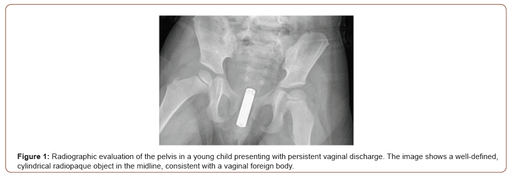
The patient was taken to the operating room for vaginoscopy under general anesthesia. Prior to any intervention, a forensic evaluation was conducted. Vaginal exploration identified an alkaline battery lodged within the vaginal cavity, causing circumferential mucosal necrosis and chemical injury to the surrounding tissues. The battery was carefully removed, followed by copious irrigation of the vaginal canal with saline solution. No vaginal perforation or deeper tissue involvement was identified.
Postoperatively, the patient recovered uneventfully. Following forensic assessment, the episode was subsequently classified as probably accidental. The patient was discharged with topical local therapy and scheduled outpatient follow-up in General Pediatrics and Pediatric Gynecology. At the three-month follow-up visit, the child was asymptomatic, with complete resolution of vaginal discharge and pain. Gynecological examination demonstrated normal healing, with no residual mucosal lesions, scarring, or functional complications.
Discussion
Vaginal foreign bodies represent a significant, albeit infrequent, clinical challenge in pediatric gynecology.1 These objects, often introduced unintentionally, may lead to a spectrum of frequently nonspecific symptoms, including vaginal discharge, bleeding, local pain, erythema, or edema [1-4]. In young children, clinical history is often limited, and physical examination may be inconclusive or poorly tolerated, frequently resulting in diagnostic delay and underscoring the importance of adjunctive diagnostic tools5. Alkaline batteries represent a particularly hazardous type of vaginal foreign body. Tissue injury occurs rapidly; therefore, prompt diagnosis and urgent removal are essential to prevent complications such as vaginal stenosis, fistula formation, or longterm reproductive sequelae [1,2,5,6].
The diagnostic approach begins with a careful historytaking and physical examination, with particular attention to the possibility of sexual abuse and associated medicolegal implications [2,7]. Imaging plays a pivotal role in the diagnostic workup when a vaginal foreign body is suspected, with ultrasonography as the preferred initial modality [2,7,8]. Plain pelvic radiography is a fast and widely available imaging modality that may aid in the detection of radiopaque objects, such as batteries, allowing prompt diagnosis, as demonstrated in our case, and facilitating timely surgical intervention and coordinated multidisciplinary management [2,7].
Vaginoscopy under anesthesia is the gold standard for both diagnosis and removal, allowing direct visualization of the foreign body and assessment of mucosal injury (burns, necrosis, or perforation) [2,9]. When the foreign body is a battery, thorough vaginal irrigation is recommended to remove residual alkaline material. In the presence of mucosal injury, topical estrogen may be considered to promote healing, and antibiotics may be indicated when there is evidence of infection or significant tissue damage [6,10].
Postoperative follow-up is essential to monitor for late complications [6].
Conclusion
This case highlights the critical role of imaging in the evaluation of genital pain and abnormal vaginal discharge in prepubertal girls. Plain pelvic radiography enabled rapid and accurate diagnosis of vaginal foreign body, directly influencing clinical management and preventing potentially severe complications. Clinicians should maintain a high index of suspicion for vaginal foreign bodies in young children with unexplained genital symptoms, and early imaging should be considered an integral part of the diagnostic approach. Prompt recognition and multidisciplinary management are essential to optimize outcomes and ensure patient safety.
Consent
Informed consent was obtained from the parent.
Funding sources
No funding was received for the research.
Disclosure
The authors declare no conflict of interest.
Author Contributions
all authors contributed equally to the conception, drafting, and revision of the manuscript and approved the final version.
Conflicts of Interests
The authors declare no conflicts of interest.
References
- Ma W, Sun YF, Liu JH, He DW, Lin T, et al. (2022) Vaginal foreign bodies in children: a single-center retrospective 10-year analysis. Pediatr Surg Int 38(4): 637-641.
- Umans E, Boogaerts M, Vergauwe B, Verest A, Van Calenbergh S (2024) Vaginal foreign body in the pediatric patient: A systematic review. Eur J Obstet Gynecol Reprod Biol 297: 153-158.
- Stricker T, Navratil F, Sennhauser FH (2004) Vaginal foreign bodies. J Paediatr Child Health 40(4): 205-7.
- Abdessamad HM, Greenfield M (2009) Vaginal foreign body presenting as bleeding with defecation in a child. J Pediatr Adolesc Gynecol 22(2): e5-7.
- Nayak S, Witchel SF, Sanfilippo JS (2014) Vaginal foreign body: a delayed diagnosis. J Pediatr Adolesc Gynecol 27(6): e127-9.
- Semaan A, Klein T, Vahdad MR, Boemers TM, Pohle R (2015) Severe Vaginal Burns in a 5-Year-Old Girl Due to an Alkaline Battery in the Vagina. J Pediatr Adolesc Gynecol 28(5): e147-8.
- Kayhan U, Liman Z, Izci A (2025) Presence of intravaginal foreign bodies in children evaluated for forensic medicine examination: Two case reports. Eur J Obstet Gynecol Reprod Biol 314: 114725.
- Yang X, Sun L, Ye J, Li X, Tao R (2017) Ultrasonography in Detection of Vaginal Foreign Bodies in Girls: A Retrospective Study. J Pediatr Adolesc Gynecol 30(6): 620-625.
- Jia Y, Shuang L, Jun W, Gang L, Hai-Tao C (2022) Small spherical foreign bodies in the genitourinary tract and their management. BMC Pediatr 22(1): 42.
- Moore E, Friedman J, Christopher D (2023) Vaginal Corrosion Due to Insertion of a 9-Volt Battery. Obstet Gynecol 141(6): 1219-1223.
-

This work is licensed under a Creative Commons Attribution-NonCommercial 4.0 International License.