 Case Report
Buttock Mass: A Typical Feature of Fetal Extraperitoneal Rectal Perforation
Anna Cuartero Gorjón1, María del Carmen Sarmiento2, José Luis Encinas2 and Raquel Corripio Collado1*
1Pediatric Department, Parc Taulí University Hospital, Autonomous University of Barcelona, Sabadell, Spain
2Pediatric Surgery Department, Hospital Infantil La Paz, Madrid, Spain
Raquel Corripio Collado, Pediatric Department, Parc Taulí University Hospital, Autonomous University of Barcelona, Sabadell, Spain
Received Date:April 24, 2024; Published Date:April 30, 2024
Brief Clinical Outline
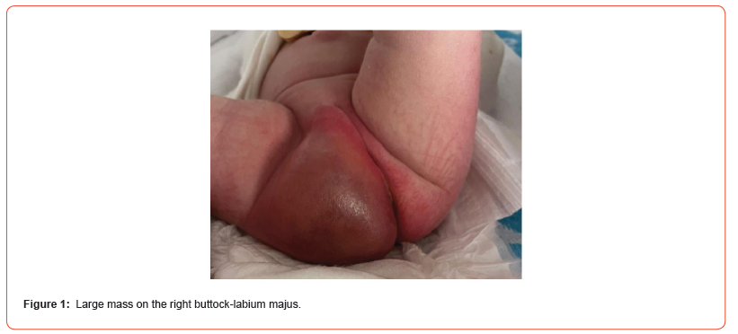
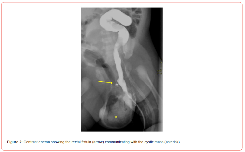
Ultrasound at 38 gestational weeks in a female fetus revealed a previously undetected cystic pelvic mass that was confirmed by fetal magnetic resonance imaging. At full-term birth, the neonate had a mass on her right buttock-labium majus (Figure 1); 48 hours after birth, she developed a fever and elevated C-reactive protein, so antibiotic therapy was started. She remained clinically stable, breastfeeding successfully and showing frequent bowel movements. Ultrasound showed the mass communicated with the pre-sacral area. CT showed air-fluid levels probably extending to the perineum. Contrast enema showed a fistula between the rectum and the cystic mass (Figure 2). On the fourth day of life, a sigmoid loop colostomy with closed mucous fistula was fashioned, and the collection was drained. Rectal biopsy ruled out Hirschsprung disease. After an uneventful postoperative course, she was discharged on day 19 after surgery. A contrast enema is planned before closure of the colostomy.


Clinical Message or Learning Point
Fetal extraperitoneal rectal perforation is rare; however, its clinical features make it easily recognizable [1]. Plain abdominal X-rays obviate the need for further investigations [2,3]. Lack of experience led us to perform additional, probably unnecessary, radiological tests to confirm the diagnosis. Delayed treatment could cause fatal septic complications. Prompt recognition followed by a simple diversion of meconium and drainage of the collection results in good outcomes with normal anorectal function expected in the long-term [1]. The colostomy should be closed after 3 to 6 months [3,4].
Funding
The authors have not declared a specific grant for this research from any funding agency in the public, commercial or not-for-profit sectors.
Patient Consent for Publication
Obtained.
Acknowledgment
None.
Conflict of Interest
None.
References
- Pitcher GJ, Davies MR, Bowley DM, Numanoglu A, Rode H, et al. (2009) Fetal extraperitoneal rectal perforation: a rare neonatal emergency. Journal of Pediatric Surgery 44(7): 1405-1409.
- Charlton R, Brisighelli G, Gabler T, Westgarth‐Taylor C (2019) Management of fetal extraperitoneal rectal perforation: a case series and review of the literature. Pediatric Surgery International 35(9): 989-997.
- Ibrahim AA, Elkadhi AS, N Nimeri (2011) Fetal Extraperitoneal Rectal Perforation in a Preterm Baby. European Journal of Pediatric Surgery 21(5): 343-345.
- Mitsudo MD, Boley SJ, Rosenzweig MJ, Campbell DE (1983) Extraperitoneal pelvic meconium extravasation in a newborn infant. The Journal of Pediatrics 103(4): 598-600.
-
Anna Cuartero Gorjón, María del Carmen Sarmiento, José Luis Encinas and Raquel Corripio Collado*. Buttock Mass: A Typical Feature of Fetal Extraperitoneal Rectal Perforation. Glob J of Ped & Neonatol Car. 4(4): 2024. GJPNC.MS.ID.000594.
Full-term birth, Air-fluid, Buttock-labium, Hirschsprung disease, Surgery, Radiological tests, Antibiotic therapy
-

This work is licensed under a Creative Commons Attribution-NonCommercial 4.0 International License.


