 Research Article
Development of an Arthrometer and Its Application in Experiments on Knee Joint Contracture
Quan-Bing Zhang, Yi Liu and Yun Zhou*
Department of Rehabilitation Medicine, The Second Affiliated Hospital of Anhui Medical University, Hefei 230601, China
Corresponding AuthorYun Zhou, Department of Rehabilitation Medicine, The Second Affiliated Hospital of Anhui Medical University, Hefei 230601, China
Received Date:March 01, 2024; Published Date:March 25, 2024
Abstract
Objectives: In order to carry out animal experimental studies on knee joint contracture, and measure the knee joint ranges of motion in experimental rabbits accurately, a mechanical arthrometer was designed, and the feasibility of the device was then discussed.
Methods:An arthrometer was designed, and the lower limbs of normal rabbits were fixed on the instrument to observe the measuring effect. The instrument was then used to measure the ranges of motion of rabbits’ contractured knee joints, and the results were compared with those measured by a protractor.
Results:The knee joint ranges of motion of New Zealand rabbits can be effectively measured using an arthrometer. Compared with the protractor, we found the arthrometer is more stable and accurate measuring knee joint ranges of motion in New Zealand rabbits.
Conclusion:The arthrometer is suitable to be used in animal experimental studies on knee joint contracture in New Zealand rabbits, providing convenience for scientific research personnel to measure knee joint ranges of motion in New Zealand rabbits and carry out related animal experimental studies.
Keywords:Arthrometer; Rabbit; Knee joint contracture; Experiment
Introduction
Joint contracture, which usually happens following the destruction of a joint and surrounding soft tissues resulting from bone and joint injury as well as long-term or inappropriate joint immobilization based on the destruction, is one of the common and challenging diseases clinically [1-3]. After the formation of joint contracture, many changes in histomorphology, molecules, biomechanics and other aspects of tissues inside and around joints may occur, such as joint capsule, muscles and ligaments around joints, leading to the reduction of active and passive joint ranges of motion [4,5]. Then, the activities of daily living of the patients may be limited, seriously affecting the qualities of life of patients with the disease and bringing about huge financial burden to families. Experimental animal studies on joint contracture can help us better understand the occurrence and development of joint contracture, so as to better prevent and treat joint-related diseases and complications. Joint range of motion is a very important index for the measurement of the severity of joint contracture in related experiments. At present, researchers at home and abroad used different methods to measure joint ranges of motion in their studies concerning joint contracture. The most commonly used method is elastic protractor [6,7]. Meanwhile, some researchers measured joint ranges of motion using self-made mechanical angulometers [8-10]. Some of these tools are difficult to measure joint ranges of motion accurately due to the involvement of many subjective factors, while the measuring procedures of others are slightly complicated to carry out large-scale animal experimental research. Extending knee joint contracture is a challenging problem in rehabilitation medicine. In order to study this type of joint contracture, we have previously established a rabbit model of extending knee joint contracture using plaster casting [1,2]. Furthermore, in order to measure the joint ranges of motion of rabbits accurately and quickly, we designed an arthrometer according to the anatomical characteristics of rabbits (utility model patent, NO. ZL 201720251124.6). The objectives of this study were to present the details of the construction and the use of the arthrometer, and to explore its reproducibility and accuracy in the experiment of rabbit knee joint contracture.
Methods
Development Of the Arthrometer
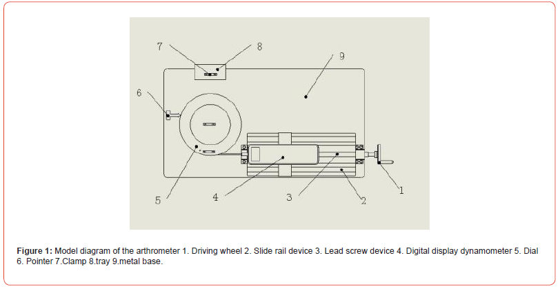
The diagrammatic figure of the arthrometer is shown in (Figure 1). The arthrometer consists of a force-measuring system and an angle-measuring system on a metal base of about 650mm×355mm. The two systems work together to measure the joint range of motion of a knee in one lower limb of a rabbit. The force-measuring system consists of a driving wheel, a slide device (about 400mm×140mm), a bearing plate (about 145mm×100mm), a digital display dynamometer (aidebao in yueqing city, HP-100) and a tension line, etc. The slide rail device is fixed on the metal base, and a bearing plate is fixed on the slide plate of the slide rail device. The digital display dynamometer is fixed on the bearing plate with one side of the digital display dynamometer connected to the dial by a tension line. When the driving wheel is rotated, the load plate can be driven by the sliding rail device, thus driving the digital display dynamometer on the load plate. One side of the tension line is connected to the digital display dynamometer, while the other side is connected to the groove of the edge of the dial. Hence, the tension line can work with force-measuring system and angle-measuring system simultaneously. When the digital display dynamometer moves, the dial of the angle-measuring system can be spined, with the digital display on the dynamometer showing the size of the force (F). The angle-measuring system consists of a dial (with a radius of approximately 0.1m), a double-bearing sleeve fixed pedestal, a shaft and a tray device (approx. 100mm×50mm), etc. The double bearing sleeve fixed pedestal is installed on the metal base, and the shaft is matched with the double bearing sleeve fixing seat by bearings. The dial is anchored to the shaft and the pointer is placed adjacent to the dial (point to 0° position of the dial initially). The tray adjoining the dial consists of a strut and a support plate. A clamp is attached to the support plate and the dial respectively. One lower limb of the rabbit can be secured by the dial and tray. When the dial turns, the pointer coordinated with the dial show the angle change dynamically between the femur and tibia of the rabbit when the dial turns.

Application Of the Arthrometer Torque selection
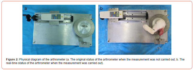
The picture of real product is shown in (Figure 2). After the execution of a New Zealand rabbit, one hind limb of the rabbit was disarticulated at the hip joint. The femur was then immobilized with a vascular clamp, two clamps and a tray. The proximal and distal ends of the tibia were fixed to the middle and edge of the dial with clamps, respectively. The tray and middle part of the dial were slightly higher than the edge part of the dial to avoid friction between the femur and the dial when the measurement was conducted. The digital display dynamometer was mounted on a base above a sliding rail device. The two ends of a tension line were tied to the digital display dynamometer and the groove of the edge of the dial. When the driving wheel was turned, the disc would be twisted. As both ends of the tibia were attached to the dial, the tibia can also be rotated with the dial. The femur was attached to the adjacent tray by a vascular forceps and thus remained fixed. During the rotation, the magnitude of the applied force (F) can be read by the digital display dynamometer with the radius of the dial remained constant (L=0.1m), so the magnitude of the applied torque (F×L) can be measured. Meanwhile, the change in the angle (θ) between the femur and tibia can be accurately calculated through the scale of the dial. During the early stage, ten knees of double hind limbs in five normal New Zealand rabbits, aged 3 ~ 4 months, were disarticulated at the hip joint, and then the knee joint ranges of motion was subsequently measured using the arthrometer. We finally found that the knee joint can be pulled to approximately 140 degrees with a torque of 0.077 Nm., and further increase of torque can result in little angle increase. Hence, 0.77N was used as the effective force, while 0.077 N.m was served as the effective torque. In our experiment, the change of knee joint ranges of motion caused by a torque of 0.077 N.m can be used as the flexion knee joint ranges of motion in rabbits, and the larger flexion angle represents the less severe degree of contracture.

Comparison Methods
Six male skeletally mature New Zealand white rabbits were used in the study (the laboratory animal center of Anhui Medical University, Hefei, China; age 3–4 months; weight 2–2.5 kg). The rabbits underwent unilateral immobilization of a knee joint at full extension using a plaster cast for 6 weeks to stimulate joint contracture. Once this was completed, joint ranges of motion in the contracted knee of the six rabbits were measured by six experimenters using a protractor and the arthrometer, respectively. Then the effects of the arthrometer with traditional protractor in the measurements of knee joint ranges of motion in rabbits were compared. The measurements were proceeded with blind methods, and the measurement orders were not known by the researchers.
Measurement methods Protractor
after 6 weeks of unilateral knee joint immobilization, the rabbits were killed with an overdose of phenobarbital. The joint ranges of motion of the immobilized knees in rabbits were then measured by 6 experimenters using elastic protractor [7] Each experimenter measured the joint range of motion of a knee for 3 times and the average value of the measurements was adopted.
Arthrometer
After the measurement with a protractor, the immobilized hind limbs of rabbits were disarticulated at the hip joint, and then the joint ranges of motion of the immobilized knees in rabbits were measured by 6 experimenters using the arthrometer. Each experimenter measured the joint range of motion of a knee for 3 times and the average value of the measurements was employed.
Statistical Analyses
The mean and standard deviation of the measuring results of the 6 researchers were calculated and presented as mean ± SEM. Data were entered in SPSS 23.0 and were analyzed by a two independent samples t-test. A value of P < 0.05 was considered statistically significant.
Results
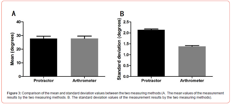
As is shown in Figure 3 there was no statistical difference between the mean values measured with the two methods (P>0.05). However, the standard deviation value measured by the arthrometer was statistically lower than that measured by a protractor (P<0.05).

Discussion
Joint contracture is a very common disease clinically at present, and the most common reason for its occurrence is trauma around the joint and joint immobilization for a long time accompanied by a lack of timely and effective rehabilitation interventions [11,12]. After the occurrence of joint contracture, the main manifestation is the decreased joint range of motion, which seriously affects the normal functions of the joint [13-15]. In clinical practice, it is necessary to test the range of motion of a joint when rehabilitation physicians and rehabilitation therapists evaluate the condition of a patient with joint contracture. At the same time, joint range of motion is also an important index to assess the severity of joint contracture in animal experiments [16-18]. At present, there are considerable number studies on joint contracture with rats or rabbits as experimental carriers using flexing knee joint contracture models [19-21]. As the most common type happened in clinical practice is extending knee joint contracture, we used New Zealand rabbits as the experimental carrier, and made an animal model of knee extending contracture using tubular plaster in our study [1,2,22]. In order to evaluate the severity of knee joint contracture in rabbits, we developed the range of motion measuring instrument based on the anatomical characteristics of rabbit to accurately measure joint ranges of motion of rabbits in the animal model. The design and development of this instrument were conducted on the basis of the anatomical characteristics of lower limbs in New Zealand rabbits. The immobilized lower limb of a rabbit was secured on the angle-measuring device and tray when the measurement was carried out. The device controls the dial by rotating the driving wheel, and the tibia fixed on the dial can be operated. The magnitude of force can be read directly through the digital display dynamometer, and the process of measurement is convenient and easy to operate. The advantage of using a scale disk is that the arm of force is constant (0.1m) when the force is applied, and the torque is only related to the force applied. Meanwhile, the scale of angle is engraved on the disk, hence the change of the knee joint range of motion of a rabbit can be readily calculated when a certain force is applied. Consequently, the device can accurately reflect the joint range of motion in a rabbit when a constant torque is used. In our study, we found that the knee joint can be pulled to approximately 140 degrees with a torque of 0.077 N.m and further increase of torque can result in little angle increase. In order to ensure the accuracy of measurement, we determined that 0.077 N.m was used as a effective torque, and the angular variation after a torque of 0.077 N.m was applied in the equipment and defined as the joint range of motion in a rabbit. In order to evaluate the effect of the joint range of motion measuring instrument, the measuring effect of the arthrometer was compared with that of elastic protractor commonly used in clinic. Six researchers measured knee joint ranges of motion of six rabbits with these two methods, the mean of the results measured between these methods showed no statistical difference, but the standard deviation value measured by the arthrometer was statistically lower than that measured by a protractor. The results indicated that the arthrometer is rarely influenced by different researchers. The arthrometer has shown to have better measurement stability and repeatability when used in the measurement of joint range of motion of a New-Zealand rabbit. To summarize, the arthrometer can reflect the torque used as well as the changes of joint range of motion in a rabbit. The data obtained in the instrument has high precision and simple operation process without the deficiencies of inaccuracy or complexity in other equipment’s. Therefore, we conclude that the arthrometer is of great importance to the measurement of knee joint ranges of motion of rabbits in the experiments of join contracture. The design principle of this experimental instrument can also be used in experiments with other animals’ carriers, such as rats [13-15].
Conclusion
The arthrometer can be used to measure knee joint ranges of motion in New Zealand rabbits with good effect and high accuracy. It can be applied in animal experiments on joint contracture or other related animal experiments for the measurement of knee joint ranges of motion in rabbits.
Conflicts of Interest
None
Funding
The present study was supported by grants from Health Research Project of Anhui Province (grant no. AHWJ2023A30077), National Natural Science Foundation Incubation Program of The Second Affiliated Hospital of Anhui Medical University (grant no. 2022GMFY05), Clinical Medicine Discipline Construction Project of Anhui Medical University in 2023 (Clinic and Preliminary Coconstruction Discipline Project) (grant no. 2023 lcxkEFY010).
Ethics approval
This experimental protocol was approved by the Institutional Animal Care and Use Committee of Anhui Medical University (LLSC20190761).
References
- Huang PP, Zhang QB, Zhou Y, Liu AY, Wang F, et al. (2021) Effect of radial extracorporeal shock wave combined with ultrashort wave diathermy on fibrosis and contracture of muscle[J]. Am J Phys Med Rehabil 100(7): 643-650.
- Zhou Y, Zhang QB, Zhong HZ, Liu Y, Li Jun, et al. (2020) Rabbit model of extending knee joint contracture: Progression of joint motion restriction and subsequent joint capsule changes after immobilization[J]. J Knee Surg 33(1): 15-21.
- Zhou CX, Wang F, Zhou Y, Zhang QB (2023) Formation progress of extension knee joint contracture following external immobilization in rats[J]. World J Orthop 14(9): 669-681.
- Dunham CL, Castile RM, Chamberlain AM, Spencer P. Lake (2019) The role of periarticular soft tissues in persistent motion loss in a rat model of posttraumatic elbow contracture[J]. J Bone Joint Surg Am 101(5): 1-7.
- Reiter AJ, Schott HR, Castile RM, Chamberlain AM, Lake SP, et al. (2023) Early joint use following elbow dislocation limits range-of-motion loss and tissue pathology in posttraumatic joint contracture[J]. J Bone Joint Surg Am 105(3): 223-230.
- Usuba M, Akai M, Shirasaki Y, Miyakawa S (2007) Experimental joint contracture correction with low torque–long duration repeated stretching[J]. Clin Orthop Relat Res 456(1): 70-78.
- Lv QY (2017) Influence of different concentrations of ozone on range of motion of knee joint and content of MMP-13 in rabbits[J]. Guoji Yiyao Weisheng Daobao 23(1): 16-17.
- Nesterenko S, Morrey ME, Abdel MP, An KN, Steinmann SP, et al. (2009) Sanchez-Sotelo J. New rabbit knee model of posttraumatic joint contracture: indirect capsular damage induces a severe contracture[J]. J Orthop Res 27(8): 1028-1032.
- Trudel G, Uhthoff HK, Goudreau L, Laneuville O (2014) Quantitative analysis of the reversibility of knee flexion contractures with time: an experimental study using the rat model.[J]. BMC Musculoskelet Disord 15(1): 338.
- Onoda Y, Hagiwara Y, Ando A, Watanabe T, Chimoto E, et al. (2014) Joint haemorrhage partly accelerated immobilization-induced synovial adhesions and capsular shortening in rats[J]. Knee Surg Sports Traumatol Arthrosc 22(11): 2874-2883.
- Jiang S, He R, Zhu L, Liang T, Wang Z, et al. (2028) Endoplasmic reticulum stress-dependent ROS production mediates synovial myofibroblastic differentiation in the immobilization-induced rat knee joint contracture model[J]. Exp Cell Res 369(2): 325-334.
- Wang F, Zhang QB, Zhou Y, Chen S, Huang PP, et al. (2019) The mechanisms and treatments of muscular pathological changes in immobilization-induced joint contracture: A literature review[J]. Chin J Traumatol 22(2): 93-98.
- Hu C, Zhang QB, Wang F, Wang H, Zhou Y (2023) The effect of extracorporeal shock wave on joint capsule fibrosis in rats with knee extension contracture: a preliminary study[J]. Connect Tissue Res 64(5): 469-478.
- Zhang R, Zhang R, Zhou T, Wang F, Zhou CX, et al. (2023) Preliminary investigation on the effect of extracorporeal shock wave combined with traction on joint contracture based on PTEN-PI3K/AKT pathway[J]. J Orthop Res 42(2): 339-348.
- Yuan H, Wang K, Zhang QB, Wang F, Zhou Y (2023) The effect of extracorporeal shock wave on joint capsule fibrosis based on A2AR-Nrf2/HO-1 pathway in a rat extending knee immobilization model[J]. J Orthop Surg Res 18(1): 930.
- Wang F, Zhang QB, Zhou Y, Liu AY, Huang PP, et al. (2020) Effect of ultrashort wave treatment on joint dysfunction and muscle atrophy in a rabbit model of extending knee joint contracture: Enhanced expression of myogenic differentiation[J]. Knee 27 (1): 795-802.
- Zhang R, Zhang QB, Zhou Y, Zhang R, Wang F (2023) Possible mechanism of static progressive stretching combined with extracorporeal shock wave therapy in reducing knee joint contracture in rats based on MAPK/ERK pathway[J]. Biomol Biomed 23(2): 277-286.
- Zhang QB, Liu AY, Fang QZ, Wang F, Wang H, et al. (2023) Effect of electrical stimulation on disuse muscular atrophy induced by immobilization: correlation with upregulation of PERK signal and Parkin-mediated mitophagy[J]. Am J Phys Med Rehabil 102(8): 692-700.
- Hagiwara Y, Saijo Y, Chimoto E, Akita H, Sasano Y, et al. (2006) Increased elasticity of capsule after immobilization in a rat knee experimental model assessed by scanning acoustic microscopy[J]. Ups J Med Sci 111(3): 303-313.
- Monument MJ, Hart DA, Befus AD, Salo PT, Zhang M, et al. (2012) The mast cell stabilizer ketotifen reduces joint capsule fbrosis in a rabbit model of post-traumatic joint contractures[J]. Inflamm Res 61(4): 285-292.
- Baranowski A, Schlemmer L, Förster K, Mattyasovszky SG, Ritz U, et al. (2018) A novel rat model of stable posttraumatic joint stiffness of the knee[J]. J Orthop Surg Res 13(1): 185.
- Zhang QB, Zhou Y, Zhong HZ, Liu Y (2018) Effect of stretching combined with ultrashort wave diathermy on joint function and it's possible mechanism in a rabbit knee contracture model[J]. Am J Phys Med Rehabil 97(5): 357-363.
-
Quan-Bing Zhang, Yi Liu and Yun Zhou*. Development of an Arthrometer and Its Application in Experiments on Knee Joint Contracture. Glob J Ortho Res. 4(5): 2024. GJOR.MS.ID.000594.
-
Arthrometer; Rabbit; Knee joint contracture; Experiment
-

This work is licensed under a Creative Commons Attribution-NonCommercial 4.0 International License.


