 Case Report
Magnetic Resonance Imaging (MRI) Diagnosis of Penile Fracture
Bryce A Baird1*, Laura Geldmaker1, Ethan J Wajswol1, Mark Sultan1, Paul R Young1 and Lauren Alexander2
1Department of Urology, Mayo Clinic Florida, USA
2Department of Radiology, Mayo Clinic Florida, USA
Bryce A Baird, Department of Urology, Mayo Clinic, Florida, 4500 San Pablo Rd Jacksonville, FL 32224, USA.
Received Date: April 29, 2022; Published Date: May 26, 2022
Abstract
Background: Penile fracture is a rare entity that can be diagnosed clinically or with imaging.
Objectives: To present a clinically inconspicuous case of penile fracture and the utilization of MRI to achieve diagnosis and determine treatment.
Case report: We report a case of penile fracture diagnosed with MRI.
Conclusion: While penile fracture often presents with signs and symptoms of pathology, in cases that are less evident, MRI can be utilized to confirm diagnosis and assist with treatment options.
Keywords: Penis; Penile fracture; MRI; Urology
Introduction
Penile fracture is a rare entity that can be diagnosed clinically or with imaging. Penile fracture is typically caused by blunt trauma that results in tunica albuginea disruption [1]. Ultrasonography (US) and magnetic resonance imaging (MRI) can both be used for penile fracture imaging. MRI can be utilized when history and physical examination or ultrasound do not provide a clear diagnosis of penile fracture.
Case Report
A 49-year-old male presented to the Emergency Room (ER) with a complaint of penile trauma. He stated that we awakened with an erection which precluded him from urination. He bent his penis slightly to try and urinate and felt a pop with some mild pain, and eventually, the erection subsided enough for him to urinate. The pain had resolved somewhat since the incident. The patient denied fevers and chills. He had not had any issues with urination since the event. The remainder of the review of systems was negative.
The past medical history was significant for Peyronie’s Disease (PD), chronic back pain, and anxiety. He had not had any prior surgery. He took gabapentin as needed, oxycodone as needed, diazepam as needed and venlafaxine daily. He had never received any medical or surgical treatment for his PD. He noted he typically had an upward, leftward erection deviation.
On physical examination, the patient’s vital signs were blood pressure 153/80 mmHg, pulse 74 beats/minute, respiratory rate 18 breaths/minute, temperature 37.0⁰C (98.6⁰F), and oxygen saturation 97% on room air. He was alert and oriented with no acute distress. Abdomen was soft and non-distended. Penis was circumcised and flaccid with a small hematoma at the left-lateral midshaft approximately 2cm in size. Patient had a normal meatus, normal glans and normal testicular size, shape, and consistency.
Laboratory testing revealed a normal urinalysis, normal basic metabolic profile, and normal complete blood count. Bedside penile ultrasound was attempted but of low quality due to soft tissue edema. In combination with the patient’s history, the diagnosis was not entirely clear.
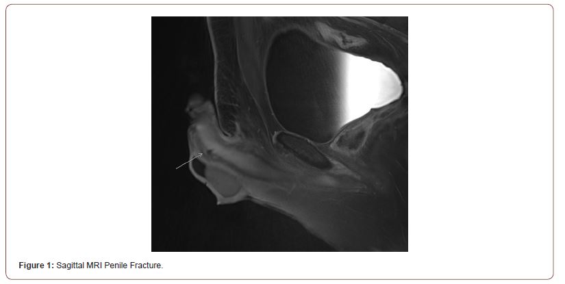
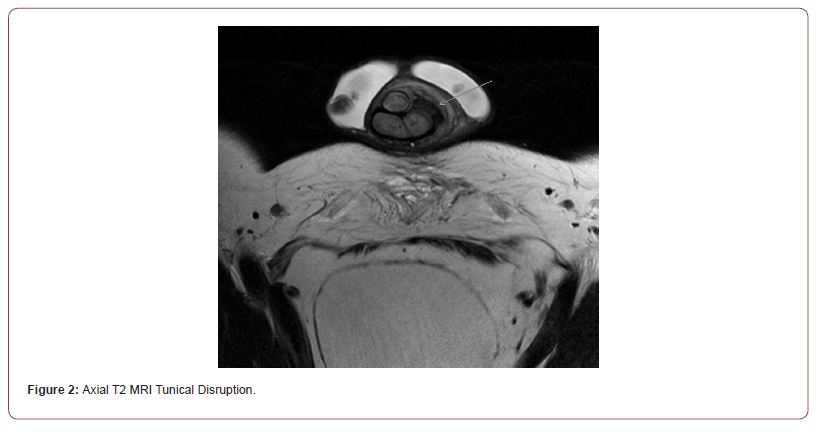
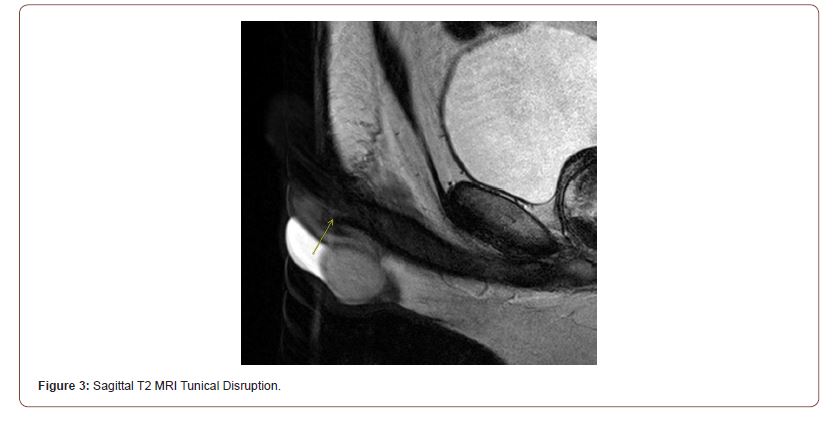
Urology was consulted for penile trauma. At this point, the decision was made to obtain a penile MRI. The MRI revealed penile fracture (tunical disruption) (Figures 1-3). Sagittal views clearly delineate a penile fracture (Figures 1&3). The axial view showed penile fracture along with penile edema and hematoma (Figure 2).


The patient was booked by urology for penile exploration and repair of penile fracture. The surgery was performed urgently, and the patient was kept overnight for observation. He was discharged in good condition with follow-up in the urology clinic.
Discussion
Penile fracture is characterized by disruption of the tunica albuginea [1]. Typically, blunt trauma leads to tunical rupture. The most common cause of penile fracture is vaginal intercourse [2]. There are a number of other causes documented, including automanipulation of the erection, which occurred with our patient.
Patients typically present with penile pain and swelling [1]. Penile vascular injuries can also present similarly to penile fractures [3,4]. This reemphasizes the need for MRI at times to ensure the correct diagnosis and obviate the need for unnecessary surgical exploration.
Some patients may not report typical signs and symptoms of penile fracture. Patients may not report rapid detumescence. If exam and ultrasonography fail to establish the diagnosis of penile fracture, MRI can be utilized. Our patient benefited from the use of penile MRI to confirm the diagnosis of penile fracture to determine the need for surgical intervention by urology.
One systematic review analyzed the most common protocols for MRI after penile trauma [5]. Similar to our case, the most commonly used protocol involved three orthogonal T2 sequences. In our case, MRI revealed a certain penile fracture, which prompted surgical intervention. While one could argue for surgical exploration regardless of MRI, the MRI not only confirmed with certainty the diagnosis, but it also gave a precise location for the tunical disruption. The MRI was paramount in the patient’s diagnosis and overall auspicious outcome.
Conclusion
Our patient presented with mild penile pain after reportedly adjusting his erection to urinate. Clinical examination by the emergency physician and urologist was indeterminate for penile fracture. MRI was used to confirm the diagnosis of penile fracture and help determine the correct treatment for the patient.
Acknowledgement
None.
Conflict of Interest
No conflict of interest.
References
- Ruckle HC, HR Hadley, PD Lui (1992) Fracture of penis: diagnosis and management. Urology 40(1): 33-35.
- Meares EM (1971) Traumatic rupture of the corpus cavernosum. J Urol 105(3): 407-408.
- Truong H, B Ferenczi, R Cleary, Kelly A Healy (2016) Superficial Dorsal Venous Rupture of the Penis: False Penile Fracture That Needs to be Treated as a True Urologic Emergency. Urology 97: e21-e22.
- Darshan K Shah, Elliot M Paul, Sanford A Meyersfield, Richard A Schoor (2003) False fracture of the penis. Urology 61(6):
- Esposito AA, C Giannitto, C Muzzupappa, Sara Maccagnoni, Franco Gadda, et al. (2016) MRI of penile fracture: what should be a tailored protocol in emergency? Radiol Med 121(9): 711-718.
-
Bryce A Baird, Laura Geldmaker, Ethan J Wajswol, Mark Sultan, Paul R Young, et al. Magnetic Resonance Imaging (MRI) Diagnosis of Penile Fracture. Annal Urol & Nephrol. 3(1): 2022. AUN.MS.ID.000556.
-
Penis, Penile fracture, MRI, Urology, Pathology, Emergency Room (ER)
-

This work is licensed under a Creative Commons Attribution-NonCommercial 4.0 International License.


