



 Research Article
The Vanderlim X-Technique as an Advanced Surgical Alternative for Immediate Loading in Severely Atrophic Maxillae: A Report of Three Clinical Cases
Received Date: March 09, 2026; Published Date: March 23, 2026
Abstract
All-on-4 procedures have revolutionized implant dentistry and promoted oral rehabilitation worldwide. However, there are clinical cases in which the All-on-4 procedure encounters limitations that make it impossible to proceed with oral rehabilitation as originally planned. Patients with severely atrophic maxillae associated with incipient zygomatic bone and highly pneumatized maxillary sinuses lack a technical solution that ensures fixed oral rehabilitation with a safety margin. In these cases, the technique reaches its limits, requiring alternative solutions. The crossed implant procedure (The Vanderlim X-technique) allows for high-quality anchorage points in zones 1 and 2, ensuring greater levels of predictability and ensuring immediate loading. However, due to the technique’s pioneering nature, the scientific literature still lacks data and information to consolidate this procedure. Therefore, this retrospective observational study aimed to present the crossed implant technique performed in three clinical cases. The results obtained, although initial, were quite positive, attesting to the functionality and safety of the crossed implant procedure “X Technique”.
Keywords:Implantology; oral rehabilitation; all-on-4; x-technique
Introduction
Implantology is a specialty of dentistry focused on oral rehabilitation through dental implants and prostheses, with the main objective of functional, aesthetic and biological restoration of the stomatognathic system. By opting for an implant-supported treatment, the patient seeks a definitive solution that allows the resumption of masticatory function, facial aesthetics and oral health, avoiding the discomfort and limitations imposed by the absence of teeth or the use of mobile or removable prostheses [1]. This avoids the discomfort of missing teeth or the use of palliative and uncomfortable solutions such as mobile or removable dentures [2]. With advancing age, the loss of dental elements is quite common, which may or may not be related to trauma, environmental factors and diseases, and edentulism is characterized by the lack of dental elements and alveolar bone atrophy, making it difficult to rehabilitate these patients [3]. It can be associated with trauma, environmental factors or periodontal diseases, and edentulism is characterized not only by the absence of teeth, but also by the progressive atrophy of the alveolar bone, especially in the maxilla, which significantly hinders oral rehabilitation with conventional implants [4,5]. Fully edentulous patients deserve special attention, as they often have severe anatomical limitations that defy the limits of traditional surgical techniques [6]. To overcome these limitations, Brånemark proposed the use of zygomatic implants as an alternative to obtain anchorage in denser bone regions distant from the atrophic maxilla [7].
From this, many edentulous patients have sought a definitive solution, abandoning old mobile or removable prostheses [8]. The most widespread concept is the All-on-4, where in this type of procedure, four implants are installed, two anterior vertical and two posterior angled, in maxillary residual bone, dispensing with grafts and supporting the immediate load of fixed prostheses, of the protocol type, with 12 or 14 elements [9]. Variations such as the hybrid All-on-4 and the zygomatic All-on-4 (QuadZygoma) have emerged, with the use of two or four zygomatic implants, and it has been common to use two or four zygomatic implants, seeking more distant anchor points that support the immediate load, called hybrid All-on-4 and zygomatic All-on-4 or QuadZygoma [10], for support of fixed dentures under immediate load [11]. However, posterior anchorage of these implants often occurs in regions of extremely thin alveolar bone, often only 1 to 2 mm thick, which increases the risk of maxillary sinusitis, bucco-sinus communication, periimplant instability, and gingival recessions, especially when the implant path is excessively positioned by buccal [12]. In cases where maxillary atrophy is associated with reduced zygomatic bones, the Vanderlim Transnasal Implant Technique emerges as an efficient alternative by providing additional anchor points in the anterior region, reducing the burden on zygomatic implants and minimizing the surgical risks associated with QuadZygoma [13]. However, in even more complex situations, characterized by severely atrophic maxillae, tiny zygomatic structures, and extensively pneumatized maxillary sinuses, even these approaches become limited, not offering a sufficiently safe and predictable surgical solution [6,14].
In this scenario, the concept of crossed implants, known as Vanderlim’s X-Technique, emerges as an advanced alternative for the rehabilitation of extreme maxillae, and with the main objective is to strategically reposition zygomatic implants, avoiding their posterior anchorage in thin alveolar bone and displacing the crestal support point to anterior regions, especially in the canine area, where there is usually greater bone volume and density, where this positioning allows the creation of a deeper channel, with a predominantly extrasinusal path, significantly reducing the risk of sinusitis and bucco-sinus communications [15,16]. In addition, the X-Technique seeks to optimize the biomechanical behaviour of the system, positioning posterior implants in an almost horizontal palatal path, while anterior implants assume a more buccal support, and with this cross configuration allows the use of long or extra-long implants, taking advantage of anterior bone regions of higher density to increase insertion torque and primary stability, especially in zone II described by Bedrossian [17-19]. In addition, the control of vestibular positioning contributes to the reduction of gingival recessions and improvement of the prosthetic emergence profile [20].
In cases of atrophic maxillae associated with tiny or reduced zygomatic bones, the option for Vanderlim Technique Transnasal Implants provides two quality anchor points in the anterior area, reducing the overload on the zygomatic bones, minimizing the risks associated with QuadZygoma surgery and thus enabling oral rehabilitation with immediate loading [21]. Finally, in the cases of patients with severely atrophic maxilla associated with reduced zygomatic structure and the presence of highly pneumatized maxillary sinuses, the technical solution reaches its newest frontier, where the available surgical options do not contemplate a safe and predictable rehabilitation procedure [18,22]. As an alternative, the procedure with crossed implants, called “X-Technique”, which makes use of two zygomatic implants associated with two or more long regular implants, in an almost horizontal palatal approach, occupying bone spaces that intersect in opposite planes, coming from anterior to posterior, one in vestibular support and the other in palatal support, enabling quality anchor points in zones 1 and 2 previously described by Bedrossian [17] and promoting oral rehabilitation on the same day. In view of this, the present study aims to present in detail the X-Implant Cross Technique, highlighting its anatomical, biomechanical and surgical foundations, as well as its main indications and clinical advantages the main characteristics, requirements and surgical qualities, using 03 complete clinical cases in immediate loading for these patients, with diagnosis, planning, procedure performed and final results followed for 3 years.
Materials and Methods
This applied, descriptive research in the area of dental implantology is classified as an observational case study, of the retrospective longitudinal type. The data of this study were collected from the clinical documentation of 03 patients, from the initial consultation, diagnosis, surgical planning, surgery, prosthetic solution and postoperative follow-up. The collected data were submitted to qualitative-quantitative analysis. In this study, the total postoperative follow-up time ranged from 1.5 to 3 years. Patients were previously consulted and consented to participate in their clinical data and radiological images for research purposes, and were treated according to the Treaty of Elsinki. All procedures were performed in a private clinic, by the same technical team. The 03 cases presented in this study involved elderly patients, edentulous, dissatisfied with the mobile prosthetic solution and desirous of the fixed solution. As a procedure, the two patients underwent imaging exams such as panoramic x-ray and ConeBean tomographic examination, and after a thorough analysis of the images in 3D planning software, taking into account the lack of bone in Zone 01 and Zone 02 for anterior anchorage [16].
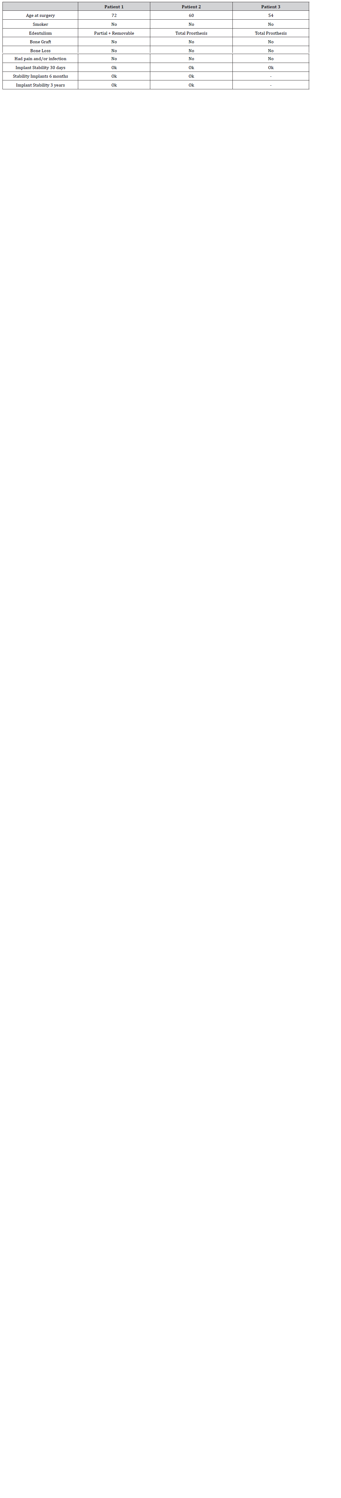
The initial imaging tests revealed the adverse clinical picture for both patients, pointing to technical difficulties to proceed with oral rehabilitation with immediate loading. Maxillary bone loss was very severe in both patients, with insufficient alveolar ridge. The surface of the zygomatic bone was small in both cases, invalidating the possibility of the QuadZygoma procedure. Finally, strong pneumatization of the maxillary sinuses was observed, ruling out all usual and known techniques, due to the high associated risk. The very adverse clinical scenario required the search for an alternative solution, which would promote oral rehabilitation in immediate load. Thus, the proposal arose to install two pairs of implants in a cross procedure, alternating zygomatic and long implants. These implants travel pathways that intersect in different planes, both buccal and palatal, as well as anterior to posterior, providing anchor points with high torque in zones 1 and 2, in the palatal approach. The technique can also be complemented with a regular implant in the vomer region, providing greater safety and prosthetic stability. Table 1 below presents data on the patients and the procedure. The absence of a nasal concha to use the transnasal technique, and having a zygomatic bone with a small size to install a double zygomatic implant was found [23] After verifying severe atrophy and little anatomical dimension, the 03 patients were indicated to perform the crossover surgical procedure “X-shaped technique”, and the patients consented to undergo the surgery.
Clinical Cases
Patient 1- This is a 72-year-old male patient, non-smoker, total edentulous. He declared to be hypertensive and diabetic, both under drug control. He has been using a mobile prosthetic solution for more than 20 years. The first appointment came complaining of discomfort when smiling and chewing, requesting a definitive solution. Two long implants were placed at the alveolar crest in the region of the first premolars. These implants were planned and executed with a three-dimensional orientation so that the apical anchorage occurred at the so-called “Point 4” of the 5 Points protocol, an anatomical concept previously described as a strategic cortical anchorage area. This approach enables maximal utilization of the available cortical bone anchorage, promotes biomechanical redistribution of functional loads in the anterior segment, and creates adequate prosthetic space for the subsequent placement of zygomatic implants. Subsequently, zygomatic implants are placed in the canine region, positioned within a previously planned osseous channel, allowing for a more favourable prosthetic emergence profile. This strategy significantly reduces the risk of gingival recession, minimizes thread exposure, and contributes to a more stable and aesthetic emergence profile. Additionally, a regular implant was placed in the midline region, increasing the safety and stability of the set.
Primary Stability and Immediate Loading
The combination of anterior cortical anchorage (Point 4) and zygomatic anchorage enables the achievement of high insertion torque values, providing adequate primary stability to support immediate loading protocols.
From a biomechanical perspective, this configuration promotes:
• Optimized distribution of occlusal forces
• Reduction of posterior cantilever length
• Greater predictability in severely pneumatized maxillae

In Image 1, below, a post-surgical panoramic examination of patient 1 reveals details of the X-Technique, where the two zygomatic implants emerge in the anterior region of the maxilla (Bedrossian region 1) and the long implants emerge in molar (Bedrossian region 2), providing the prosthetic stability necessary to support a protocol prosthesis with 12 elements.
Patient 2 - This is a 60-year-old male patient, non-smoker, with partial edentulousness. He stated that he has been using a removable prosthesis for more than 20 years, but that he does not feel comfortable. The first appointment came complaining of discomfort, pain when chewing and requesting a definitive solution. Two long implants were placed at the alveolar crest in the region of the first premolars. These implants were planned and executed with a three-dimensional orientation so that the apical anchorage occurred at the so-called Point 5 of the 5 Points protocol, an anatomical concept described as a strategic posterior cortical anchorage area.
Subsequently, two zygomatic implants were placed in the canine region, positioned within a previously planned osseous channel, allowing for a more favorable prosthetic emergence profile. In this configuration, the zygomatic implants emerge in the anterior region of the maxilla (Bedrossian region 1), whereas the long implants emerge in the molar region (Bedrossian region 2), providing the prosthetic stability necessary to support a 12-unit fixed full-arch protocol prosthesis. Additionally, an implant was placed in the right pterygoid region for potential delayed use, if necessary, thereby expanding the possibilities for posterior prosthetic support. The combination of posterior cortical anchorage (Point 5) and zygomatic anchorage enables the achievement of high insertion torque values, providing adequate primary stability for immediate loading protocols.


In Image 1, below, a post-surgical panoramic examination of patient 1 reveals details of the X-Technique, where the two zygomatic implants emerge in the anterior region of the maxilla (Bedrossian region 1) and the long implants emerge in premolar (Bedrossian region 2),
Paciente 3- This was a case of severely atrophic maxilla, in which adequate anchorage was achieved through the placement of two zygomatic implants, with prosthetic emergence in the anterior region of the maxilla (Bedrossian region 1). In addition, two long implants were placed at the alveolar crest in the region of the first premolars, planned and executed with a three-dimensional orientation so that apical anchorage was achieved at the so-called Point 5 of the 5 Points protocol, described as a strategic area of posterior cortical anchorage.
In this configuration, the long implants present prosthetic emergence in the premolar region (Bedrossian region 2), contributing to a more favorable biomechanical distribution of masticatory loads and providing the prosthetic stability required to support a 12-unit fixed full-arch protocol prosthesis. The combination of posterior cortical anchorage at Point 5 and zygomatic anchorage allowed the achievement of high insertion torque values, ensuring adequate primary stability for immediate loading protocols, even in the context of severe maxillary atrophy.


In Image 1, below, a post-surgical panoramic examination of patient 1 reveals details of the X-Technique, where the two zygomatic implants emerge in the anterior region of the maxilla (Bedrossian region 1) and the long implants emerge in molar (Bedrossian region 2), providing the prosthetic stability necessary to support a protocol prosthesis with 12 elements.
Datas of Patients
Table 1:Characteristics of the arches and Type of Prosthesis.

Table 2:General Patient Age and Health Status and Habits.

Table 3:Anatomical region and characteristics of the implants installed.

Table 4:Intraoral or extraoral surgical complications.

Table 5:Intraoral prosthetic complications.

Table 6:Final General Clinical Status Cases in Vanderlim Technique X.
From a technical point of view, the osseointegration of the implants was uneventful for both patients, with very firm and stable implants. The palatine approach proved to be the most appropriate, promoting surgical precision, more efficient healing and, thus, avoiding unnecessary risks. The prosthetic solution remains stable and the tissues healthy, attesting to the success of the procedure as a whole. As a result, it was observed in this study that the crossimplant procedure appears as an option to be evaluated for patients with unfavorable anatomical conditions, and the “The Vanderlim X-technique” provided oral rehabilitation of these patients with the use of fixed prostheses of 12 elements in immediate loading, supported by implants geometrically well distributed and with high torque, there was no need for bone grafting, and no complications were observed over the regular 3-year follow-ups, where the implants remained stable and functional. Finally, the patients declared satisfaction and, when questioned, demonstrated total confidence in the prosthetic solution and in the results of the procedure.
Discussion
The oral rehabilitation of edentulous patients with severe maxillary atrophy has always imposed a challenging level for implant dentistry, and technological innovations have provided greater safety and more adjusted solutions, seeking safety and shorter recovery time, opening the opportunity for the rehabilitation of an important portion of patients [13,24]. Wellknown technical solutions such as All-on-4, All-on-4 hybrid and QuadZygoma procedures are recommended and can rehabilitate the vast majority of patients, including cases of atrophic jaws, and Transnasal implants the Vanderlim X-technique emerge as an option in the oral rehabilitation of patients with adverse anatomical conditions, providing two quality anchor points in zone 1, reducing the overload on incipient zygomatic bones and, With this, they minimize the associated risks [21,22,25]. However, patients who have unfavorable anatomical characteristics that are difficult to solve technically end up instigating the limits of technique, opening up the possibility of questioning the limits of science and suggesting new procedures [16,26,27].The first point concerns the steps that precede the surgical procedure itself, ensuring the safety of the procedure, it is understood that the professional must be aware of all the steps that precede the surgery, as well as the initial consultation of the patient, the correct diagnosis, the surgical planning, as well as the learning curve, are equally important and necessary steps, for the delivery of a good surgical result, which is the sum of all the steps that precede [19,20,23,28,29].
With the imaging exams (tomography), the diagnosis of the case can be made, at which time the anatomical characteristics and residual bone tissue must be evaluated in their details, and at this stage the surgeon must use 3D software to better evaluate the different possibilities of rehabilitation and decide which technical solution to be proposed [14]. 3D software simulations are indicated to determine the exact model and anchorage site of the implants and the use of resin prototypes can help visualize the anatomy and anchor points, simulating and anticipating the procedure in a full-size model, as well as surgical guides are advisable and avoid deviations in milling [30,31]. The X-Implant Cross Technique represents a conceptual evolution in the rehabilitation of severely atrophic maxillae, based on solid biomechanical principles and the strategic reorganization of implant anchor points. By repositioning the zygomatic implant to anterior regions of higher bone density, adopting predominantly extrasinusal paths, and associating long implants in a cross-configuration, the technique allows avoiding posterior support in extremely thin alveolar bone, reducing flexion moments, biomechanical overload, and the risk of sinus complications.
From a biomechanical perspective, the The Vanderlim X-technique promotes a more favorable three-dimensional distribution of force vectors, aligning the occlusal load center to the longitudinal axes of the implants and creating an integrated load containment system. This configuration contributes to greater primary stability, increased insertion torque, and predictability of immediate loading, even in anatomical scenarios considered borderline for conventional zygomatic protocols. Regarding the learning curve, it is important for the professional to be very familiar with the various technical approaches, such as regular implants, long implants, transinusal and transnasal implants, zygomatic implants and pterygoids, and it is important to know the indications of each procedure as well as its technical limitations and associated risks [15,25,32].
However, it is important to note that the cross implant procedure uses surgical techniques known in implantology, with small adjustments to fix the implants in bone sites located in different dimensional planes, promoting the distribution of support forces in a cross-way manner, and this perception is clear when analyzing the imaging exam, where the implants visually cross each other without touching, promoting a unique characteristic for the technique [13,15,24]. However, the benefits of the X-Implant Cross Technique are intrinsically associated with strict indication criteria. This is an advanced approach, indicated mainly for patients with severely atrophic maxillae associated with extremely small posterior alveolar bone, reduced zygomatic bones, and widely pneumatized maxillary sinuses, in which traditional techniques present an increased risk. The need for longer zygomatic implants
and the complexity of the surgical path impose a high learning curve, restricting their application to surgeons experienced in advanced zygomatic surgery. In addition, the long-term success of the The Vanderlim X-technique depends on detailed three-dimensional planning, precise surgical execution, hygiene-friendly prosthetic design, and strict clinical follow-up. Patients with poor adherence to maintenance protocols or with risk factors for peri-implant disease should be carefully evaluated, since these conditions constitute contraindications related to the technique.
Conclusion
Thus, when correctly indicated and performed within welldefined biomechanical, anatomical and clinical criteria, the X-Implant Cross Technique safely and predictably extends the limits of oral rehabilitation with immediate loading in extreme maxilla’s. Its use, however, must be judicious and based on scientific evidence, avoiding indiscriminate applications that may compromise the predictability of the treatment. As presented in this study, the cross-implant procedure called “The Vanderlim X-technique”, as a result, the patients in this study were rehabilitated without any intercurrences, with a fixed prosthesis screwed with 12 elements. In a 3-year follow-up, this technique offers the possibility of oral rehabilitation with fixed prosthesis in immediate loading for some patients who have unfavorable anatomical conditions, such as severe maxillary atrophy, small or incipient zygomatic bone and wide pneumatization of the maxillary sinuses, with little bone remaining only in Zone 01.
With these very promising results, we continue to encourage the exploration of the cross-implant technique, as well as the production of new and more complete studies on the subject, broadening the horizons of implantology. Another point to be discussed concerns the pioneering and possible novelty of the cross-implant procedure “X-shaped technique”, from the point of view of this literature, there are no references to the identical or similar procedure in the specialized literature, indicating the novelty of the cross-technique in oral rehabilitation in extremely atrophic edentulous jaws without bone grafts and in immediate loading.
Acknowledgement
None.
Conflict of Interest
No conflict of interest.
References
- Albrektsson T, Dahlin C, Jemt T, Sennerby L, Turri A, et al. (2014) Is marginal bone loss around oral implants the result of a provoked foreign body reaction?. Clin Implant Dent Relat Res 16(2): 155-165.
- Aparicio C, Lopez-Piriz R, Albrektsson T (2020) ORIS Criteria of Success for the Zygoma-Related Rehabilitation: The (Revisited) Zygoma Success Code. Int J Oral Maxillofac Implants 35(2): 366-378.
- Aparicio C, Manresa C, Francisco K, Claros P, Alandez J, et al. (2014) Zygomatic implants: indications, techniques and outcomes, and the zygomatic success code. Periodontol 66(1): 41-58.
- Aparicio C, Ouazzani W, Garcia R, Arevalo X, Muela R, et al. (2006) A prospective clinical study on titanium implants in the zygomatic arch for prosthetic rehabilitation of the atrophic edentulous maxilla with a follow-up of 6 months to 5 years. Clin Implant Dent Relat Res 8(3): 114-122.
- Aparicio C, Ouazzani W, Hatano N (2008) The use of zygomatic implants for prosthetic rehabilitation of the severely resorbed maxilla. Periodontol 47: 162-171.
- Aparicio C, Sanz A, Tahmasebi S, Broumand V (2025) Incision and Flap Design in Zygomatic Implant Surgery: A ZAGA-Based Decision Tree. Int J Periodontics Restorative Dent 0(0): 1-44.
- Bedrossian E, Bedrossian EA (2019) Fundamental Principles for Immediate Implant Stability and Loading. Compend Contin Educ Dent 40(9): i1-i8.
- Bedrossian E, Bedrossian EA (2025) Full Mouth Reconstruction with Dental Implants: Planning, Surgical, and Prosthetic Phase. Dent Clin North Am 69(2): 257-274.
- Branemark PI, Adell R, Albrektsson T, Lekholm U, Lundkvist S, et al. (1983) Osseointegrated titanium fixtures in the treatment of edentulousness. Biomaterials 4(1): 25-28.
- Branemark PI, Grondahl K, Ohrnell LO, Nilsson P, Petruson B, et al. (2004) Zygoma fixture in the management of advanced atrophy of the maxilla: technique and long-term results. Scand J Plast Reconstr Surg Hand Surg 38(2): 70-85.
- Branemark PI, Hansson BO, Adell R, Breine U, Lindstrom J, et al. (1977) Osseointegrated implants in the treatment of the edentulous jaw. Experience from a 10-year period. Scand J Plast Reconstr Surg Suppl 16: 1-132.
- Chrcanovic BR, Albrektsson T, Wennerberg A (2014a) Flapless versus conventional flapped dental implant surgery: a meta-analysis. PLoS One 9(6): e100624.
- Chrcanovic BR, Albrektsson T, Wennerberg A (2014b) Periodontally compromised vs. periodontally healthy patients and dental implants: a systematic review and meta-analysis. J Dent 42(12): 1509-1527.
- Eshoiee N, Aalam AA, Zelig D, Oh S, Kar K, Bakshalian N (2025) Trans-nasal dental implants: indication and the report of 10 cases. Ann Med Surg (Lond) 87(4): 1814-1822.
- Halldin A, Jimbo R, Johansson CB, Wennerberg A, Jacobsson M, et al. (2014) Implant stability and bone remodeling after 3 and 13 days of implantation with an initial static strain. Clin Implant Dent Relat Res 16(3): 383-393.
- Jensen OT (2014) Complete arch site classification for all-on-4 immediate function. J Prosthet Dent 112(4): 741-751 e742.
- Jensen OT, Oh S, Butura CC (2025) Maxillary Complete Arch Oral Implant Treatment for Fixed-Denture Immediate Function. Atlas Oral Maxillofac Surg Clin North Am 33(2): 105-115.
- Kammerer PW, Fan S, Aparicio C, Bedrossian E, Davo R, et al. (2023) Evaluation of surgical techniques in survival rate and complications of zygomatic implants for the rehabilitation of the atrophic edentulous maxilla: a systematic review. Int J Implant Dent 9(1): 11.
- Malo P, Machado A, Medeiros R (2025) The All on 4 Concept Revisited. Atlas Oral Maxillofac Surg Clin North Am 33(2): 97-104.
- Malo P, Nobre Mde A, Lopes A (2012) Immediate rehabilitation of completely edentulous arches with a four-implant prosthesis concept in difficult conditions: an open cohort study with a mean follow-up of 2 years. Int J Oral Maxillofac Implants 27(5): 1177-1190.
- Malo P, Rangert B, Nobre M (2003) "All-on-Four" immediate-function concept with Branemark System implants for completely edentulous mandibles: a retrospective clinical study. Clin Implant Dent Relat Res 5 Suppl 1: 2-9.
- Mitrani R, Papaspyridakos P, Bedrossian EA, Goldberg J, Tsigarida A, et al. (2025) Treatment planning algorithm for patients with a terminal dentition. J Prosthet Dent 134(5): 1391-1397.
- Nobre MA, Lopes A, Ferro A, Nunes M, Gouveia M, et al. (2025) Immediate full-arch mandibular rehabilitation supported by four implants: A retrospective study with 20 to 25 years of follow-up. J Dent 106286.
- Nunes M, De Araujo Nobre M, Camargo V (2024) All-on-4 Hybrid with Extra-Long Transnasal Implants: Descriptions of the Technique and Short-Term Outcomes in Three Cases. J Clin Med 13(11): 3348.
- Orentlicher G, Jensen O, Horowitz A, Adams M, Abboud M (2013) Combining All-on-4 treatment with CT-guided technology: technique and report of three cases. Compend Contin Educ Dent 34(7): 534-544.
- Papaspyridakos P, Vazouras K, Gotsis S, Bokhary A, Sicilia E, et al. (2023) Complete digital workflow for prosthesis prototype fabrication with double digital scanning: A retrospective study with 45 edentulous jaws. J Prosthodont 32(7): 571-578.
- Sahin O, Odabasi O, Rizaj X, Akan E (2025) Three-dimensional Finite Element Analysis of Quad Zygomatic, Transnasal and Pterygoid Implants for Rehabilitation of Patients with a Severely Atrophic Maxilla. Int J Oral Maxillofac Implants 0(0): 1-23.
- Tannyhill RJ 3rd, Jensen OT (2019) Computer Simulation and Maxillary All-on-Four Surgery. Oral Maxillofac Surg Clin North Am 31(3): 497-504.
- Tartsch J, Chrcanovic B, Wennerberg A, Kohal R, Albrektsson T (2025) Two-piece Zirconia Implants: An Office based Retrospective Study with up to 7 and mean 3 Year Follow-Up. Int J Oral Maxillofac Implants 0(0): 1-36.
- Togac E, Ayranci F, Tapci O (2025) Finite Element Analysis of Transnasal, Zygomatic and Pterygoid Implants in the Rehabilitation of the Atrophic Maxilla. J Stomatol Oral Maxillofac Surg 127(3): 102674.
- Tomaselli L, Giammarinaro E, Grassi R (2025) Basal bone anchorage for immediate loading in the atrophic maxilla: A technical note with surgical case series. Clin Adv Periodontics.
- Wadhwani CPK, Albrektsson T, Schoenbaum TR, Chung KH (2025) Residual Debris Within Internal Features of As-Received New Dental Implants. Int J Oral Maxillofac Implants 40(2): 257-261.




