 Case Report
Paratesticular Schwannoma In A 13-Year-Old Boy: A Case Report And Literature Review
Volkan Sarper Erikci*
Visceral surgery, Moulay Ismail Military Hospital, Morocco
Department of Pediatric Surgery, Sağlik Bilimleri University, İzmir Faculty of Medicine,Tepecik Training Hospital, Izmir, Turkey
Received Date: January 18, 2023; Published Date: January 27, 2023
Summary
Paratesticular schwannoma is a rare neoplasm of mesenchymal origin and there are a few reports in the literature describing this entity. In this study, with a painless scrotal lesion for 11 years, a 13-year-old boy with intrascrotal extratesticular schwannoma is presented. The tumoral mass was totally excised in a testis sparing manner. The medical history of the patient, radiological and histopathological findings are also given together with a brief literature review on this issue.
Keywords:Paratesticular tumor; Children; Schwannoma
Introduction
Paratesticular tumors are uncommon neoplasms of mesenchymal origin. These tumors are usually seen in ductus deferens, epididymis, tunica vaginalis, lymphatics, vessels and other supportive tissues and most common of these are leiomyoma, lipoma and adenomatoid tumor [1]. Derived from schwann cells, schwannomas are the most common tumor of the peripheral nerves and these tumors are commonly seen in head, neck and limbs [2]. To our knowledge there are less than 10 cases of intrascrotal extratesticular schwannoma in the literature including a 16-yearold child with paratesticular schwannoma [2] In this study a 13-year-old boy with intrascrotal extratesticular schwannoma is presented. The medical history of the patient, radiological and histopathological findings are also given and the topic is discussed under the light of relevant literature.
Case Report
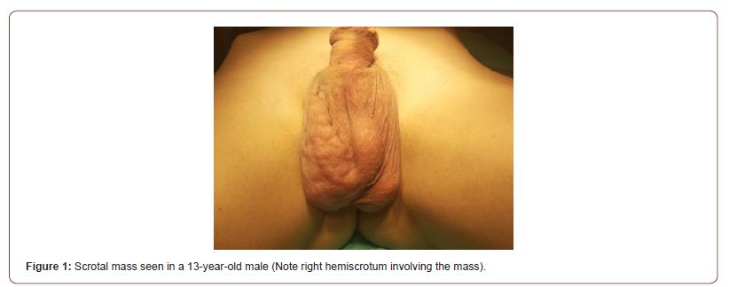
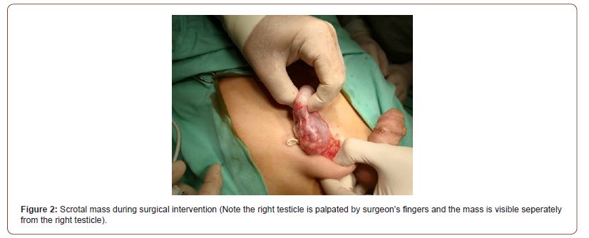
A 13 year-old male presented with painless and slowly growing scrotal mass. At the age of 2 years he was diagnosed as hydrocele and did not come to regular controls. He was admitted to our hospital with the symptoms of straining while walking. On physical examination an irregular, firm, mobile non-reducible was palpabl separately from the right testicle [Figure 1]. There was not hyperemie, tenderness or edema on the surface of the mass. Both testes were normal in size and shape and there was no palpabl lymph node upon physical examination. Systemic physical examination was otherwise normal. Ultrasonography [US] showed a solid, volimunious, heterogeneous and lobulated mass measuring 5.5x2.0 cm in size with a mixed echogenicity and internal vascularity seperately from right testis and epididymis with normal testicles and an increase in size of right epididymis. Magnetic resonance imaging [MRI] showed similar findings and tumor markes including beta-HCG, AFP and LDH levels were within normal age. After consultation with pediatric oncology surgical treatment was planned. With a trans-inguinal approach, after spermatic cord was clamped, biopsy revealed a benign mass on frozen study. Using a testis sparing surgery, total excision of the mass was performed [Figure 2]. The spermatic cord was then released and right testicle was pushed back into the right hemiscrotum. He was discharged 24 hours after surgery without complications. After 3 months of follow up physical examination and scrotal USG were negative for recurrence of the disease.


Discussion
Also named as neurilemmomas or neurinomas, schwannomas are benign schwann cels derived-tumor. Mostly affected sites are head and neck in these tumors. Although it can occur in all age groups effecting male and female equally peak patient age is between 20-50 years old [3-5]. Majority of these tumors are usually seen sporadic but an association with neurofibromatosis type 2, schwannomatosis and meningiomatosis has also been reported [2, 6]. Most of patients with schwannoma are asymptomatic and if symptomatic typical symptoms include symptoms of dysthesia, sensory loss, weakness or radicular type pain [7].
Although majority of schwannomas are seen in head and neck, schwannoma is a rare finding in the differential diagnosis of scrotal neoplasms and paratesticular lesions. Literature review on this topic clearly reveals that there are less then 10 cases of intrascrotal extratesticular schwannoma including a case of 16 year-old male with this tumor who was reported to be the first pediatric case of paratesticular schwannoma [2, 3, 8-10]. The presented case in this study is probably the second pediatric case of benign intrascrotal extratesticular schwannoma. Besides, to our knowledge, the presented case is probably the youngest male with this paratesticular tumor reported in the English language literature. The reported history of the previous cases include painless scrotal swelling which is similar finding in our case also. There is no pathognomonic radiological finding for this disease. US and MRI are commonly used in identifying these tumors. On US schwannoma generally appears as a well-circumscribed mass with hypoechoic pattern with poor hypervascularity on colour Doppler imaging [11]. On the other hand, MRI is more capable of identifying the tumor and distinguishing it from testicle parenchyme which shows a peripheral rim enhancement in T2 weighted sequences [3, 12]. US and MRI were also used in identifying the tumor in the presented case with similar radiological findings. Testicular tumor markers were found to be normal in range in the previously reported cases which is also a similar finding to our case in this study.
Surgical excision is the mainstay of treatment for scrotal schwannomas. With the help of frozen study during surgical intervention in addition to clamping of spermatic cord, after discovery of benign nature of tumoral mass, testis sparing surgery with total excision of the mass is not also curative but also important for preserving future fertility of these cases especially if presented bilateral involving both testes. Malignant degeneration of schwannoma is extremely rare if present it may be as a sarcomatoid like behaviour [13].
In conclusion, diagnosing paratesticular schwannoma is a challenge for the first liners of medical providers especially before surgical treatment. Clinicians should keep in their minds the rare diagnosis of schwannoma also in cases with paratesticular tumor in pediatric patients and a prompt pediatric surgical consultation is recommended.
Acknowledgments
None.
Conflict of Interest
None.
References
- Akbar SA, Sayyed TA, Jafri SZA, Hasteh F, Neill JSA (2003) Multimodality imaging of paratesticular neoplasms and their rare mimics. RadioGraphics 23: 1461-1476.
- Alsunbul A, Alenezi M, Alsuhaibani S, AlAli H, Al-Zaid T, et al. (2020) Intra-scrotal extra-testicular schwannoma: a case report and literature review. Urology Case Reports 32: 101205.
- Chan PT, Tripathi S, Low SE, Robinson LQ (2007) Case report-ancient schwannoma of the scrotum. BMC Urol 7: 1.
- Jiang R, Chen JH, Chen M, Meng Li Q (2003) Male genital schwannoma, review of 5 cases. Asian J Androl 5: 251-254.
- Pilavaki M, Chourmouzi D, Kiziridou A, Skordalaki A, Zarampoukas T, et al. (2004) Imaging of peripheral nerve sheath tumors with pathologic correlation: Pictorial review. Eur J Radiol 52: 229-239.
- Bergeron M, Bolduc S, Labonté S, Moore K (2014) Intrascrotal extratesticular schwannoma: a first pediatric case. Can Urol Assoc 8: 3-4.
- El-Sherif Y, Sarva H, Valsamis H (2012) Clinical reasoning: an unusual lung mass causing focal weakness. Neurology 78: e4-7.
- Barde NG, Sachidanand S, Madura C, Chugh V (2013) Intrascrotal non-testicular schwannoma: a rare case report. J Cutan Aesthet Surg 6: 170-171.
- Shamsa A, Omidi AA (2004) Huge primary intrascrotal schwannoma: a case report and review of the literature. Med J Islamic Rep Iran 18: 85-86.
- Bhanvadia V, Santwani PM (2010) Intrascrotal extratesticular schwannoma. J Cytol 27: 37-39.
- Sighinolfi MC, Mofferdin A, De Stefani S, Celia A, Micali S, et al. (2006) Benign intratesticular schwannoma: a rare finding. Asian J Androl 8(1): 101-103.
- Ikari R, Okamoto K, Tetsuya YT, Johnin K, Okabe H, et al. (2010) A rare case of multiple Schwannomas presenting with scrotal mass: a probable case of Schwannomatosis. Int J Urol 17: 734-736.
- Palleschi G, Carbone A, Cacciotti J, Manfredonia G, Porta N, et al. (2014) Scrotal extratesticular schwannoma: a case report and review of the literature. BMC Urology 14: 32.
-
Volkan Sarper Erikci*. Paratesticular Schwannoma In A 13-Year-Old Boy: A Case Report And Literature Review. Anaest & Sur Open Access J. 3(5): 2023. ASOAJ.MS.ID.000572.
-
Paratesticular Schwannoma, Schwann cells, testicle, epididymis, biopsy, surgery, Pediatric
-

This work is licensed under a Creative Commons Attribution-NonCommercial 4.0 International License.


