

 Case Report
Fully Digital Rehabilitation of a Quad Zygoma Maxilla and Mandibular All-on-4 Arch with Photogrammetry: A Clinical Case Report with Literature Review
Received Date:October 12, 2026; Published Date:October 26, 2026
Abstract
The rehabilitation of severely atrophic maxillae remains one of the most demanding challenges in implant prosthodontics. Quad zygoma protocols have become a predictable graftless alternative for advanced maxillary atrophy, while photogrammetry has emerged as a highly accurate method for transferring implant positions in full-arch rehabilitations. To describe a fully digital workflow utilizing photogrammetry, intraoral scanning, CAD/ CAM design, and titanium-zirconia hybrid prostheses for the rehabilitation of a patient treated with Quad Zygoma implants in the maxilla and an All-on-4 protocol in the mandible. A completely edentulous patient presenting with severe maxillary atrophy underwent rehabilitation in a hospital environment under general anesthesia. The maxilla was restored using four zygomatic implants according to the exteriorized technique described by Migliorança, while the mandible was rehabilitated using four conventional implants following the All-on-4 concept. Implant positions were recorded using the X-Gram photogrammetry system and merged with intraoral scanning data within Exocad. CAD/CAM-manufactured titaniumzirconia hybrid restorations were fabricated following immediate loading protocols. The digital workflow enabled precise implant position transfer, efficient prosthetic planning, and clinically passive prosthetic adaptation despite significant implant divergence. Immediate loading was achieved successfully and the final restorations demonstrated favorable esthetic and functional outcomes. Photogrammetry combined with intraoral scanning represents a promising strategy for highly complex full-arch rehabilitations involving zygomatic implants. The workflow may enhance implant position accuracy, reduce laboratory remakes, and improve prosthetic predictability.
Keywords: Photogrammetry; quad zygoma; zygomatic implants; all-on-4; digital dentistry; CAD/CAM; immediate loading; titanium-zirconia hybrid prosthesis
Introduction
Rehabilitation of the severely atrophic maxilla remains a major challenge in contemporary prosthodontics and implant dentistry and historically, extensive bone grafting procedures were considered necessary to allow implant-supported restorations in patients presenting with advanced maxillary resorption [1]. However, these procedures are associated with increased morbidity, treatment time, cost, and patient discomfort [2]. The introduction of zygomatic implants by Brånemark in 1998 represented a paradigm shift in the treatment of severe maxillary atrophy, utilizing the zygomatic bone as anchorage and making implant-supported rehabilitation possible without extensive grafting procedures. Long-term clinical evidence has since demonstrated high survival rates and predictable outcomes for zygomatic implant-supported prostheses [3]. Computer-aided design and manufacturing (CAD/CAM), virtual treatment planning, and guided surgery have significantly improved treatment efficiency, and subsequent developments led to modifications of the original intrusions technique and the extra sinus and exteriorized approaches improved prosthetic emergence profiles and reduced sinus-related complications [4].
Among these developments, the exteriorized technique proposed by Migliorança has gained international recognition due to its prosthetic advantages and simplified anatomical management [5]. Simultaneously, digital dentistry has transformed implant rehabilitation workflows by the intraoral scanners, CAD/CAM manufacturing, virtual treatment planning, and guided surgery have significantly improved treatment efficiency and the integration of digital workflows has further refined surgical and prosthetic protocols, enabling more precise and reproducible outcomes [6]. Nevertheless, the accurate transfer of implant positions in fullarch rehabilitations remains challenging, particularly in cases involving highly divergent implants such as zygomatic fixtures [7]. The passive fit of implant-supported full-arch prostheses remains one of the most critical factors influencing long-term biological and mechanical success and errors during implant position transfer may result in framework misfit, generating stresses that can contribute to screw loosening, component fracture, prosthetic complications, and peri-implant bone remodelling [8].
Although conventional splinted impression techniques have historically been considered the reference standard for full-arch implant impressions, digital workflows have gained widespread acceptance due to their efficiency and patient comfort [9]. Nevertheless, the challenge of accurately transferring implant positions becomes increasingly significant as the number of implants and the distance between them increase and the digital workflows have gained widespread acceptance due to their efficiency and patient comfort; however, the challenge of accurately capturing implant positions across extended arches remains a significant clinical consideration [10,11]. Recent investigations have demonstrated that stereophotogrammetry may provide superior trueness and when compared with both conventional impressions and intraoral scanning in complete-arch implant rehabilitations [12]. Unlike sequential image acquisition systems, photogrammetry records the spatial relationships among implant scan bodies simultaneously, minimizing the cumulative stitching errors that may occur during complete-arch scans [13].
This characteristic becomes particularly relevant in highly complex rehabilitations involving zygomatic implants, where the divergence of implant trajectories may amplify the clinical consequences of small positional inaccuracies [14]. The increasing adoption of immediate loading protocols has further emphasized the importance of obtaining highly accurate implant position records [15]. Unlike delayed loading approaches, immediate full-arch rehabilitations require the fabrication of provisional restorations within a very limited timeframe while maintaining optimal prosthetic adaptation [16]. Any discrepancy between the clinical implant position and the digital or laboratory model may compromise passive fit and negatively affect the biomechanical behavior of the restoration during the critical healing phase [17]. Consequently, the precision of implant position transfer has become a fundamental component of contemporary digital workflows [18].
In a Quad Zygoma approach, in which two zygomatic implants are placed bilaterally, the complexity of implant distribution introduces additional restorative challenges in the complex zygoma rehabilitations, also the complexity of implant distribution introduces additional restorative challenges and the zygomatic implants often exhibit substantial differences in angulation and spatial orientation when compared with conventional implants, creating a unique prosthetic scenario that demands exceptional accuracy during impression procedures [11]. Small deviations in implant position registration may be amplified over the length of a complete-arch framework, potentially affecting prosthetic adaptation, screw access channel positioning, occlusion, and longterm mechanical stability [19]. Therefore, technologies capable of reducing distortion and improving implant position capture have become increasingly relevant in the treatment of severely atrophic patients [12].
Beyond the surgical advantages associated with graftless rehabilitation, the exteriorized zygomatic implant technique offers important prosthetic benefits that align closely with modern digital workflows [20]. By allowing more favorable implant emergence profiles and positioning restorative components closer to the ideal prosthetic envelope, the technique facilitates CAD/ CAM framework design and contributes to improved prosthetic access for maintenance and hygiene procedures [19]. These characteristics are particularly important in full-arch restorations, where the relationship between surgical implant placement and prosthetic design directly influences the predictability, longevity, and overall success of the rehabilitation [21]. Photogrammetry has recently emerged as a highly accurate method for recording implant positions and unlike conventional intraoral scanning, photogrammetry captures implant relationships simultaneously, minimizing cumulative stitching errors frequently associated with complete-arch scans [22]. The purpose of this report is to describe a fully digital rehabilitation involving a Quad Zygoma maxilla and mandibular All-on-4 restoration using X-Gram photogrammetry, intraoral scanning, Exocad planning, CAD/CAM manufacturing, and titanium-zirconia hybrid prostheses.
Literature review
To contextualize the present case, a focused literature search was conducted in the PubMed database. The search strategy included the keywords and combinations “photogrammetry”, “Quad Zygoma”, “zygomatic implants”, “All-on-4”, and “full-arch rehabilitation”, using the Boolean operators AND and OR. Particular attention was given to publications containing the terms “photogrammetry AND Quad Zygoma” and “photogrammetry AND All-on-4”. The search revealed an increasing number of studies evaluating photogrammetry for complete-arch implant rehabilitations; however, no publications specifically addressed workflows combining photogrammetry and Quad Zygoma rehabilitation. Likewise, although only four reports described the use of photogrammetry in All-on-4 and Allon- X treatments [23,24,16, 25]. The available evidence remains relatively limited and is primarily composed of technical reports, in vitro investigations, case reports, and a small number of clinical studies.
Although the literature suggests that photogrammetry may improve the accuracy of implant position transfer and facilitate the fabrication of passively fitting frameworks, and a review of the available literature revealed a growing interest in photogrammetry for complete-arch implant rehabilitations [19]. However, publications specifically combining photogrammetry and Quad Zygoma are remarkably scarce and robust clinical evidence on complex rehabilitations combining photogrammetry and Quad Zygoma particularly in cases involving immediate loading and severe maxillary atrophy is still lacking.Although several studies have reported the accuracy and clinical applicability of photogrammetric systems for full-arch implant-supported prostheses and All-on-4 rehabilitations, no reports have investigated their use in conjunction with Quad Zygoma protocols and consequently, evidence regarding the clinical performance of workflows combining photogrammetry and Quad Zygoma remains rare. Similarly, while photogrammetry has gained increasing acceptance in All-on-4 and All-on-X rehabilitations, most available studies consist of technical reports, in vitro investigations, and a relatively small number of clinical studies. The literature suggests that photogrammetry may improve the accuracy of implant position transfer and facilitate the fabrication of passively fitting frameworks [19]. Nevertheless, there is still a lack of robust clinical evidence evaluating complex rehabilitations combining photogrammetry and Quad Zygoma, particularly in cases involving immediate loading and severe maxillary atrophy.
Considering the severity of the maxillary atrophy and the extensive implant divergence anticipated in the zygomatic rehabilitation, a digital workflow based on photogrammetry was selected to maximize the accuracy of implant position transfer [26]. The use of the X-Gram system (X-Gram, Runyes Medical Instruments Co., Ltd.) was intended to reduce potential distortions associated with conventional impressions and full-arch digital stitching processes. Particular attention was directed toward obtaining an accurate representation of the implant coordinates to facilitate the fabrication of a passively fitting prosthetic framework capable of supporting immediate loading. The treatment was planned through a multidisciplinary approach involving surgeons, prosthodontists, and digital dental technicians. This collaboration enabled the integration of surgical planning with prosthetic objectives from the earliest stages of treatment.
Therefore, the present clinical report contributes to the emerging body of evidence on digital workflows combining photogrammetry and Quad Zygoma, demonstrating its application in the rehabilitation of a severely atrophic maxilla restored with four zygomatic implants and an opposing mandibular Allon- 4 prosthesis. This report highlights the clinical feasibility of this approach and underscores the need for further clinical investigations to validate the long-term outcomes of treatments combining photogrammetry and Quad Zygoma in advanced implant rehabilitation. The purpose of this report is therefore to describe a full-mouth rehabilitation involving a Quad Zygoma maxilla and a mandibular All-on-4 restoration, with implant position transfer performed using a novel extraoral photogrammetry system, and to contribute to the emerging body of evidence on digital workflows combining photogrammetry and Quad Zygoma in advanced implant rehabilitation.
Case Report
Diagnosis and Treatment Planning
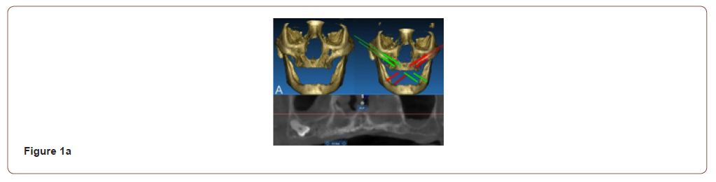
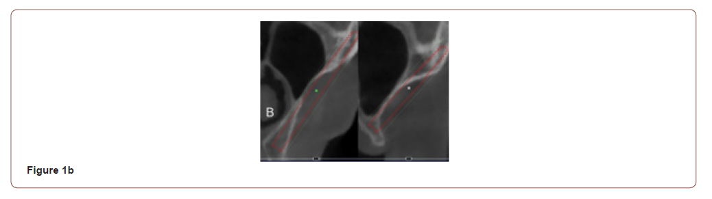
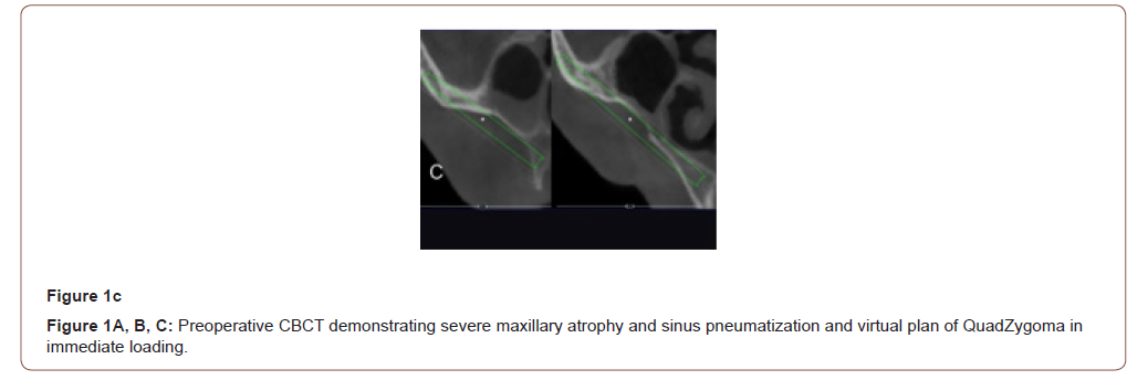
A completely edentulous patient presented seeking fixed implant-supported rehabilitation. Clinical examination and conebeam computed tomography (CTBCT) revealed severe maxillary atrophy characterized by advanced posterior bone loss and extensive sinus pneumatization (fig01). The mandibular arch exhibited sufficient residual bone volume to support rehabilitation according to the All-on-4 treatment concept. Following interdisciplinary planning, the following treatment strategy was selected: A Quad Zygoma rehabilitation of the maxilla and a mandibular rehabilitation using the All-on-4 protocol in immediate loading and fully digital prosthetic workflow with a titanium-zirconia hybrid definitive prosthesis. All procedures were performed in a hospital environment under general anesthesia.
A complete digital impression of both arches was first obtained with an intraoral scanner (IOS) (Neoscanner, 1000 Neodent-to be specified) to enable the design of prototype restorations in CAD/ CAM software. An indexing material was used to reline the intaglio surfaces of the patient’s existing maxillary and mandibular complete dentures, capturing the soft tissue morphology of the edentulous ridges. The dentures were then removed and scanned extraorally, including the intaglio surfaces, to facilitate the subsequent digital alignments.
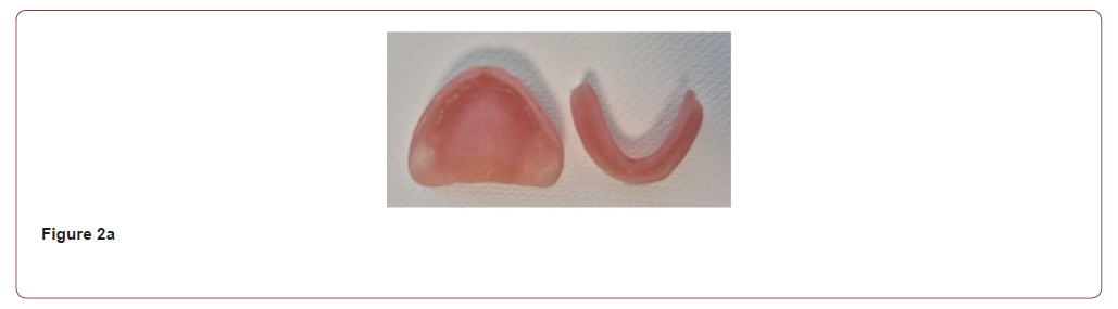
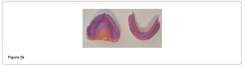
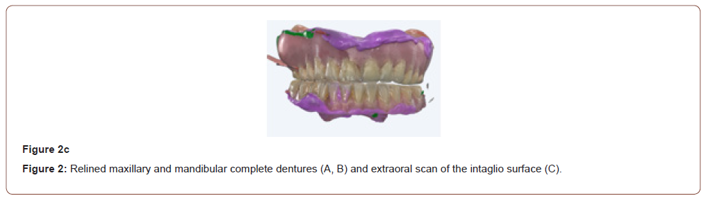
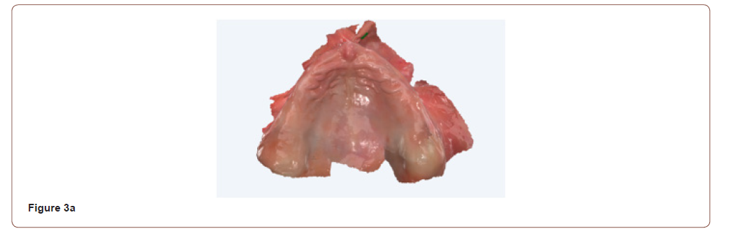
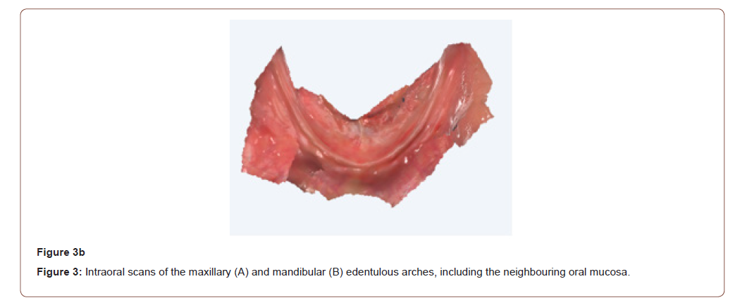
The dentures were repositioned in the patient’s mouth, and a digital bite registration was recorded with the dentures in situ at the planned occlusal vertical dimension and midline, in centric relation. The maxillary and mandibular arches were then scanned, including the neighboring oral mucosa (vestibule, tuberosity, hard palate, and retromolar trigone) (Figure 3).
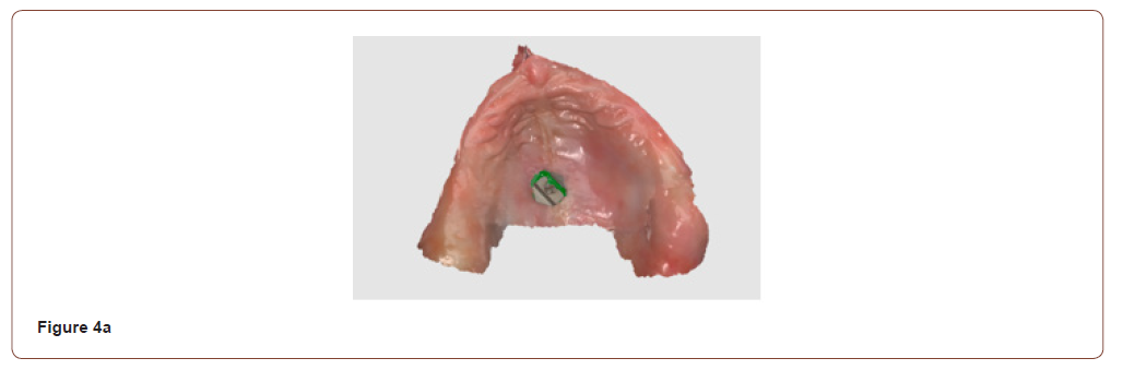
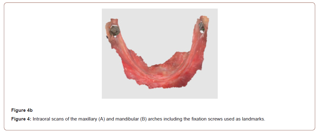
Before surgery, fixation screws (Arch Tracers, Digital Arches LLC) were inserted under local anesthesia: one in the center of the hard palate and one on each mandibular retromolar pad. The edentulous arches were rescanned including the fixation screws, which served as stable reference landmarks for the alignment of subsequent datasets (Figure 4).
Surgical Phase
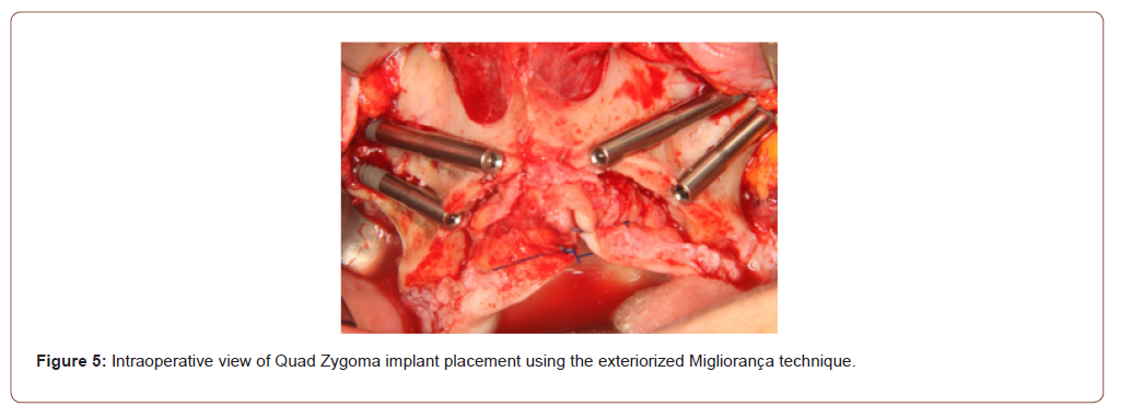
All surgical procedures were performed in a hospital environment under general anesthesia. The maxilla was rehabilitated with four zygomatic implants (JDZygoma, J Dental Care) placed according to the exteriorized technique described by Migliorança [5]. Implant trajectories were planned to maximize zygomatic anchorage while optimizing prosthetic emergence through the alveolar crest. The exteriorized approach allowed improved prosthetic positioning and reduced dependence on extensive cantilever extensions (Figure 05).
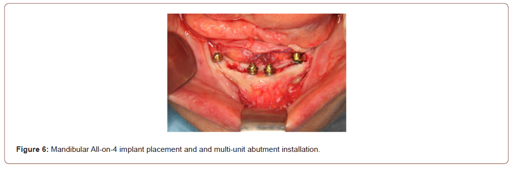
The mandible was rehabilitated with four conventional implants (J Evolution, J Dental Care) placed according to the Allon- 4 concept: two anterior implants were positioned axially, and two posterior implants were tilted to maximize the anteroposterior spread. Primary stability compatible with immediate loading was achieved for all implants, and multi-unit abutments (Conical, J Dental Care) were connected according to the restorative plan (Figure 6).
Photogrammetry and Intraoral Scanning
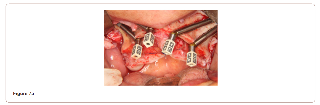
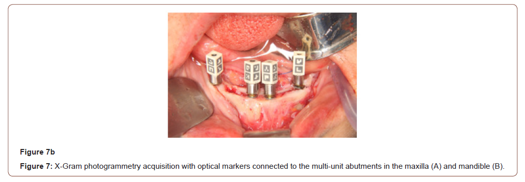
Following connection of the multi-unit abutments, optical markers (X-Gram, Runyes Medical Instrument Co., Ltd.) were fitted onto the abutments with the flaps retracted, and an extraoral photogrammetry scan was acquired (X-Gram, Runyes Medical Instrument Co., Ltd.). The photogrammetry system recorded the three-dimensional position and orientation of all implants simultaneously in less than 10 seconds, enabling accurate capture of the implant relationships despite the severe divergence associated with zygomatic implant placement (Figure 7).
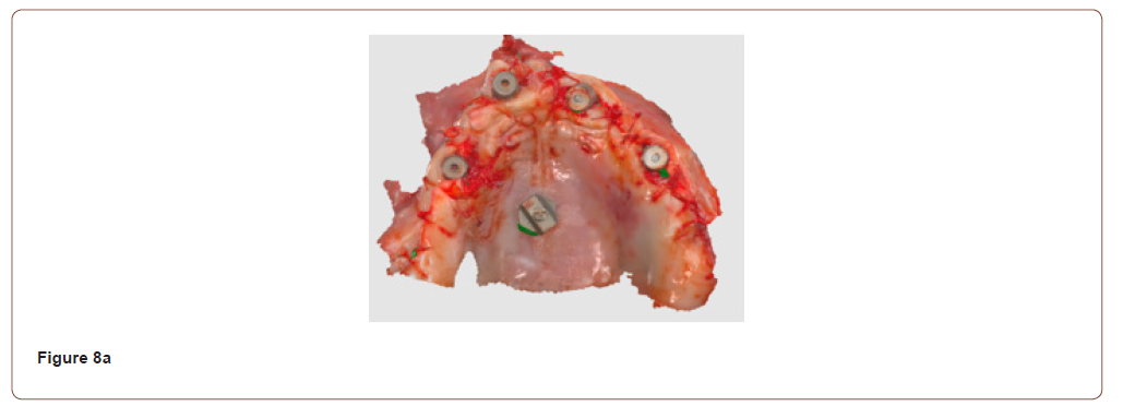
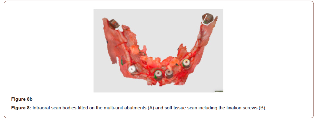
Intraoral scan bodies (X-Gram, Runyes Medical Instrument Co., Ltd.) were then fitted on the multi-unit abutments, the flaps were sutured, and a soft tissue scan was obtained with the intraoral scanner, including the fixation screws (Figure 8). The scan captured the soft tissue morphology, vestibular anatomy, occlusal relationships, and opposing arch information.
Data Integration and CAD/CAM Design
The standard tessellation language (STL) data from the intraoral scans and the implant positions recorded by the photogrammetry system were imported into a CAD software (Exocad, Exocad GmbH, Darmstadt, Germany) and aligned using the fixation screws and common geometric landmarks to generate a complete virtual patient model (Figure 9). This approach combined the implant positional accuracy of photogrammetry with the soft tissue anatomy and functional occlusal information of the intraoral scans, providing the foundation for all subsequent prosthetic design steps.
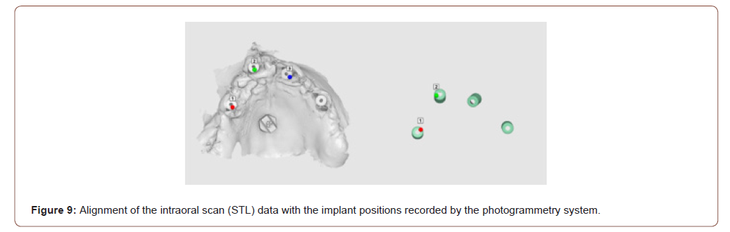
Photogrammetry Acquisition after following installation of the abutments, and the photogrammetry scan bodies were connected to all implants, in less than 10 seconds the X-Gram photogrammetry system was utilized to record the three-dimensional spatial relationships among all implants and this technology enabled simultaneous capture of implant positions despite the severe divergence associated with zygomatic implant placement, needing after these step, the acquisition of the intraoral images by intraoral Scanning and a complete intraoral scan was subsequently obtained using a digital scanner with the objective to the scan captured the soft tissue morphology, the vestibular anatomy, the occlusal relationships and the opposing arch information.
These data integration of photogrammetry and intraoral scanning datasets were imported into Exocad. Using common geometrical landmarks, both datasets were merged to generate a complete virtual patient model, and this approach combined all implant positional accuracy from photogrammetry, the soft tissue anatomy from intraoral scanning and the functional occlusal information, delivering in an CAD/CAM Design to perform a digital prosthetic planning including, the verification prostheses, the immediate provisional restorations, the definitive titanium framework design and finally the zirconia prosthetic superstructure design all the frameworks were manufactured using CAD/CAM technology.
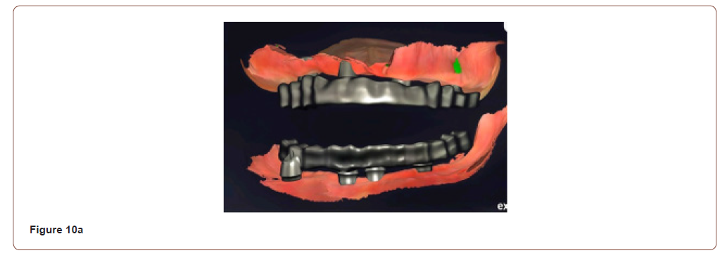
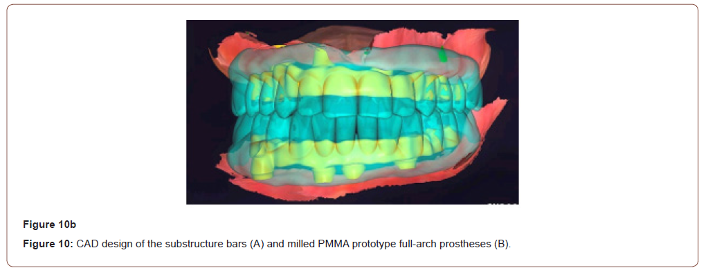
Substructure bars were designed in the CAD software, and maxillary and mandibular prototype full-arch fixed prostheses were designed and milled with a 5-axis milling machine (S2, vhf camfacture AG, Ammerbuch, Germany) from prefabricated polymethyl methacrylate (PMMA) discs (Multilayer PMMA Block A2, Huge Dental Material Co., Rizhao City, China) (Figure 10).
Prototype Try-in and Definitive Prostheses
Five days after surgery, the maxillary and mandibular prototype full-arch fixed prostheses designed in CAD (Figure 11) were secured in the patient’s mouth to assess occlusion, aesthetics, and phonetics, and minor occlusal adjustments were made.
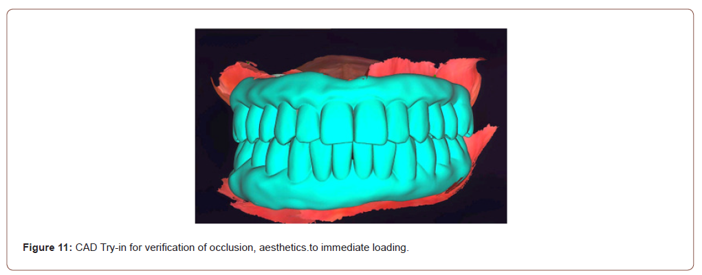
The adjusted prototypes were then rescanned with the intraoral scanner, including the neighbouring oral mucosa, and a new digital bite registration was recorded. The STL files were sent to the dental laboratory, and the definitive restorations were designed virtually in the CAD software.
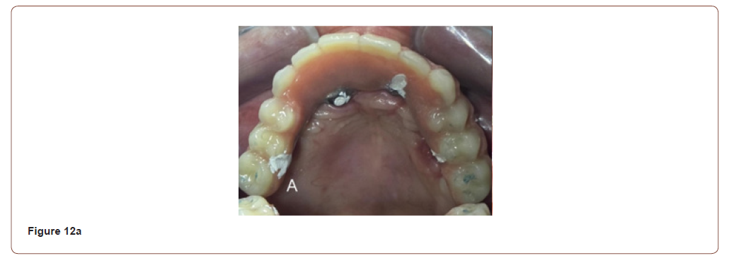
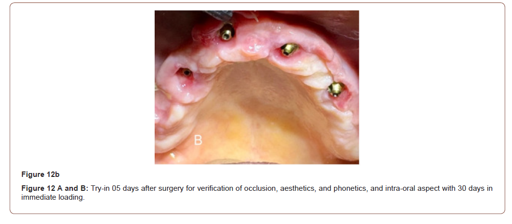
The definitive prostheses were milled with the 5-axis milling machine from prefabricated zirconia discs [zirconia disc brand, manufacturer, city, country-to be specified] and reinforced with milled titanium bars. The definitive prostheses were fitted at the manufacturer-recommended torque of 20 Ncm (Figure 13a), and a postoperative panoramic radiograph confirmed adequate fit of the prostheses and fully seated prosthetic screws (Figure 13b).
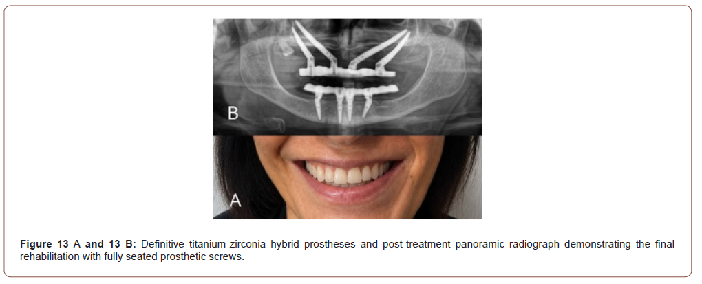
Post-treatment panoramic radiograph demonstrating the final rehabilitation with fully seated prosthetic screws
Passive adaptation of the prostheses was clinically verified, and immediate loading was successfully achieved. The final titaniumzirconia hybrid prostheses demonstrated stable occlusion, favorable aesthetics, and functional rehabilitation, with high patient satisfaction. No biological or prosthetic complications were observed [follow-up period-to be specified]. Special consideration was given to the emergence profile of the zygomatic implants, prosthetic screw access trajectory, framework design, and future maintenance requirements. The objective was not only to achieve implant survival but also to optimize long-term prosthetic function, hygiene accessibility, and patient satisfaction. One of the major advantages of the photogrammetry-based workflow was the ability to accurately record implant positions despite the extreme divergence between the anterior and posterior zygomatic implants. In complete-arch rehabilitations, particularly those involving Quad Zygoma configurations, the cumulative effect of small registration errors may become clinically significant due to the long-span nature of the prosthetic framework.
Discussion
The use of photogrammetry reduced dependence on sequential image stitching and provided a highly reliable digital representation of the implant platform positions. The subsequent integration of intraoral scanning and photogrammetry data created a hybrid digital workflow capable of combining the strengths of both technologies. While photogrammetry provided highly accurate implant coordinates, intraoral scanning contributed soft tissue morphology, occlusal information, and prosthetically relevant anatomical details. The resulting virtual patient model served as the foundation for CAD/CAM framework design and facilitated communication between the surgical and laboratory teams throughout the restorative process. The photogrammetry workflow enabled accurate recording of implant positions in both arches. The digital merge process facilitated reliable integration between implant coordinates and soft tissue anatomy. Passive adaptation of the provisional prostheses was clinically verified. Immediate loading was successfully achieved. The final titanium-zirconia hybrid prostheses demonstrated stable occlusion, favorable esthetics, functional rehabilitation, passive adaptation and patient satisfaction. No immediate biological or prosthetic complications were observed. The accurate transfer of implant positions remains one of the most critical determinants of success in full-arch implant-supported prosthodontics and the challenge becomes considerably greater in Quad Zygoma rehabilitations due to long implant lengths, extreme implant divergence, extended prosthetic spans and complex emergence profiles [27]. While contemporary intraoral scanners have demonstrated excellent performance in short-span rehabilitations, concerns remain regarding cumulative stitching errors in complete-arch implant scans, in which the increased distance between scan bodies and the complexity of the scanning path may lead to clinically relevant discrepancies in framework adaptation [28]. This distinction becomes particularly relevant when treating zygomatic implant cases [19]. The integration of photogrammetry and intraoral scanning creates a hybrid workflow that combines implant accuracy with detailed soft tissue representation [29].
Photogrammetry offers a fundamentally different approach by recording implant relationships simultaneously rather than sequentially, thereby reducing the risk of distortion associated with complete-arch image stitching [22]. In the present case, one of the major advantages of the photogrammetry-based workflow was the ability to accurately record implant positions despite the extreme divergence between the anterior and posterior zygomatic implants, and the exteriorized zygomatic technique contributed to favourable prosthetic emergence and framework design, reinforcing the restorative advantages previously described in the literature [30]. Although limited by the nature of a singlecase report, the findings support the growing body of evidence suggesting that photogrammetry may become an important component of advanced digital workflows for full-arch implant rehabilitation [7]. This illustrates how surgical design and prosthetic planning become increasingly interconnected within a fully digital workflow, where implant positioning directly influences restorative predictability [16]. The advantages of the exteriorized zygomatic technique extend beyond surgical execution and have direct implications for prosthetic rehabilitation by improving the prosthetic emergence profile and positioning implant platforms closer to the ideal restorative envelope, the technique facilitates CAD/CAM framework design and may reduce cantilever length [20,19]. Furthermore, improved implant emergence often results in more favourable prosthetic contours, enhanced oral hygiene access, and simplified long-term maintenance [9].
This illustrates how surgical design and prosthetic planning become increasingly interconnected within a fully digital workflow, where implant positioning directly influences restorative predictability [8], while the addition of a zirconia superstructure provides enhanced esthetics, wear resistance, and biocompatibility without compromising the structural stability of the metallic framework (26). Within a digital workflow, CAD/CAM manufacturing allows precise control of framework dimensions and restorative contours, supporting the objective of achieving passive fit and biomechanical stability across the entire prosthetic arch [19]. The present case illustrates how surgical design and prosthetic planning become increasingly interconnected within a fully digital workflow, where implant positioning directly influences restorative predictability [16]. An additional consideration involves the relationship between implant divergence and digital impression accuracy, while modern intraoral scanners have demonstrated excellent performance in short-span applications, complete-arch rehabilitations involving zygomatic implants present unique challenges due to the increased distance between scan bodies and the complexity of the scanning path [13]. Another relevant aspect of the present case is the role of digital technologies in improving communication among the multidisciplinary team involved in treatment planning and execution [7]. Small cumulative registration errors may lead to clinically relevant discrepancies in framework adaptation and photogrammetry addresses this limitation by capturing implant relationships independently of scanning sequence, thereby reducing the risk of distortion associated with complete-arch image stitching [22].
The findings of the present report are consistent with the growing trend toward hybrid digital workflows in implant dentistry [31]. The integration of photogrammetry, intraoral scanning, and CAD/CAM design enabled surgeons, prosthodontists, and dental technicians to work from a common digital dataset throughout all treatment phases (7), reducing the need for multiple analog verification steps and facilitating a predictable transition from surgical implant placement to definitive prosthetic rehabilitation [15]. Rather than replacing intraoral scanning, photogrammetry complements it by providing highly accurate implant position information while allowing the scanner to focus on soft tissue anatomy and occlusal relationships [7]. This combination appears particularly advantageous in cases involving Quad Zygoma rehabilitation, where passive fit and prosthetic precision are essential determinants of long-term clinical success [32]. In complex cases involving zygomatic implants, where prosthetic limitations may directly influence surgical planning, the availability of accurate digital information becomes particularly valuable, and this report describes a single clinical case and therefore does not permit statistical analysis and long-term clinical follow-up and controlled prospective investigations remain necessary to establish the precise impact of photogrammetry on prosthetic accuracy, complication rates, and patient-centered outcomes [19].
The concept of passive fit has long been considered a fundamental requirement for the longevity of implant-supported restorations [28]. In full-arch rehabilitations, inaccuracies during implant position transfer may be amplified by the length of the prosthetic framework, increasing the risk of biomechanical complications [22]. Recent systematic reviews have suggested that stereophotogrammetry may achieve superior trueness and precision compared with conventional and intraoral digital impression techniques in complete-arch implant cases [31,27,33]. These findings support the growing adoption of photogrammetry as a valuable adjunct in advanced implant prosthodontics, particularly when multiple implants and complex angulations are present [34]. Another relevant aspect of the present case is the role of digital technologies in improving communication among the multidisciplinary team involved in treatment planning and execution [9]. The integration of photogrammetry, intraoral scanning, and CAD/CAM design enabled surgeons, prosthodontists, and dental technicians to work from a common digital dataset throughout all treatment phases [7].
This approach reduced the need for multiple analog verification steps and facilitated a more predictable transition from surgical implant placement to definitive prosthetic rehabilitation [15]. In complex cases involving zygomatic implants, where prosthetic limitations may directly influence surgical planning, the availability of accurate digital information becomes particularly valuable [19]. The use of a titanium-zirconia hybrid restoration may also have contributed to the favourable clinical outcome observed in this case because the titanium frameworks continue to be considered the benchmark for full-arch implant-supported prostheses due to their excellent mechanical properties, passive adaptation potential, and long-term clinical performance [8]. The addition of a zirconia superstructure provides enhanced esthetics, wear resistance, and biocompatibility while maintaining the structural stability of the metallic framework [26]. Within a digital workflow, CAD/CAM manufacturing allows precise control of framework dimensions and restorative contours, supporting the objective of achieving passive fit and biomechanical stability across the entire prosthetic arch [8,19]. The findings of this report are consistent with the growing trend toward hybrid digital workflows in implant dentistry [26]. Rather than replacing intraoral scanning, photogrammetry complements it by providing highly accurate implant position information while allowing the scanner to focus on soft tissue anatomy and occlusal relationships [7]. This combination appears particularly advantageous in Quad Zygoma rehabilitations, where passive fit and prosthetic precision are essential determinants of long-term clinical success [32].
From a clinical perspective, the combination of Quad Zygoma rehabilitation and mandibular All-on-4 treatment represents one of the most demanding scenarios in implant prosthodontics [35]. The simultaneous management of long-span restorations, implant divergence, immediate loading requirements, and esthetic demands requires a highly coordinated workflow [32]. The present case suggests that photogrammetry may serve as an important tool for reducing uncertainty during implant position transfer and framework fabrication [7]. Although additional prospective clinical studies are necessary to validate these findings, the integration of photogrammetry into full-arch digital protocols appears to align with the ongoing evolution of implant dentistry toward increasingly accurate, efficient, and patient-centred treatment solutions. Nevertheless, this report describes a single clinical case and therefore does not permit statistical analysis. Long-term clinical follow-up and controlled prospective investigations remain necessary to establish the precise impact of photogrammetry on prosthetic accuracy, complication rates, and patient-centred outcomes.
Conclusion
The combination of extraoral photogrammetry, intraoral scanning, CAD planning, and CAD/CAM manufacturing enabled a fully digital rehabilitation of a patient treated with Quad Zygoma implants in the maxilla and an All-on-4 protocol in the mandible. Photogrammetry facilitated accurate implant position transfer despite significant implant divergence and contributed to the fabrication of passively fitting titanium-zirconia hybrid restorations. This digital workflow appears to represent a promising strategy for highly complex implant-supported full-arch rehabilitations, although prospective clinical studies with long-term follow-up are required to validate these findings.
Acknowledgement
None.
Conflict of interest
No conflict of interest.
References
- Sbordone C, P Toti, F Guidetti, L Califano, A Santoro, et al. (2012) Volume changes of iliac crest autogenous bone grafts after vertical and horizontal alveolar ridge augmentation of atrophic maxillas and mandibles: a 6-year computerized tomographic follow-up. J Oral Maxillofac Surg 70(11): 2559-2565.
- Widmark G, B Andersson, GE Carlsson, AM Lindvall, CJ Ivanoff (2001) Rehabilitation of patients with severely resorbed maxillae by means of implants with or without bone grafts: a 3- to 5-year follow-up clinical report. Int J Oral Maxillofac Implants 16(1): 73-79.
- Aparicio C, W Ouazzani, R Garcia, X Arevalo, R Muela, et al. (2006). A prospective clinical study on titanium implants in the zygomatic arch for prosthetic rehabilitation of the atrophic edentulous maxilla with a follow-up of 6 months to 5 years. Clin Implant Dent Relat Res 8(3): 114-122.
- Aparicio C, W Ouazzani, A Aparicio, V Fortes, R Muela, et al. (2010) Extrasinus zygomatic implants: three-year experience from a new surgical approach for patients with pronounced buccal concavities in the edentulous maxilla. Clin Implant Dent Relat Res 12(1): 55-61.
- Migliorança R, JP Ilg, AS Serrano, RP Souza, MS Zamperlini (2006) Sinus exteriorization of the zygoma fixtures: a new surgical protocol. Implant News 3(5): 30-35.
- Grossi JRA, L Perussolo, E Meurer, C Dalmolin, CVB (2023) Digital Panning and Printed Guides for Zygomatic implants. Brazilian Digital Dentistry Society Book. F. Andretti. Brazil Brazilian Digital Dentistry Society Book. 04.
- Clozza E (2024) Intraoral scanning and dental photogrammetry for full-arch implant-supported prosthesis: A technique. Clin Adv Periodontics 14(4): 244-249.
- Conejo J, M Ordway, TH Yoo, HP Fraiman, PJ Atria, et al. (2025) In Vitro Comparison Between Complete-Arch Implant Supported Frameworks Using Intraoral Scanning and Photogrammetry. Int J Prosthodont 39(7): 95-101.
- Bedrossian E, E A Bedrossian (2025) Full Mouth Reconstruction with Dental Implants: Planning, Surgical, and Prosthetic Phase. Dent Clin North Am 69(2): 257-274.
- Revilla-Leon M, M Gomez-Polo, M Drone, AB Barmak, C Guinot-Barona, et al. (2024) Impact of scanning distance on the accuracy of a photogrammetry system. J Dent 142: 104854.
- Gomez-Polo M, R Cascos, R Ortega, AB Barmak, JC Kois, et al. (2024) Influence of scanning pattern on accuracy, time, and number of photograms of complete-arch implant scans: A clinical study. J Dent 150: 105310.
- Negreiros WM, TC Sun, S Jain, M Finkelman, GO Gallucci, et al. (2025) Precision of complete-arch digital implant scans using photogrammetry and intra-oral scanning. An in vivo cross-over study: Complete-Arch Implant Scans: Photogrammetry vs IOS. J Dent 161: 105928.
- Lyu M, Y Li, D Xu, Q Xing, S Zhang, et al. (2025) Accuracy of Photogrammetry, Intraoral Scanning, and Conventional Impression for Multiple Implants: An In Vitro Study. Clin Implant Dent Relat Res 27(1): e13419.
- Muslu T, S Kurtulmus-Yilmaz (2025) Effect of implant angulation on the trueness and precision of intraoral scanning and photogrammetry. BMC Oral Health 25(1): 1739.
- Yao S, X Yang, X Han, Y Xue, L He, et al. (2025) Immediate full-arch rehabilitation of edentulous jaws on 4 or 6 implants using a photogrammetry system: A retrospective study up to 2 years of follow-up. J Stomatol Oral Maxillofac Surg 126(4S): 102202.
- De Araujo Nobre M, R Almeida, C Moura Guedes, G Alvarez, C Antunes, et al. (2025) Digital Workflow for Interim Prosthetic Rehabilitation Through the All-on-4 Concept Using 3D Printing Additive Process. J Clin Med 14(23): 8353.
- Nulty AB, C Kelly, O Ambridge, M Ambridge, R Ferguson, et al. (2025) In Vitro Comparison of Trueness and Precision of an AI-Driven Real-Time Library Matching Protocol with Irregular Geometry Scan Bodies for Full-Arch Implant Scanning. Dent J (Basel) 13(11): 533.
- Eldabe AK, D Adel-Khattab, KH Botros (2026) Accuracy of intraoral photogrammetry in complete arch digital implant scanning: An in vivo prospective comparative study. J Prosthet Dent 135(2): 342-351.
- Gercek ABK, AO Gercek (2026) How do zygomatic implant configurations and prosthetic framework materials influence the biomechanics of quad zygoma rehabilitation in severely atrophic maxilla? J Prosthodont.
- Miglioranca RM, BS Sotto-Maior, PM Senna, CE Francischone, AA Del Bel Cury (2012) Immediate occlusal loading of extrasinus zygomatic implants: a prospective cohort study with a follow-up period of 8 years. Int J Oral Maxillofac Surg 41(9): 1072-1076.
- Yuan Z, Z Zhou, S Ge, Y Yang, J Zhu, et al. (2026) Comparing the accuracy of three digital impression techniques with conventional impression technique for implant-supported fixed full-arch restorations: A prospective clinical study. Am J Dent 39(1): 25-30.
- Mangano FG, H Lerner, G Valle, F Biaggini, C Mangano, et al. (2026) Trueness of extraoral photogrammetry, intraoral photogrammetry and direct intraoral scanning in the full-arch digital implant impression: a comparative in vitro study. J Dent 165: 106269.
- Zhou Y, L You, Z Fan (2024) Accuracy of implant abutment level digital impressions using stereophotogrammetry in edentulous jaws: an in vitro pilot study. BMC Oral Health 24(1): 1167.
- Betita T, DJ Rusthoven (2025) The Digital Full-Arch Solution Plus Photogrammetry for Guided Patient Success in All-On-X Cases. Oral Maxillofac Surg Clin North Am 37(2): 213-233.
- Dawoud Y, HS Eid, OE Sadat, D Essam, RM Ghali (2026) Evaluation of Passivity of Co-Cr Complete Arch Screw Retained Maxillary Prostheses Using Different Intraoral Scanners with the Aid of Auxiliary Geometric Device. Int J Oral Maxillofac Implants 0(0): 1-22.
- Pozzi A, E Agliardi, F Lio, K Nagy, A Nardi, et al. (2024) Accuracy of intraoral optical scan versus stereophotogrammetry for complete-arch digital implant impression: An in vitro study. J Prosthodont Res 68(1): 172-180.
- Alfaraj AF, Alqudaihi Z, Khurshid O, Qadiri, WS Lin (2026) Comparative analyses of accuracy between digital and conventional impressions for complete-arch implant-supported fixed dental prostheses-A systematic review and meta-analysis. J Prosthodont 35(3): 252-274.
- DeFee M, W Renne, MA Shazib, A Queiroz, A Mennito, et al. (2026) Virtual-fit assessment of full-arch implant photogrammetry accuracy in a human cadaver model. J Dent 170: 106668.
- Cheng J, H Zhang, H Liu, J Li, H L Wang, et al. (2024) Accuracy of edentulous full-arch implant impression: An in vitro comparison between conventional impression, intraoral scan with and without splinting, and photogrammetry. Clin Oral Implants Res 35(5): 560-572.
- Fu XJ, ZZ Cai, JY Shi, SC Qiao, MS Tonetti, et al. (2025) Accuracy of a Novel Intraoral Photogrammetry Technique for Complete-Arch Implant Impressions: An In Vitro Study. Clin Oral Implants Res 36(8): 991-999.
- Pozzi A, L Arcuri, P Carosi, A Laureti, J Londono, et al. (2025) Photogrammetry Versus Intraoral Scanning in Complete-Arch Digital Implant Impression: A Systematic Review and Meta-Analysis. Clin Implant Dent Relat Res 27(3): e70059.
- Tezerisener HA, O Ozalp, MA Altay, A Sindel (2024) Comparison of stress distribution around all-on-four implants of different angulations and zygoma implants: a 7-model finite element analysis. BMC Oral Health 24(1): 176.
- Noronha CP, C Ferreira de Souza, M Dantas Dos Santos Oliveira, E Silva, N Sesma, et al. (2026) Accuracy of Stereophotogrammetry in Implant Scanning Compared to Intraoral Scanning in Completely Edentulous Patients-A Systematic Review and Meta-analysis. Int J Prosthodont 39(3): 397-404.
- Del Fabbro M, A Pozzi, D Romeo, M De Araujo Nobre, E Agliardi (2022) Outcomes of Fixed Full-Arch Rehabilitations Supported by Tilted and Axially Placed Implants: A Systematic Review and Meta-Analysis. Int J Oral Maxillofac Implants 37(5): 1003-1025.
- Yolal MS, A Ozkan, O Yildirim, M Sencimen, U Karacayli (2026) Biomechanical evaluation of All-on-4, M-4, V-4, and zygomatic implant concepts in the rehabilitation of the atrophic maxilla: a finite element analysis. J Stomatol Oral Maxillofac Surg 127(6): 102857.
-

This work is licensed under a Creative Commons Attribution-NonCommercial 4.0 International License.